Skip to main content Accessibility help
×
  • Cited by 4
Publisher:
Cambridge University Press
Online publication date:
October 2013
Print publication year:
2013
Online ISBN:
9781139207294

Book description

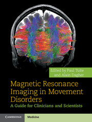
Magnetic Resonance Imaging in Movement Disorders is the first book to focus in detail on MRI in a range of movement disorders. Since MRI was first employed in imaging Parkinson's disease, the number of imaging techniques and their application in diagnosis and management has extended widely. The book shows various imaging strategies ranging from functional, structural and chemical methods as they relate to both motor and non-motor aspects of Parkinson's disease and other conditions such as Huntington's disease and dystonia. Chapters on MRI in surgery and using MRI as a potential outcome measure in clinical trials show the clinical relevance of methods. Novel methods including DTI, tractography and resting case studies are described in detail. The book also summarises the relevance of fMRI to various aspects of movement disorders. Magnetic Resonance Imaging in Movement Disorders is essential reading for neurologists, radiologists and movement disorder specialists.

Refine List

Actions for selected content:

Select all | Deselect all
  • View selected items
  • Export citations
  • Download PDF (zip)
  • Save to Kindle
  • Save to Dropbox
  • Save to Google Drive

Save Search

You can save your searches here and later view and run them again in "My saved searches".

Please provide a title, maximum of 40 characters.
×

Contents

  • Chapter 8 - MRI for targeting in surgical treatment of movement disorders
    pp 94-105
  • View abstract

    Summary

    This chapter provides an overview of magnetic resonance imaging (MRI) methods. The focus on iron in Parkinson's disease (PD) imaging has remained an important topic and researchers have often utilized T2*, or its reciprocal R2*, in nigral imaging protocols. Some iron-sensitive methods have been recently developed. These include adiabatic T2ρ, magnetization transfer (MT) imaging, and susceptibility-weighted imaging (SWI). The authors have developed a novel rotating frame relaxation experiment called relaxation along a fictitious field (RAFF). There has been greater refinement with the utilization of methods that do not employ a-priori regions of interest (ROIs). One such method is voxel-based morphometry (VBM), in which there is standardization of data and then voxel-by-voxel comparison between group data to determine if there are differences in signal intensity. Diffusion tensor imaging (DTI) provides structural data based on the directionally restrained diffusion of water within fiber tracts.
  • Chapter 9 - Magnetic resonance imaging and spectroscopy in Huntington’s disease
    pp 106-120
  • View abstract

    Summary

    Iron content is one of the physiological variables that can be estimated with magnetic resonance imaging (MRI) in the basal ganglia of patients with Parkinson's disease (PD). Brain iron is relatively independent from total body iron content since it is excluded by the blood-brain barrier. The microstructural and physiological organization of tissue plays an important role in determining the local magnetic field behavior of a given region in the brain. Recent work has extended previous observations by using a multimodal approach that combines MRI techniques for imaging iron content with other MRI sequences. MR-based measurements that are directly related to magnetic susceptibility changes should be closely related to iron content and less dependent on its microscopic spatial distribution. Susceptibility-weighted imaging (SWI) is a technique that uses magnetic susceptibility differences between different regions to generate image contrast. Deep brain stimulation (DBS) plays an important role in the treatment of PD.
  • Chapter 10 - Neuroimaging of Gilles de la Tourette syndrome
    pp 121-133
  • View abstract

    Summary

    This chapter reviews magnetic resonance (MR) techniques from conventional structural MRI to advanced MRI (volumetry, magnetization transfer (MT), neuromelanin imaging, diffusion imaging, and rs-fMRI). It outlines the ways in which these techniques may be used to detect changes in the brain of Parkinson's disease (PD) patients and their relationships with Parkinsonian symptoms. Functional connectivity (FC) methods that take advantage of intrinsic signal fluctuations have demonstrated that the interactions of brain networks are abnormal in PD at the resting state. MRI has proven useful in the differential diagnosis of the various atypical Parkinsonian disorders such as progressive supranuclear palsy (PSP) and the Parkinson variant of multiple system atrophy (MSA-P). In PD, changes in the basal ganglia and brainstem are subtle and restricted to nuclei such as the substantia nigra (SN) and locus coeruleus (LC). PSP patients present extensive changes in the brainstem, basal ganglia, and cortical regions.
  • Chapter 11 - Dystonia
    pp 134-146
  • View abstract

    Summary

    This chapter reviews relevant magnetic resonance imaging (MRI) methods and their application to various stages of disease including early or incident Parkinson's disease (PD), treated patients, including those with mild cognitive impairment (MCI), and those with Parkinson's disease with dementia (PDD) and dementia with Lewy bodies (DLB). In PD pathological changes have been found in the hippocampus, parahippocampus, amygdala, and the entorhinal cortex. Several groups of investigators have shown that the substantia innominata is atrophic in PD, with progressive loss in PD-MCI and PDD. The chapter also reviews longitudinal studies and the impact of white matter pathology. A study that compared progression of white matter changes between Alzheimer's disease (AD), PDD, and DLB and older controls found that AD patients had more white matter changes than controls; there was no difference in progression of white matter changes between the dementia groups.
  • Chapter 12 - Tauopathies
    pp 147-166
  • View abstract

    Summary

    This chapter reviews functional imaging studies, including those with functional magnetic resonance imaging (fMRI), and nuclear tracing studies such as positron emission tomography (PET) and single-photon emission computed tomography (SPECT) scanning as they relate to motor signs of Parkinson's disease (PD). fMRI of healthy controls (HC) performing unilateral hand movements typically identifies cerebral correlates in the motor system. To examine the functional changes occurring with learning of automatic finger movements, T. Wu and M. Hallett performed fMRI on a group of individuals with moderate or advanced PD before and after practicing a finger-sequence. To study cerebral correlates of increasing demand by dual-task performance, they also introduced two secondary distractor tests in addition to the sequential finger movements during the same experimental session. A whole brain analysis demonstrated a significant increase of the blood oxygen level dependent (BOLD) signal in the posterior mid-mesencephalon in PD with freezing.
  • Chapter 13 - Magnetic resonance imaging of multiple system atrophy
    pp 167-203
  • View abstract

    Summary

    Parkinson's disease (PD) is associated with cognitive and emotional disturbances, whose response to dopamine therapy is either absent or, occasionally, deleterious to the patient. By measuring cerebral blood flow (CBF), first with positron emission tomography (PET) and now with functional magnetic resonance imaging (fMRI), researchers have mapped the neurobiological substrates of cognitive and behavioral symptoms in PD. Basal ganglia processing is based on the anatomy of parallel cortico-striatal loops, each of which has a functional role. Dopaminergic overmedication may lead to cognitive impairment via an action on the relatively preserved mesocortical or mesolimbic systems. The overmedication hypothesis has been rekindled recently with the description of disorders of motivation and impulse control in PD. fMRI provides evidence that dopamine may contribute to impulse control disorders (ICDs) in PD via its role as an energizing or activating agent that assigns incentive value to stimuli and actions.
  • Chapter 14 - Ataxia
    pp 204-228
  • View abstract

    Summary

    Appropriate analysis of resting-state functional magnetic resonance imaging (rs-fMRI) allows the description of spontaneous networks of interaction known as resting-state networks (RSNs). Number of studies has revealed that the normally observed RSNs are frequently and significantly disrupted in neurological disorders. Several analysis methods have been proposed to assess functional connectivity from rs-fMRI. Assessing the functional connectivity between two brain loci can either occur at the individual voxel or the region of interest (ROI) level. The most straightforward method of analysis for functional connectivity is the seed-based or ROI-based method. Despite the extensive application of resting-state imaging modalities to brain disorders, relatively fewer studies have assessed altered connectivity in Parkinson's disease (PD). rs-fMRI studies have described RSNs not only related to motor symptoms, but also to non-motor features of PD, such as cognitive dysfunction. Apathy and depression have been assessed in PD using resting state.
  • Chapter 15 - Magnetic resonance spectroscopy in Parkinson’s disease
    pp 229-237
  • View abstract

    Summary

    Three specific deep brain stimulation (DBS) targets are currently in widespread use for the treatment of specific movement disorders. These are ventral intermediate nucleus (VIM) for the treatment of tremor (Parkinsonian or essential), subthalamic nucleus (STN) for Parkinson's disease (PD), and globus pallidus internus (GPi) for PD and for dystonia. All DBS surgeries currently require some form of stereotaxy, which is a means of translating information from two-dimensional (2D) brain images to the three-dimensional (3D) operative field. Two types of stereotactic approaches are available for DBS surgery. The success of DBS surgery is critically dependent on accurate localization of the target structure of interest. Two different approaches to initial target localization exist, and frequently are combined in the operating room: indirect and direct targeting. Ultra-high-field magnetic resonance imaging (MRI) systems are proving invaluable for basic science research and neuroscience applications.
  • Chapter 16 - The image of essential tremor: current neuroimaging and clues to disease localization, pathogenesis, and diagnosis
    pp 238-245
  • View abstract

    Summary

    This chapter focuses on the main findings and advances brought by magnetic resonance imaging (MRI) and magnetic resonance spectroscopy (MRS) to the understanding of prodromal and early-stage patients. In Huntington's disease (HD), as in many neurodegenerative disorders, accurate markers of disease progression that reflect pathogenic mechanisms are currently lacking, and therefore are an important focus of current research. MR techniques are a particularly promising tool in the identification of such biomarkers. Using MRI, cerebral blood flow (CBF) or perfusion can be quantitatively measured using a technique named arterial spin labeling (ASL). For measuring brain activity, another technique can be used, namely blood oxygen level-dependent (BOLD) imaging. MRI is based on the detection of the proton signal within water molecules. Finally, the chapter reviews new approaches used in animal models that will hopefully be translated in clinical studies in the near future.
  • Chapter 17 - Clinical applications
    pp 246-263
  • View abstract

    Summary

    This chapter describes the clinical phenomenology and pathophysiology of Gilles de la Tourette syndrome (GTS) and reviews current structural and functional neuroimaging data of this fascinating neuropsychiatric condition. Significant neuroimaging evidence exists for a primary cortical dysfunction in GTS. Structural changes were reported in the basal ganglia, including both the striatum and the globus pallidus. Diffusion tensor imaging (DTI) has pointed to the microstructural abnormalities in white matter in GTS patients, including the corpus callosum and anterior and posterior limb of the internal capsule. Defects in brain maturation could be one of the pathophysiological mechanisms that lead to emergence of the GTS symptoms in childhood and their persistence into adulthood. Evidence from pharmacological trials, especially the fact that dopamine receptor blockers are the most effective treatment for tics to date, and postmortem analyses suggested that abnormalities of dopaminergic neurotransmission play a key role in the pathogenesis of GTS.
  • Chapter 18 - MRI in clinical trials
    pp 264-270
  • View abstract

    Summary

    This chapter focuses on various functional magnetic resonance imaging (fMRI), voxel-based morphometry (VBM), and diffusion tensor imaging (DTI) studies in patients with (focal) dystonia, which have shown functional and structural changes within subcortical-cortical sensorimotor networks. An important role of a dysfunction of somatosensory processing has been more and more recognized over the past years and has become one of the main focuses of research in dystonia. One important issue when investigating the somatosensory system with fMRI is the standardization of sensory stimuli. Resting-state fMRI studies of patients with action-induced dystonia can further add to the understanding of the disorder. A recent resting-state MRI study investigated patients with writer's cramp (WC) and applied independent component analyses (ICA) to study functional connectivity of different functional networks at rest. Based on possible genetic predisposition these functional and structural changes could lead to the clinical presentation of dedifferentiated motor programs in dystonia.

Metrics

Full text views

Total number of HTML views: 0
Total number of PDF views: 0 *
Loading metrics...

Book summary page views

Total views: 0 *
Loading metrics...

* Views captured on Cambridge Core between #date#. This data will be updated every 24 hours.

Usage data cannot currently be displayed.