

Abstract
Copper nanoparticles, due to their interesting properties, low cost preparation and many potential applications in catalysis, cooling fluid or conductive inks, have attracted a lot of interest in recent years. In this study, copper nanoparticles were synthesized through the chemical reduction of copper sulfate with sodium borohydride in water without inert gas protection. In our synthesis route, ascorbic acid (natural vitamin C) was employed as a protective agent to prevent the nascent Cu nanoparticles from oxidation during the synthesis process and in storage. Polyethylene glycol (PEG) was added and worked both as a size controller and as a capping agent. Cu nanoparticles were characterized by Fourier transform infrared (FT-IR) spectroscopy to investigate the coordination between Cu nanoparticles and PEG. Transmission electron microscopy (TEM) and UV–vis spectrometry contributed to the analysis of size and optical properties of the nanoparticles, respectively. The average crystal sizes of the particles at room temperature were less than 10 nm. It was observed that the surface plasmon resonance phenomenon can be controlled during synthesis by varying the reaction time, pH, and relative ratio of copper sulfate to the surfactant. The surface plasmon resonance peak shifts from 561 to 572 nm, while the apparent color changes from red to black, which is partly related to the change in particle size. Upon oxidation, the color of the solution changes from red to violet and ultimately a blue solution appears.
Export citation and abstract BibTeX RIS

Content from this work may be used under the terms of the Creative Commons Attribution-NonCommercial-ShareAlike 3.0 licence. Any further distribution of this work must maintain attribution to the author(s) and the title of the work, journal citation and DOI.
1. Introduction
Interest in copper nanoparticles arises from the useful properties of this metal such as the good thermal and electrical conductivity at a cost much less than silver, for example. This leads to potential application in cooling fluids for electronic systems [1] and conductive inks [2]. Due to plasmon surface resonance, copper nanoparticles exhibit enhanced nonlinear optical properties, which could result in many applications in optical devices and nonlinear optical materials, such as optical switches or photochromic glasses [3]. Furthermore, in this last case, it is possible to expect an interesting effect coming from the depression of the melting temperature of a metal when it has the form of nanoparticles [4]. In this work, we carry out the synthesis of copper nanoparticles with specific attention to their future use in conducting inks.
Most of relevant recent studies on conductive inks have focused on noble metals exempt from significant oxidation, such as silver and gold nanoparticles (silver slightly oxidizes but its oxide is still a good conductor). In particular, silver with its high conductivity is of great interest [5] and has led to much development with commercially available products. However, these noble metals are too expensive to be used in large quantities. In this context, copper is a good candidate material because it is highly conductive but significantly cheaper than Au and Ag. However, copper nanoparticles synthesized in ambient atmospheric temperature and pressure inevitably have surface oxide layers because the Cu oxide phases are thermodynamically more stable than pure Cu. Further, copper particles are found to aggregate severely without proper protection. The problems of aggregation and oxidation can be circumvented by the use of various protecting agents, such as polymers [6–8] and organic ligands [9, 10].
Currently developed synthesis methods for copper nanoparticles include chemical reduction [7–11], thermal decomposition [12, 13], polyol [5, 14], laser ablation [15], electron beam irradiation [16] and an in situ chemical synthetic route [17]. Among these methods, chemical reduction is the most preferred, because this method is simple and economical, and it can realize better size and size distribution control by optimizing the experimental parameters, such as the molar ratio of the capping agent with the precursor salt and the ratio of reducing agent with the precursor salt. A chemical reduction method usually involves the reduction of metal salts in some type of solvent and a separate reducing agent.
We are working on a simple and rapid approach to pure copper nanoparticle preparation via a natural antioxidant—ascorbic acid, with no gas protection. Ascorbic acid is essential to avoid the oxidation of copper nanoparticles during the synthesis process and in storage. The antioxidant properties of ascorbic acid come from its ability to scavenge free radicals and reactive oxygen molecules [11], accompanying the donation of electrons to give the semi-dehydroascorbate radical and dehydroascorbic acid.
2. Experimental
2.1. Material
All of the chemicals were analytical grade and used as purchased without further purification. Copper (II) sulfate pentahydrate salt, CuSO 4·5H 2 O, of 98% purity (Merck), was dissolved in high purity water. Polyethylene glycol 6000 (PEG 6000—Merck) was used as the capping agent. Sodium borohydride (NaBH 4-Reagent Plus 99%, Sigma-Aldrich) was used as the main reducing agent, while ascorbic acid (99.7%, Prolabo) was used as the antioxidant of colloidal copper. Sodium hydroxide NaOH (>98%, China) was also used to adjust the pH and accelerate the reduction reaction in water.
2.2. Synthesis of copper nanoparticles
The four-step preparation scheme for copper nanoparticles starts with dissolving copper (II) sulfate pentahydrate salt, CuSO 4·5H 2 O (0.01 M), in deionized water to obtain a blue solution. Next, polyethylene glycol 6000, PEG 6000 (0.02 M) was dissolved in water and added to the aqueous solution containing the copper salt while vigorously stirring. In this step, the solution changed from blue to white. In the third step, ascorbic acid (0.02 M) and sodium hydroxide (0.1 M) were dissolved in water and added to the synthesis solution. Color change occurred in the aqueous phase from white to yellow. Finally, a solution of NaBH 4 (0.1 M) in deionized water was prepared and added to the solution under continuous rapid stirring. An instant color change occurred in the aqueous phase from yellow to black/red. The appearance of this dark color indicated that the reduction reaction had started. The source of electrons for the reaction was BH − 4. The mixture was further stirred rapidly for around 10 min in ambient atmosphere, to allow the reaction to complete.
2.3. Characterization
Synthesized samples were studied by use of UV–vis absorption spectroscopy from a double beam spectrophotometer (Jasco UV–vis V530) in the wavelength range from 190 to 1100 nm. Transmission electron microscopy (TEM) was used to study particle size. Samples for TEM measurements were suspended in ethanol and ultrasonically dispersed. Drops of the suspensions were placed on a copper grid coated with carbon. Finally FT-IR spectra were recorded (Brucker TENSOR 37 FT-IR spectrophotometer) between 400 and 4000 cm −1 both in solution and after KBr pellets were formed.
3. Results and discussion
3.1. Optical characterization
Small metal nanoparticles exhibit the absorption of visible electromagnetic waves by the collective oscillation of conduction electrons at the surface [18]. This is known as the surface plasmon resonance effect. The interest in this effect is the possibility of using it as a tracer for the presence of metal nanoparticles with a simple UV-visible spectrometer. The size dependence of the plasmon resonance for particles smaller than 20 nm (for gold [18]) is a complex phenomenon. One interesting feature is the increase in the bandwidth of the resonance with the decrease in the size of the particles due to electron scattering enhancement at the surface. The shift in the resonance and the variation in its bandwidth are thus interesting parameters to characterize the metal nanoparticles.
Several samples were taken from the synthesis solution over time: one just after pouring the ascorbic acid solution, the second just before pouring the NaBH 4 solution and then at 5 and 10 min afterwards, as shown in figure 1. Plasmon absorbance (562 nm) appears only when the solution is red (roughly 10 min after the strong reducing agent was added), although absorption already increased after 5 min, which suggests the appearance of small clusters or nanoparticles.

Figure 1 UV–vis spectra of copper nanoparticle synthesis solution at different steps: just after ascorbic acid addition (yellow), 60 min later just before NaBH4 addition (orange), 5 min (light red) and 10 min (red) after NaBH 4 addition.
Before the addition of NaBH 4, the yellow and orange solutions did not show plasmon resonance. Upon the addition of NaBH 4, a quick increase in the absorbance at low wavelengths occurred that probably indicated the onset of particle formation (light red). The plasmon resonance of the Cu nanoparticles appeared at 562 nm when the solution turned red. The reaction was allowed to proceed in air. After the end of synthesis, the solution was kept under ambient atmosphere and the oxidation was qualitatively monitored with time by observation its color change. Within 8 h, the solution turned black to violet and ultimately blue particles appeared (figure 2).
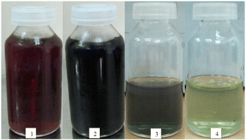
Figure 2 Freshly prepared red Cu sol (1), black (2), violet (3) upon onset oxidation (4).
3.2. Effect of reaction time
Time is a very important parameter in nanoparticle synthesis. As an empirical rule, the availability of a larger number of nuclei at a given time induces a decrease in the nanoparticle size, because smaller metal nuclei grow and consume metal ions at the same time.
To study the effect of the reaction time during synthesis on the formation of product and the stability of copper nanoparticles, all of the samples in table 1 were prepared according to the procedure described, with the only variable being the duration of stirring with ascorbic acid before pouring the sodium borohydride.
Table 1. Plasmon resonance after synthesis and qualitative stability duration as a function of reaction time during synthesis.
| 1 | 15 | 572 | Black | 4 days |
| 2 | 30 | 573 | Black | 2 days |
| 3 | 60 | 562 | Red | 5 days |
| 4 | 90 | – | Black | 4 days |
It can be seen from figure 3 that a reaction time with ascorbic acid of up to 60 min led to well-defined nanoparticles with a decrease in the mean particle size with time. This suggests a homogenization mechanism, which provides a larger number of nuclei with time. For 90 min, no clear resonance was visible despite a clear absorption in an even lower range of wavelength. This could indicate an even smaller particle size.

Figure 3 UV/vis absorption spectra of solution formed with different reaction times.
At the moment, the mechanism associated with this phenomenon is not well understood. Ascorbic acid is well known to scavenge free radicals and thus provide an antioxidant action during copper nuclei formation. This provides the right conditions for subsequent rapid reduction by NaBH 4 and copper nanoparticle completion. The red color characteristic of well-defined copper metal nanoparticles is essentially obtained at 60 min and is much darker at other times. It also appears that these particles present the longest time for stability under ambient atmosphere. The mechanism responsible for the change in color remains unclear: oxidation, redissolution of the particles, or both at the same time.
3.3. Effect of pH
The work reported in [8, 14] showed that the pH in aqueous media has an influence on the progress of the copper reduction reaction. The probable kinetic enhancement could also be conducive to a reduction in crystallite size because of the enhancement of the nucleation rate. The use of higher concentrations of ascorbic acid induced a reduction in the solution pH, which was adjusted back in the range from 6 to 14 with the dropwise addition of 0.1 M NaOH solution.
Figure 4 shows the UV–vis absorption spectra of five colloidal solutions synthesized under otherwise the same conditions except for the pH ranging from 6 to 14. The plasmon absorption of copper colloids for each solution can be extracted from all spectra except at pH 6. This probably indicates very small particles at this low pH. Plasmon resonance is clearly visible for pHs from 8 to 12. At pH 14, the peak is still detectable but much weaker. The measured values are 566, 575, 573 and 554 nm for pHs from 8 to 14. The decrease in the intensity of the peak around the maximum value at pH 10 could be attributed to the decrease in particle size [8], but the exact position of the plasmon absorption may depend on several factors (including particle size, shape, solvent type and capping agent) and, in this case, there might be some variation in the arrangement of the capping molecules around the copper particles as a consequence of the variation in pH.

Figure 4 UV–vis absorption spectra at different pH.
3.4. Effects of [PEG] to [Cu2+] molar ratio
PEG is frequently used as the surfactant to prepare nanomaterials and as the stabilizer of metal colloids, because of its availability, low cost and non toxicity. It has already been shown [9, 10] that the size and shape of nanomaterials strongly depends on the solution concentration of PEG.
Once nuclei are formed, they tend to aggregate in order to decrease the total surface energy. This aggregation, which can be a consequence of attractive Van der Waals forces between crystals, should be inhibited or limited to restrict the final particle size at the nanometric scale. One way to prevent nanoparticles from aggregation is the use of substances that lead to steric repulsion between individuals. PEG is an example of this type of growth and aggregation inhibitors. Our investigation shows that it depends on the molar ratio (PEG [mol]/Cu 2+[mol]), as shown in table 2 and figure 5.

Figure 5 Transmission electron micrographs of Cu nanoparticles with variable PEG to copper mole ratios (w=6:1, 7:1 and 9:1).
Table 2. Effects of PEG to Cu 2+ molar ratio on the particle size.
| a | 6:1 | 28 | 572 |
| b | 7:1 | 18 | 564 |
| c | 9:1 | 4 | 561 |
The shape and size distribution of colloidal particles were characterized by transmission electron microscopy (TEM) two days after preparation. Figure 5(a) illustrates a TEM image and the size distribution of colloidal copper particles with a PEG to copper ratio w=6:1. With a size range between 14 and 50 nm we can say that those particles are large and widely dispersed. The strong aggregation observed on this figure may be partly a consequence of the oxidation of colloidal copper in water, which enhances electrostatic attraction between particles. As shown in figures 5(b) and (c), the size distribution of colloidal copper particles tends to narrow while the mean diameter significantly decreases with the increase in PEG concentration. Furthermore, aggregation seems to be diminished as well. Overall, it clearly shows that the influence of the capping molecule concentration is crucial to the control of mean diameter and particle size distribution of our copper nanoparticles.
Figure 6 shows the UV–vis spectra of copper colloids of the previous solutions. The peak positions are reported in table 2. For a molar ratio of 6:1 where particles are large, we observe a peak position at a longer wavelength. However, for higher molar ratios, the plasmon resonance seems stabilized in between 561 and 564 nm. It is rather difficult to determine at this stage which part is due to the influence of the particle size and which is linked to extrinsic phenomena, such as the arrangement of the capping layer around the smaller particles [18].

Figure 6 UV–vis spectra of the different samples with varying PEG to copper molar ratios.
3.5. Infrared spectroscopic studies
To examine the interaction between the PEG 6000 and Cu nanoparticles, FT-IR spectra were recorded for PEG 6000 alone and when copper nanoparticle were formed (figure 7).

Figure 7 The FT-IR absorption spectra Cu nanoparticle dispersed in (a) PEG 6000 aqueous solution (w=9:1) and (b) PEG 6000 powder.
A coordination through the ester bond of PEG to the copper is expected [8] due to electrostatic attraction. This tends to stabilize the copper nanoparticle and also prevent copper oxide formation. This ester bond is located at 1086 cm -1 and is expected to shift to a lower wavenumber when coordinated to the copper nanoparticle surface. In our case, only a decrease in the bandwidth can be seen. This means that it may not be the main mechanism of action for our particular system. However, two absorption peaks appear with copper nanoparticles at 1690 and 1760 cm -1. The corresponding bonds clearly seem to be involved in the interaction with the copper nanoparticles. Further study is under way to understand their role and the possible way to engineer this coordination for enhanced resistance to oxidation.
4. Conclusion
In this paper, copper nanoparticles were successfully synthesized by a chemical reduction method in water. The presence of non-oxidized metal nanoparticles is proved by the appearance of the surface plasmon resonance on these colloids. Synthesis parameters were shown to influence particle size and oxidation resistance like reaction time, pH of the solution and relative ratio of PEG 6000 to copper sulfate. The particle size decreases with increasing reducing agent concentration and relative concentration of capping molecules. The smallest average diameter obtained is 4 nm, which is suitable for future use as the base of a conductive ink. Future work will include increasing the resistance of nanoparticles to oxidation.
Acknowledgment
The authors appreciate the financial support of Vietnam National University in Ho Chi Minh City.