Multimodal Magnetic-Plasmonic Nanoparticles for Biomedical Applications


School of Chemistry, University of Dublin, Trinity College, Dublin 2, Ireland
*
Author to whom correspondence should be addressed.
Appl. Sci. 2018, 8(1), 97; https://doi.org/10.3390/app8010097
Submission received: 17 December 2017
/
Revised: 7 January 2018
/
Accepted: 8 January 2018
/
Published: 11 January 2018
(This article belongs to the Special Issue Biological Applications of Magnetic Nanoparticles)
Abstract
:Magnetic plasmonic nanomaterials are of great interest in the field of biomedicine due to their vast number of potential applications, for example, in molecular imaging, photothermal therapy, magnetic hyperthermia and as drug delivery vehicles. The multimodal nature of these nanoparticles means that they are potentially ideal theranostic agents—i.e., they can be used both as therapeutic and diagnostic tools. This review details progress in the field of magnetic-plasmonic nanomaterials over the past ten years, focusing on significant developments that have been made and outlining the future work that still needs to be done in this fast emerging area. The review describes the main synthetic approaches to each type of magnetic plasmonic nanomaterial and the potential biomedical applications of these hybrid nanomaterials.
1. Introduction
Over recent years the development of new magnetic nanomaterials for biomedical applications has attracted great attention. For example, magnetic nanoparticles may be used as contrast agents in magnetic resonance imaging (MRI). The use of an external magnetic fields could also bring magnetic particles to a site of interest, thereby enabling site-specific drug delivery in the body. Magnetic nanoparticles can also heat up once subjected to an external magnetic AC (alternating current) field, which opens up possibilities in hyperthermia cancer treatment. The area of magnetic nanoparticles is therefore not only enticing in terms of applications, but it also represents an exciting and rapidly expanding field. One of the attractive possibilities of magnetic nanoparticles is the fact that they can be relatively easily functionalised with molecules which may bestow new properties on the particles. These include drug molecules, fluorescent compounds and various hydrophobic and hydrophilic coatings. Moreover, magnetic nanoparticles can be coated with a shell of a different functional material such as a metal, semiconductor, dielectric etc. The combination of magnetic and other properties in one nanocomposite can provide new multifunctional nanomaterials with unique multimodal properties opening up great prospects for application of these nanomaterials both in nano- and biotechnologies. In particular, there is great interest in development of new materials for multimodal imaging applications, biosensing, drug targeting and therapy in nanomedicine. Multimodal magnetic nanomaterials could be utilised as diagnostic, surgical and drug delivery tools, which could help in the diagnosis and treatment of cancer and many other diseases. Because of their small size, optional functionalisation with specific biomarkers (e.g., antibodies, receptors, etc.) and combination of magnetic and fluorescent or plasmonic properties, these nanocomposites open up the unique possibility of controlled target-directed applications. For example, plasmonic nanoparticles have unique properties involving surface plasmon resonance phenomena and enabling to achieve the intensive selective absorption or scattering of light that is widely used in sensing, bioassays, diagnostic tools, photothermal and photodynamic therapy and other biomedical applications [1,2,3,4,5,6,7,8,9].
Thus, multimodal nanoparticles are of particularly great importance in medicine, as one major drawback of any therapy is the problem of getting the drug in the site of interest. An external magnetic field and targeted functionalisation can be used to deliver the multimodal magnetic nanocomposites to the desired area, to hold them there until the diagnostic or treatment is complete and to eradicate them at the end. Importantly, all of these steps and processes can be monitored by MRI, CT (computed tomography) and other imaging techniques potentially allowing full control over the medical procedures [10,11,12]. New multimodal magnetic nanostructures have also found applications in molecular-imaging [13,14], as PET-MRI (positron emission tomography-magnetic resonance imaging) contrast agents [15], thermal therapy [16,17] targeted drug delivery and many others [18]. Our review will focus only of the recent developments of magnetic-plasmonic nanocomposites and their biological applications.
2. Synthetic Methods
Nowadays, a range of synthetic methods are utilised to produce monodisperse, stable suspensions of magnetic-plasmonic nanocomposites. Generally, magnetic-plasmonic nanomaterials consist of an inner magnetic core, and an outer plasmonic layer. For the purpose of this review, the synthesis of the central magnetic core will be used to differentiate between the resultant materials, with subsections on the attachment of the plasmonic or magnetic moiety involved in each section. Among the most common are chemical coprecipitation, thermal decomposition, and the solvent-thermal method; all of which are utilised in the preparation of magnetic-plasmonic nanoparticles for potential biomedical applications.
2.1. Coprecipitation Techniques
Chemical coprecipitation is a common method of synthesis for magnetic nanoparticles, and involves mixing chosen metal precursors in a specific ratio, then using a base, such as NH4OH, to precipitate out the nanoparticles. Ordinarily, coprecipitation is carried out in aqueous media, which lends itself to biomedical applications—phase transfer reactions are not usually necessary, and the stabilising agent and coating material can be easily chosen to fit a specific purpose. A common approach involves precipitating magnetic nanoparticles and stabilising them in situ, then further functionalisation with another outer layer on the nanoparticles that can facilitate either the attachment or targeting of another species.
2.1.1. Synthesis of Magnetic Core/Silica Shell/Plasmonic Nanoparticles
Silica is a common choice for coating the magnetic nanoparticles as it is biocompatible, can be functionalised with a variety of surface functional groups and aids internalisation of the nanoparticles into cells [10]. Silica can act as in important mediator layer between an inner magnetic core (e.g., Fe3O4) and an outer plasmonic layer (e.g., gold)—the silica allows the seeding of small gold nanoparticles onto its surface, thus allowing for the further reduction of a gold layer, negating the difficulty of reducing gold directly onto magnetite which is difficult due to the mismatch between the crystal lattices of the two materials [11].
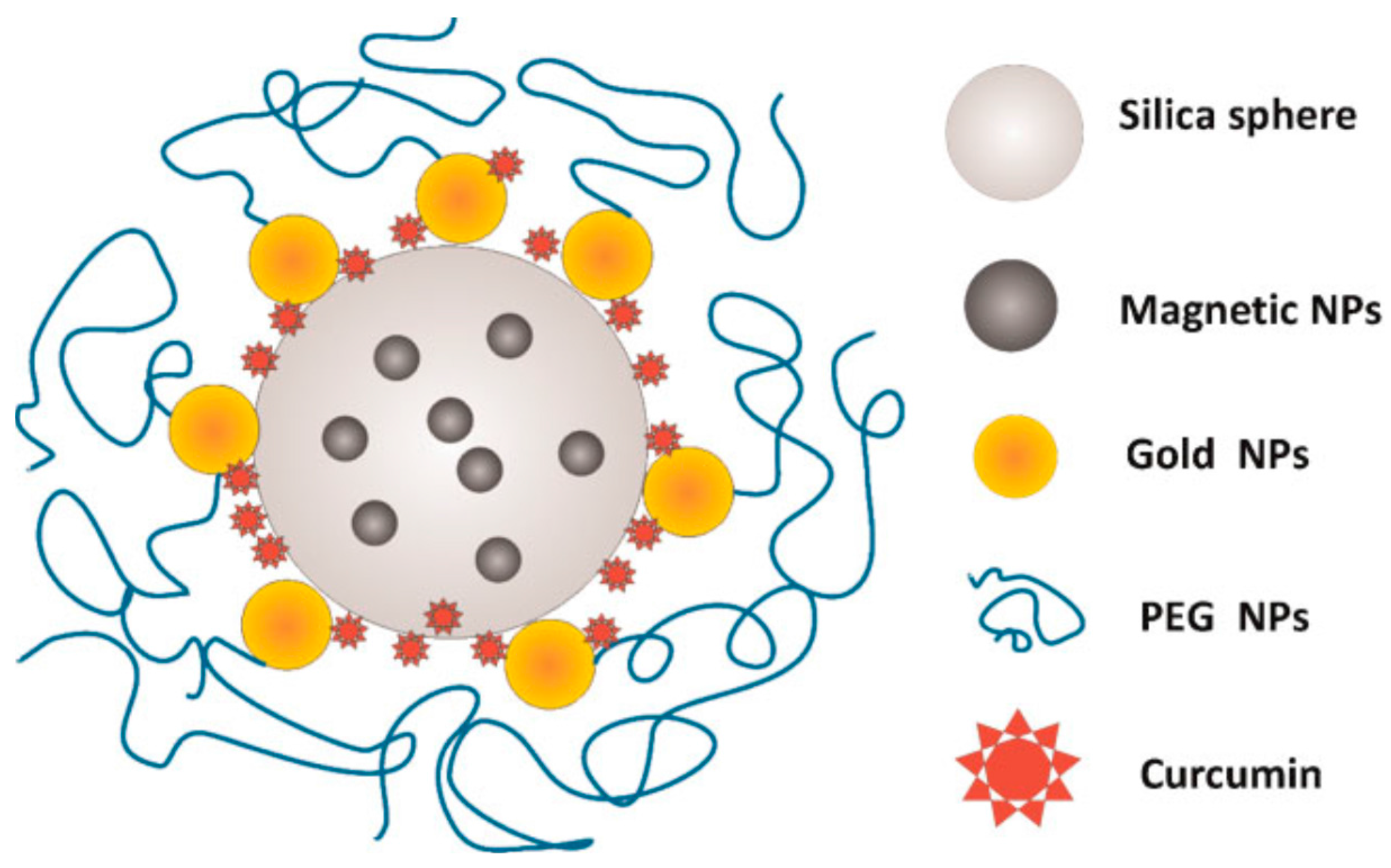
A range of Fe3O4@Ag magnetic plasmonic materials can be synthesised using an intermediate silica layer. Such Ag-coated magnetic nanoparticles can be produced through addition of a silica layer using a Stober synthesis. The silica provides a basis for further functionalisation, for example, in a number of cases, APTES ((3-aminopropyl)triethoxysilane) has been used to endow the substrate with amino groups which help facilitate the attachment of the plasmonic metal nanoparticles [19], in this case through sonication of the Fe3O4@SiO2@APTES nanocomposites with AgNO3—this ensured the attachment of the AgNO3 to the surface of the nanoparticles. Following this, the pH was re-adjusted to 11 and an outer silver shell was obtained by reduction of the nitrate with NaBH4. A similar approach using Fe3O4@SiO2@APTES was used to obtain a gold coated nanocomposite—in this case, however, the functionalised nanoparticles were dissolved in phosphate buffer to increase the positive charge; thus driving the electrostatic adsorption of the gold nanoparticles onto the surface of the magnetite core nanoparticles [20]. The raspberry-structured resultant Fe3O4@Au nanoparticles were loaded with curcumin and were then coated with polyethylene glycol (PEG), which acts as a cloaking agent to increase blood circulation time (Figure 1).
2.1.2. Synthesis of Core/Polymer/Shell Magnetic-Plasmonic Nanoparticles
Conductive polymers have been used to aid the fabrication of magnetic plasmonic nanocomposites. This approach was demonstrated by Feng et al., who prepared polypyrrole encapsulated gold nanorods decorated with Fe3O4 nanoparticles (Au NR@PPY@Fe3O4) [21]. Polypyrrole (PPY) is a conductive polymer that has been demonstrated to induce photothermal effects, and is already widely used in many biomedical applications [22,23]. The core gold nanorods were synthesised using a modified seed mediated approach in which gold seeds synthesised using NaBH4 and chloroauric acid in CTAB (hexadecyltrimethylammonium bromide), and a growth solution of AgNO3, HAuCl4 and ascorbic acid. Iron cation mediated polymerisation was used to fabricate the PPY coating on the gold nanorods—this gave rise to an abundance of Fe3+ and Fe2+ ions in the resulting PPY@Au nanorod product. This solution was then diluted with water and ethanol and the Fe3O4 nanoparticles were precipitated directly out of this solution using ammonia, and adhesion of the Fe3O4 nanoparticles was driven electrostatically to the conductive polymer shell.
Aqueous based syntheses are of increasing interest, and of particular importance when the material is being considered for biomedical applications. Eliminating the use of organic solvents throughout the synthetic process and using only biocompatible, non-toxic materials and reagents streamlines the synthesis for biological purposes. An aqueous synthetic method and biocompatible polymer was utilised in the synthesis of heterostructured Fe3O4@PVP@Au nanocomposites [24]. In this synthesis, PVP (polyvinylpyrrolidone) polymer was used to stabilise the Fe3O4 nanoparticles, and also to form a homogenous layer surrounding them that has a negative charge. PVP is amphiphilic, non-toxic and non-ionic and already has a broad spectrum of biomedical applications [25]. Gold seeds were prepared through the reduction of chloroauric acid using NaBH4 in the presence of CTAB, and these seeds assemble on the PVP-coated Fe3O4 through electrostatic interaction. The outer gold shells were formed through the use of hydroxylamine hydrochloride. Polyethyleneimine (PEI) can also be used as a reducing agent to cause the formation of an outer plasmonic layer. Fe3O4@Au structures were synthesised by Ahmed et al.; coprecipitated Fe3O4 nanoparticles were dispersed in chloroauric acid solution and placed in a shaking incubator to allow the adsorption of Au3+ onto the surface of the Fe3O4 nanoparticles. PEI was then added to the shaking mixture to reduce the gold, causing the formation of a gold shell on the Fe3O4 nanoparticles [26]. Hydroxylamine was utilised by Wang et al. in the preparation of their multimodal magnetic plasmonic Fe3O4@Au nanoparticles [27]. They first modified their Fe3O4 nanoparticles with chitosan, post synthesis and stabilisation with oleic acid. Acetic acid is then added to the Fe3O4-Chitosan NPs and the mixture is sonicated, any unreacted chitosan is then removed using magnetic separation. Preformed gold colloids are then introduced to these particles and sonicated, and hydroxylamine hydrochloride is used to reduce the gold colloids on the surface of the Fe3O4 to form a gold shell. Magnetic separation is then utilised to ensure no unreacted gold remains in the solution.
2.1.3. Synthesis of Magnetic-Plasmonic Nanoparticles without Intermediate Layers
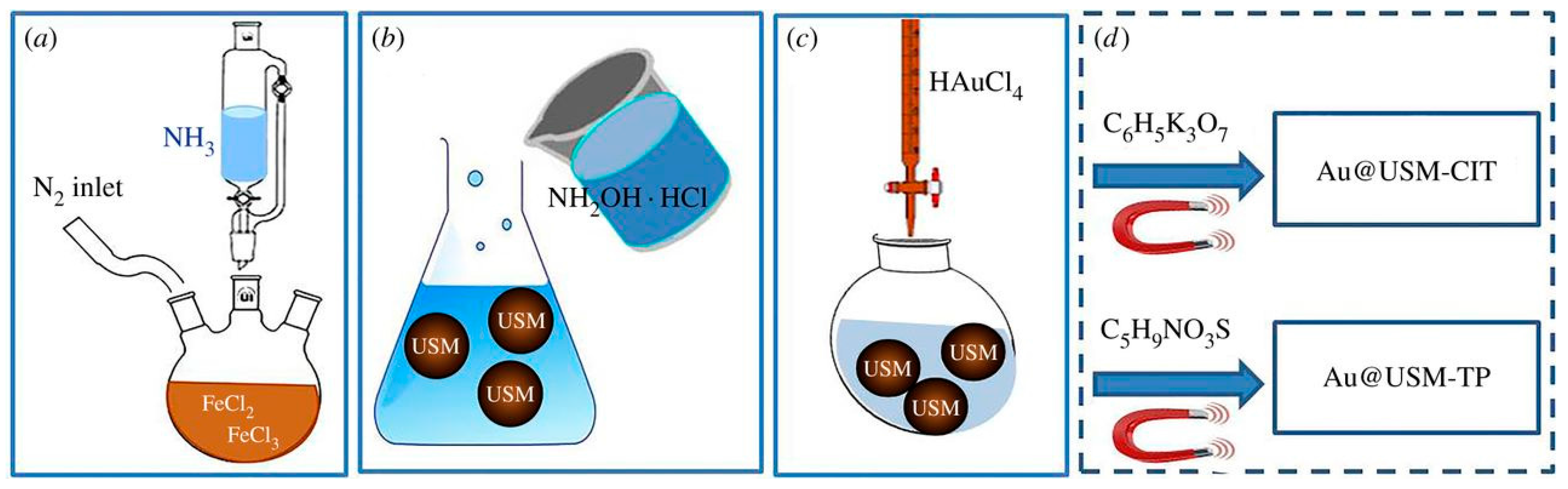
Multimodal magnetic plasmonic structures can also be synthesised by co-precipitation followed by direct attachment of the plasmonic moiety to the magnetic particles. Hydroxylamine has frequently been utilised as a seeding and reducing agent in a number of cases for coating of magnetic nanoparticles with a plasmonic shell [24,27,28]. For example, Fantechi et al. first synthesised ultra-small Fe3O4 NPs using Massart’s coprecipitation; then, hydroxylamine was used as a seeding agent for the gold NPs (Figure 2) [28].
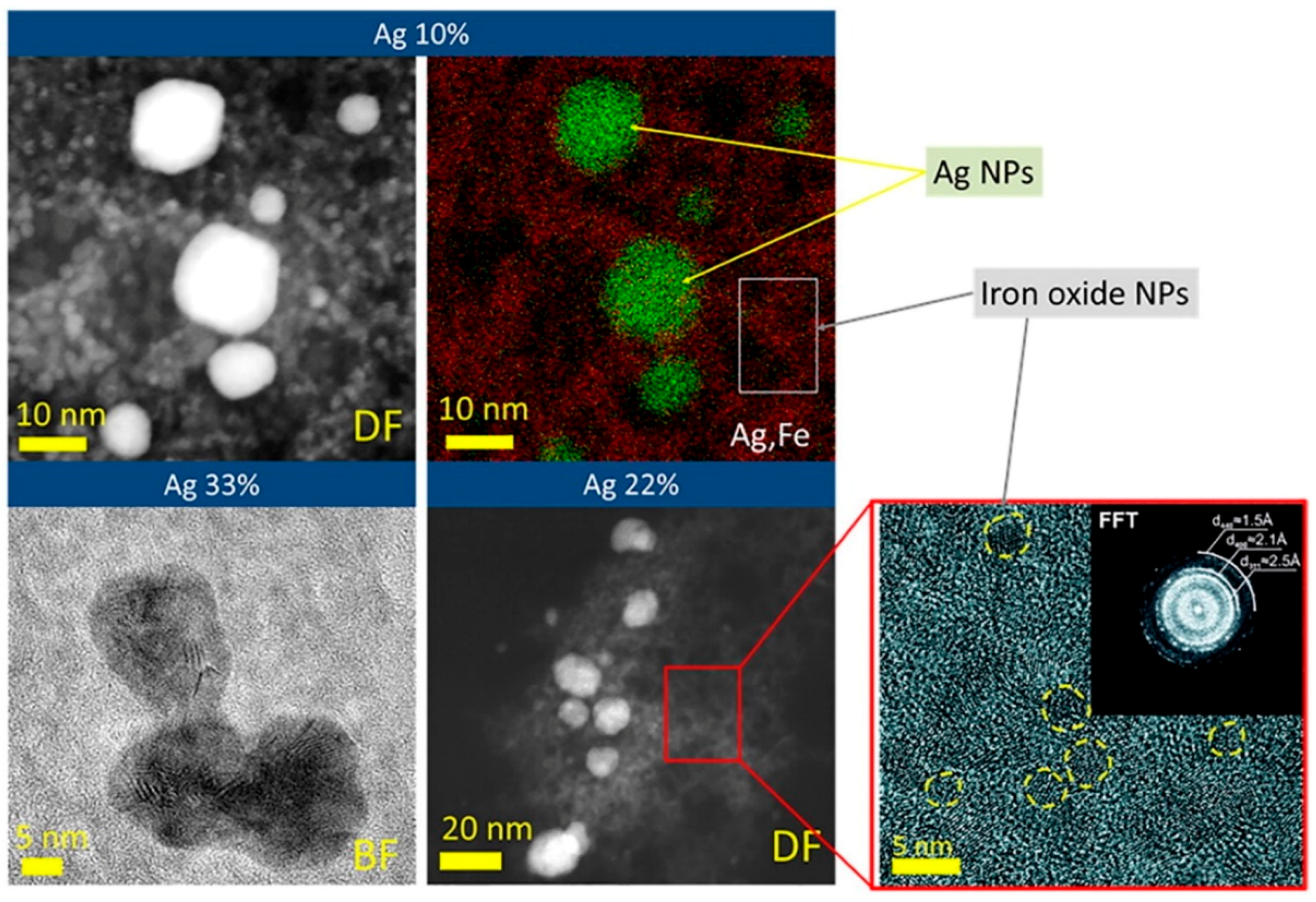
Self-organising silver and ultra-small iron oxide nanoparticles were recently made using a green synthetic approach [29]. The coprecipitation method was slightly modified in this case—ginger rhizome extract was used as a stabilising agent, as well as serving as an antioxidant to prevent oxidation of the Fe2+ ions. To the iron salts and ginger route extract, silver nitrate was added dropwise, followed by NaOH, and the resultant mixture was heated under reflux. The entire process takes place exclusively in aqueous solution, with magnetic separation utilised to clean and purify the Ag-Fe3O4 nanocomposite product (Figure 3).
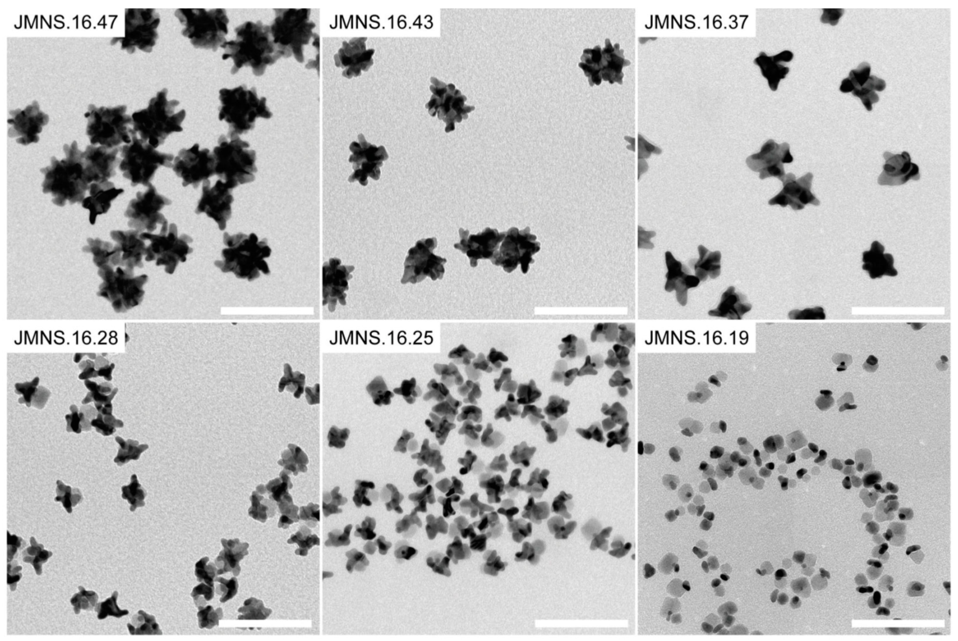
The synthesis of magnetic plasmonic nanostructures with non-platonic geometries is a growing area of interest within this field, as these novel structures have a large surface area, strong NIR (Near Infrared) absorbance, plasmonic hot spots and interesting interactions with cells and cell components [30,31,32]. For example, an Fe3O4 inner core was coated by a smooth layer of gold which then acted as a basis for the growth of the gold spikes to produce star-like Fe3O4@Au magneto-plasmonic supraparticles (Figure 4) [33]. Variation in the redox potential of the gold reducing agent is used to vary the morphology of the gold layer—the smooth layer of gold on the surface of the Fe3O4 nanoparticle is grown through introducing the pre-formed citrate stabilised Fe3O4 NPs into the boiling HAuCl4 solution (citrate acts as both the stabilising and the reducing agent in this case). Hydroquinone is then used as the reducing agent in the formation of the gold spikes, as it has a more negative redox potential than citrate [33].
2.2. Thermal Decomposition Techniques
Thermal decomposition, initially used to synthesise high quality semiconductor nanocrystals, is now commonly employed to synthesise monodisperse magnetic nanocrystals [34,35,36,37]. Thermal decomposition involves the heating (often to temperatures in the region of 300 °C) of organometallic precursors in organic solvents that contain a stabilising surfactant. Examples of such organometallic precursors include, but are not limited to metal acetylacetonates (e.g., Fe(acac)3) and metal carbonyls (e.g., Fe(CO)5) [38].
2.2.1. Simultaneous Thermal Decomposition
Among the simpler methodologies involving thermal decomposition as a synthetic route for magnetic-plasmonic structures is the thermal decomposition of both the magnetic and plasmonic moieties, either together or in tandem. Au-Fe3O4 dumbbells were obtained through the simultaneous thermal decomposition of Fe(CO)5 and HAuCl4·3H2O in oleylamine and 1-octadecane [39]. The precursor solution was heated to 300 °C, and the resultant nanoparticles were exposed to air for 30 min to allow oxidation of the iron. Both 16 nm and 20 nm iron oxide dumbbell nanostructures were formed through variation in the amounts of precursor added. PEG and DMF (dimethylformamide) were used to produce interesting morphologies through further reduction of the Au3+ ions onto the surface of the gold dumbbells. The Au-Fe3O4 dumbbells are then used to achieve directional growth of asymmetric gold nanostars using PEG and DMF (Figure 5).
Thermal decomposition was used as the basis for making magnetic-plasmonic nanoclusters by Wu et al. [40]. Fe(acac)3 was used as the organometallic precursor in this case. Iron oxide nanoparticles were first synthesised, and these iron oxide nanoparticles were then added to gold acetate and further heated to 180 °C in organic solvents with vigorous stirring. Nanoclusters were then formed using SDS and the water in oil microemulsion method. However, the resultant nanoclusters had a broad size distribution.
2.2.2. Thermal Decomposition for Synthesis of Magnetic-Plasmonic Core/Silica/Shell Nanoparticles
Similar to the approaches used in coprecipitated nanoparticles, often an intermediate layer is introduced onto the either the magnetic or plasmonic moiety in order to facilitate easier adhesion of the two components, and silica remains a popular choice. Flower-like Au@Fe3O4 nanoparticles were prepared by Fantechi et al. using thermal decomposition followed by a seeded growth approach in which the Fe3O4 nanoparticles are grown onto the gold nanoparticles to form heteronanocrystals [41]. Two iron precursors, Fe(acac)3 and Fe(CO)5 were used in the presence of pre-formed gold nanoparticles to investigate the effect that different reaction parameters have on the morphology of the hetero-nanocrystals. A silica coating is then applied to illustrate the ease with which the resultant nanoparticles can be transferred from the organic to the aqueous phase, and hence their applicability for biomedical applications. Silica is also utilised in work carried out by Huang et al., where the Fe3O4 nanoparticles are coated with silica and then further functionalised with APTES to obtain surface amino groups [42]. Gold nanoseeds are prepared in situ with the pre-formed Fe3O4@SiO2 and NaBH4 as a reducing agent; the Au seeds adhere to the silica coating electrostatically. Nanorattles are then achieved through etching of the silica using potassium carbonate.
Acetylacetonates can be used in the thermal decomposition method to prepare magnetic alloys, as is demonstrated by Kostevsek et al. through their synthesis of multimodal hybrid FePt@SiO2@Au nanoparticles [43]. Fe(acac)3 and Pt(acac)3 were added to a mixture of benzyl ether, oleic acid and oleylamine, and heated to 200 °C for 30 min, and then heated to 280 °C for a further 30 min. The FePt magnetic-plasmonic alloy was used as it has a much higher saturation magnetisation than iron oxides; it also is thought to be a better contrast agent. A silica shell was added using TEOS and further functionalised with APTES. The gold nanoparticles were added using a two-step seeding approach. This was realised through adding HAuCl4·3H2O and NaOH to the FePt@SiO2 mixture and stirring at 70 °C for 5 min, allowing the seeding of Au(OH)3 onto the silica surface. Ice-cold NaBH4 was then used to reduce the gold onto the surface of the nanoparticles.
2.2.3. Thermal Decomposition for Synthesis of Magnetic Core/Polymer Shell/Plasmonic Nanoparticles
Green approaches to the syntheses of magnetic plasmonic approach based on thermal decomposition are also of interest, although these can be more difficult as organic solvents are generally a prerequisite for this type of approach. However, as demonstrated by Yu et al., an aqueous based thermal decomposition reaction in benzyl alcohol can be carried out through the use of a microwave [44]. The Fe3O4 nanoparticles are synthesised in the presence of PVP; and the resultant nanoparticles are then dispersed in an ethylene glycol solution containing HAuCl4 and PVP—again, these are placed in the microwave in order to obtain triangular gold nanoparticles studded with Fe3O4 nanoparticles.
PEG is a popular choice as it is biocompatible and can be readily functionalised with a number of desirable functional groups. In some cases, this intermediate layer can also aid the formation of a novel morphology, such as in work carried out by Li et al. [45]. In this work, the Fe3O4 first underwent a ligand exchange to obtain an outer layer of PEG. The resultant nanoparticles were then coated with poly-l-lysine, which aids the seeding and growth of a trisoctahedral gold shell. PEG is also used in the ligand mediated self-assembly of hybrid plasmonic superparamagnetic nanostructures [46]. Au nanorods, initially synthesised with CTAB as an outer stabilising layer, are replaced with bifunctional PEG groups that have both surface carboxylic acid and thiol groups. This replacement of the CTAB with the bifunctional PEG layer drastically improves the biocompatibility of the Au nanorods. Fe3O4 TREG (triethylene glycol) functionalised nanoparticles were then added to the Au nanorod solution; and, as the TREG groups are easily displaced by the –COOH terminal groups on the bifunctional PEG coated Au nanorods, the product is Fe3O4 studded Au nanorods.
2.3. Solvothermal Methods
The solvothermal method is now routinely used to prepare magnetic iron oxide nanoparticles [47]. In the first studies on this synthetic approach, cupferron transition metal ion complexes were used as precursors to numerous metal oxide nanoparticles [48]. Nowadays, the solvent thermal method can also be extended to the synthesis of magnetite-gold nanocomposites, as is now commonly carried out. Li et al. utilised a solvothermal approach to synthesise Fe3O4 nanoparticles in a single step—using FeCl3 salts dissolved in gelatin in ethylene glycol with sodium acetate reacted at 200 °C for 6 h [49]. Gold seeds were prepared using chloroauric acid and tetrakis-hydroxymethylphophonium chloride (THPC), and seeded onto the gelatin coated Fe3O4 nanoparticles through stirring. A gold shell was obtained through seeding of the gold nanoparticles using K2CO3. PEGylation was utilised to make the resultant nanoclusters more bio-compatible. Gelatin was used as a mediator as it can be degraded by a subclass of matrix metalloproteinases, and superparamagnetic Fe3O4 nanoparticles have been shown to be biodegradable in vivo. A similar approach was used by Jin et al.—except in this case an APTES functionalised silica coating is used in place of gelatin, and THPC is also used to reduce a layer of gold onto the Fe3O4 nanoparticles [50]. An interesting method of obtaining concave magneto-plasmonic core-shell supraparticles is used by Lee et al. [51]. The Fe3O4 cores were prepared by the solvothermal method using PEI as a capping ligand. Electrostatics are then exploited to seed the gold nanoparticles onto the surface of the nanoparticles—this is done by sonication of the Au seeds followed by shaking for two hours. The thickness of the gold shell can be carefully controlled through layer by layer deposition of gold seeds. In this case, graphene quantum dots were anchored to the surface in order to produce an enhanced Raman signal.
2.4. Brust’s Method
Brust’s method of nanoparticle synthesis involves reduction of a metal precursor in a two-phase (water-toluene) system by NaBH4 in the presence of an alkane thiol, and is a good method of preparing highly stable thiol functionalised nanoparticles. The metallic clusters are grown by the simultaneous attachment of self-assembled thiol monolayers on the growing nuclei [52]. The synthesis proceeds at room temperature, and first involves the phase transfer of chloroauric acid (Au3+) from the aqueous phase to the organic phase using a phase transfer catalyst. The two phases are then separated and the organic phase is first treated with dodecanethiol to cause the reduction of Au3+ to Au1+. Sodium borohydride is then added to reduce the Au1+ to Au0, and this reduction is indicated by a colour change [53]. This method was utilised in the synthesis of Fe3O4@Au nanoparticles; in this case the Brust Schiffrin method was used to obtain 2 nm Au nanoparticles, and the shell was obtained through a modified thermally activated processing protocol [54]. While the Brust method was first used to synthesise gold nanoparticles, the methodology can be extended to other metal based systems. For example, Brusts method was used by Bhattarai et al. to produce bimetallic magnetic plasmonic Au/Co. nanoparticles (Figure 6) [55].
2.5. Flame Aerosol Synthesis
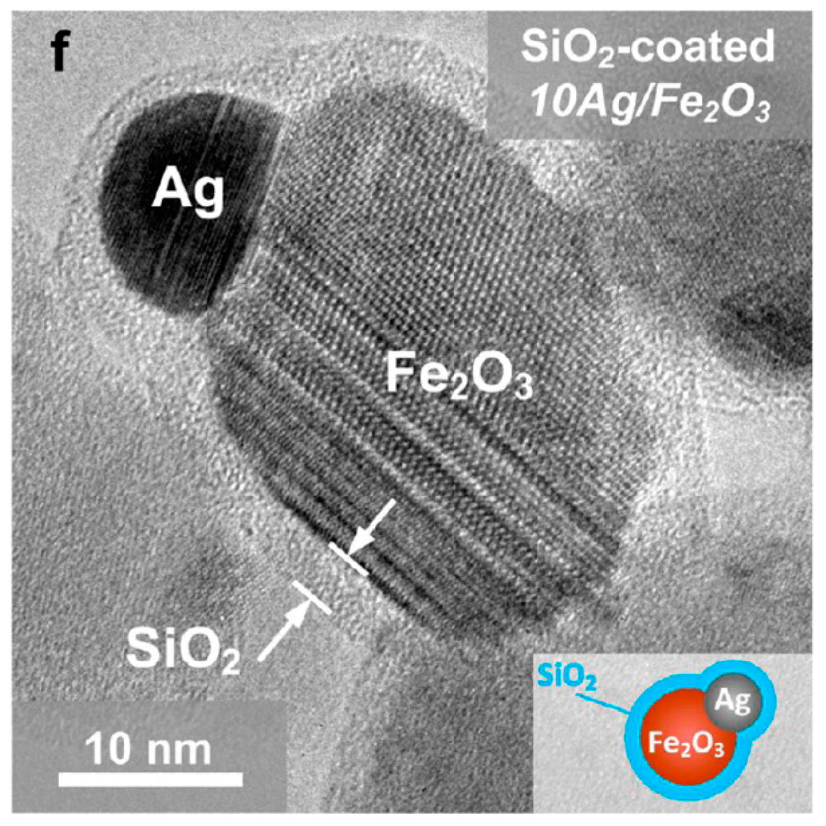
Flame spray pyrolysis is an advanced one-step synthetic approach to produce magnetic plasmonic nanoparticles. Briefly, two metal precursors are used in a 1:1 ratio and fed to a flame in a spray pyrolysis set up. This method was used to synthesise Fe2O3@Ag bimetallic magnetic nanoparticles using Fe(acac)3 and Ag(OAc) as starting materials (Figure 7). A silica coating on the nanoparticles was then obtained through coating in flight through the swirling injection of hexamethyldisiloxan (HDMSO) in nitrogen [56]. The same method was used to produce Janus-like Fe2O3@Ag nanoparticles with a nano-thin SiO2 coating to prevent leaching of Ag+ ions, which are toxic. These nanomaterials were then used as biomarkers [57].
3. Biomedical Applications of Magnetic-Plasmonic Nanocomposites
The dual modalities of a magnetic plasmonic nano-system open up a range of potential biomedical applications. The multimodal nature of these nanoparticles paves the way for their use as theranostic agents—i.e., materials that can be used to both diagnose, or quantify a disease, and also simultaneously provide treatment. In an ideal world, a single nanoparticle with these theranostic properties should be easy to functionise and the size and properties should be tunable.
When designing nanoparticles for use in biomedical applications, a number of factors must be taken into consideration. The nanoparticles should be taken up only by targeted cells, and should not be present in any significant concentration in non-targeted tissues. Efficient renal clearance from the body is necessary to minimise toxicity, and for any drug to be considered feasible for widespread use by the FDA (US Food and Drug Administration), it must be cleared from the body within a reasonable timeframe [58,59]. Hydrodynamic diameter is also of utmost importance, as the nanoparticles must be small enough to not be cleared through the mononuclear phagocyte system, but large enough so that they do not become coated with serum proteins, thus increasing the hydrodynamic diameter and hindering renal elimination [60].
3.1. Photothermal Therapy
Photothermal therapy is an attractive approach to cancer therapy as it is can be a highly selective theranostic approach if nanoparticles (most commonly gold) are delivered to tumour sites. An external NIR laser can be applied, causing the oscillation of the plasmons and resulting in the nanoparticles generating heat, which has been shown to be highly effective in the thermal ablation of cancer cells [61,62]. Nanoparticles based on gold are the most popular choice for photothermal agents as it is biocompatible enabling a thiol functionalisation which allows the easy attachment of antibodies and other targeting agents. Gold nanoparticles also have an effective light to heat conversion, and small sizes which can be tuned to efficiently absorb NIR light, which penetrates tissue better than other wavelengths of light. Utilising multimodal magnetic plasmonic nanoparticles means that additional functionalities are now possible. Not only can the gold component be used to facilitate photothermal therapy, but the magnetic component can be used to direct the nanoparticles to the tumour site using an external magnet, the gold nanoparticles can be used for dark field imaging and the magnetite could potentially act as an MRI agent during the process. Targeting moieties could be conjugated to the surface of the gold to endow extra cell specificity. The litany of possible applications for these multimodal nanosystems ensures that they are the subject of intensive research efforts. Some magnetic plasmonic nanoparticle systems proposed for use in photothermal therapy are discussed below.
Gelatin was used as a mediator in the synthesis of Fe3O4@Au nanoparticles proposed for use as photothermal agents. This is of particular importance as gelatin can be readily degraded by a subclass of metalloproteinases in the body, which further ensures that the system is suitable for use in vivo [49]. The biocompatibility and bioelimination of this system was closely examined, and it was found that in the acidic tumour microenvironment the Fe3O4@Au nanoparticle assemblies were successfully broken in conjunction with the metalloproteinases. An 808 nm NIR laser was found to cause significant temperature increases, and high doses of the nanoparticle composites were found to be nontoxic, thus illustrating its potential for use in vivo as a photothermal agent. This material also showed potential for uses in MRI, CT and PAT (photoacoustic therapy). A similar chitosan Fe3O4@Au nanomaterial showed a temperature increase at various concentrations under illumination of a NIR laser [27]. Chitosan is a polysaccharide often used due to its interesting biopharmaceutical properties; for example, it’s high mucoadhesion and suitability for facilitating drug delivery, and is the subject of many reviews [63,64,65,66]. The authors also highlighted the multiple uses of the synthesised nanoparticles—not only as photothermal agents but for use in simultaneous MRI and dark field imaging. While these proposed photothermal agents have under-gone heating tests using NIR lasers ex vivo, more comprehensive biocompatibility and in vivo testing on these multimodal magnetic photothermal agents is urgently required.
3.2. Drug Delivery and Targeting
Multimodal magnetic nanoparticles have long been the subject of investigation for use as drug delivery vehicles, and many strategies for their synthesis, drug loading and release have been examined [67]. However, comparatively, few comprehensive studies on magnetic-plasmonic based drug delivery systems have been carried out, despite many potential theranostic applications which these systems would have. This could be due in part to a number of difficulties in the complexity of making these materials, and how biocompatible the resulting systems may be. For example, a balance must be drawn between the complexity of the nanomaterial and the time and cost of its synthesis with the increase in efficacy of the cancer treatment it can provide. Not only this, but the size and morphology of the magnetic plasmonic nanoparticles needs to be precisely controlled in order to optimise uptake in vivo—too small (<50 nm) and the nanoparticles are washed out of the body, too large (>300 nm) and they accumulate in the liver and spleen [18].
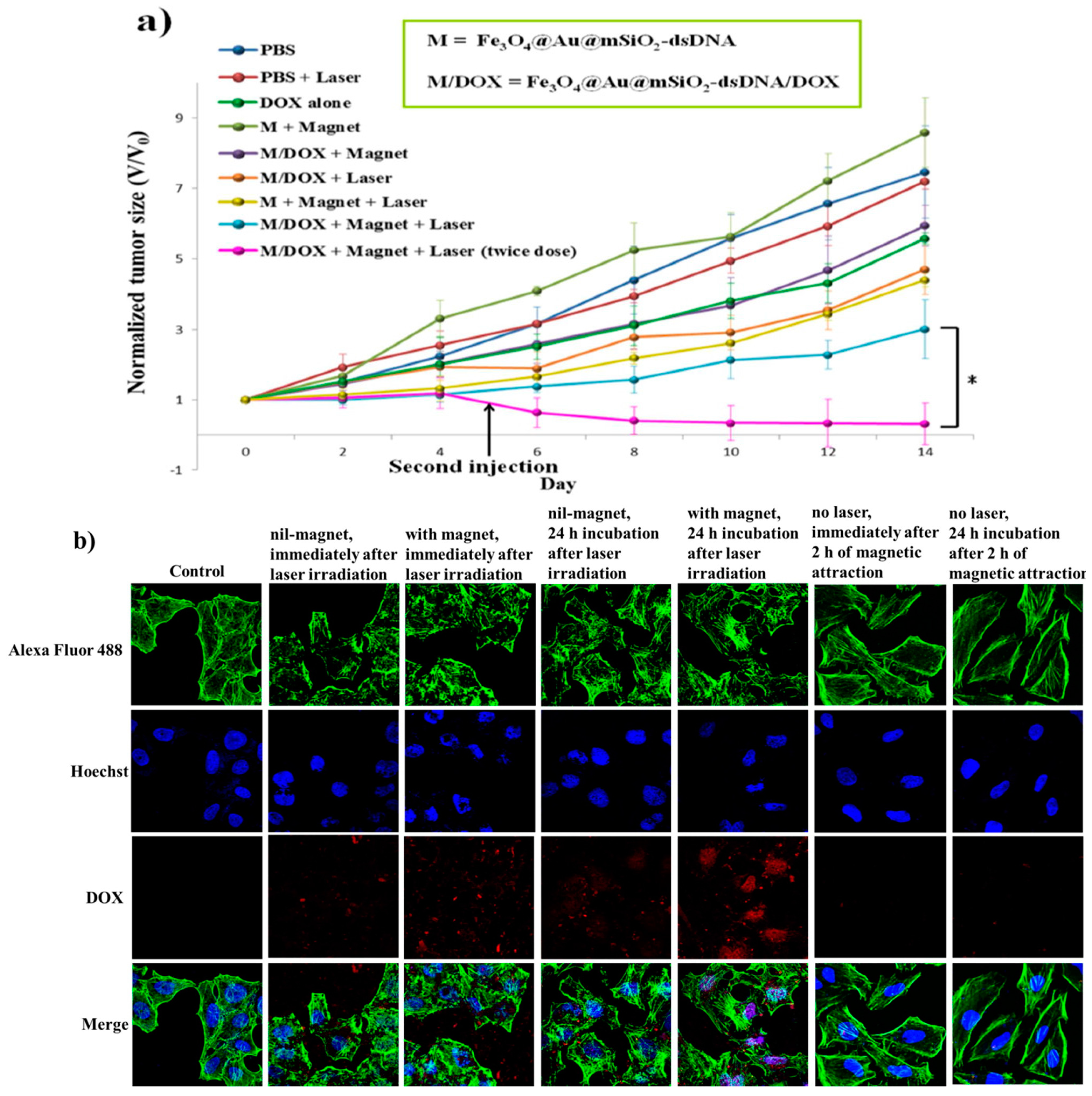
In spite of these challenges, such multimodal nanoparticles comprising of anisotropically shaped Fe3O4@Au were prepared with a biocompatible mesoporous silica shell, which also allowed anticancer drug delivery [45]. The doxorubicin containing pores were capped with double stranded DNA, which decomposed under NIR light, causing the heating of the gold component of the nanoparticle. The magnetite, in this case, provided magnetic guidance, which was shown to increase uptake of the nanoparticles into HeLa cells (Figure 8).
3.3. Imaging and Contrast Agents
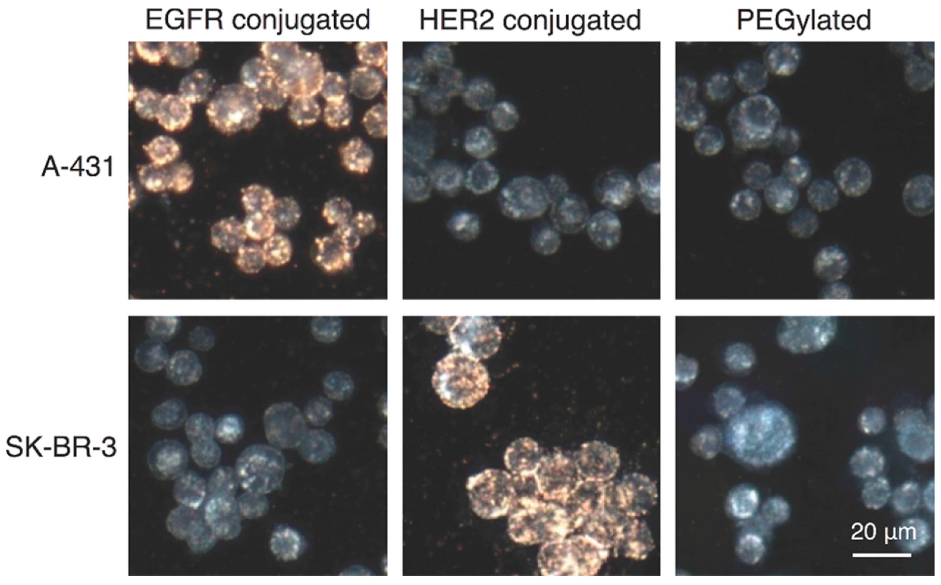
Magnetite nanoparticles have been of great interest as new MRI contrast agents, largely due to their ease of synthesis, biocompatibility and superparamagnetism [68]. The magnetic plasmonic nanoclusters synthesised by Wu et al. are proposed for use as multimodal imaging agents, utilising magnetic guidance under monitoring of the image in situ [40]. In this work, the molecular specificity of the composite nanoparticles is tested on SK-BR-3 breast cancer cells and A-431 keratinocytes, and it was shown that the cell targeting agent demonstrates high cell specificity and was preferentially taken up by cells with the complementary antigen (Figure 9).
Another important consideration made by these authors was the choice of surfactant—this can have a major influence on the viability of the resultant nanoparticles use in biomedical applications. SDS (sodium dodecyl sulfate) is used in this synthesis in place of CTAB (which is cytotoxic). This SDS was easily replaced with methylated PEG thiol molecules, which enhance biocompatibility.
3.4. In Vitro Toxicity Studies
While many of the current nanomaterials propose potential biomedical applications, such as multimodal imaging, photothermal therapy, magnetically guided drug delivery etc., very few comprehensive studies have been carried out. While the nanoparticles can be used to treat or diagnose disease, we must also consider any possible side-effects that the nanoparticles themselves may have in vivo. A number of toxicity studies have been carried out on different cell lines. Pariti et al. carried out toxicity evaluations on Au@Fe3O4 nanoparticles capped with l-cysteine on Chinese Hamster Ovary (CHO) cell lines [69]. These nanoparticles, proposed as drug delivery vehicles and hyperthermia agents, showed no significant cytotoxicity to the CHO cells over 48 h even up to concentrations of 1 mg/mL. The toxicity of similar nanoparticles was tested using an MTT (3-(4,5-dimethyltetrazolium bromide)) assay and it was found that the nanoparticles were non-toxic at 50 μg/mL [26]. However, it was noted by the authors that the toxicity pathways of the Au@Fe3O4 composites could be different due to size and morphology variations.
4. Conclusions and Future Outlook
In this review, we have shown that new types of magnetic-plasmonic nanocomposites of different varieties have been developed over the last 10 years. There is a great need and potential demand for these materials. From the above discussion, it is clear that magnetic-plasmonic nanocomposites can offer new opportunities in biology and medicine. However, despite all the recent progress that has been made, the magnetic-plasmonic nanocomposite area is still in its early development stage, and significant efforts are needed for further development of these materials and their applications. We believe that new magnetic-plasmonic nanomaterials could serve as all in one nano-sized drug delivery tools, which could help in the diagnosis and treatment of cancer, HIV and many other diseases. Because of their small size and combination of magnetic and plasmonic properties, these nanocomposites open up the unique possibility of controlled target-directed applications. This is of particularly great importance in medicine, as one major disadvantage of any nanoparticle therapy is the problem of getting the particle to the site of interest. An external magnetic field could be used to attract the multi-modal magnetic-plasmonic nanocomposites to the desired area, to hold them there until the diagnosis or treatment is complete and finally to remove them. All steps can be monitored by MRI and CT allowing the full control over the processes. Thus, further development and utilisation of magnetic-plasmonic nanoprobes could revolutionise many aspects of modern medicine. However, potential toxicity is of major concern when advocating multimodal nanoparticles for any biomedical use. Unfortunately, toxicity of magnetic-plasmonic nanomaterials is still very poorly studied and understood. We believe that a significant part of the future work in this area must be focused on the investigation of the toxicity and improvement of biocompatibility of multi-modal magnetic-plasmonic nanocomposites. This will be crucial for further development of relevant areas of nano-biotechnology and nanomedicine.
Acknowledgments
This work was supported by Irish Research Council (IRC) postgraduate scheme and the EU-ITN network Mag(net)icFun (PITN-GA-2012-290248).
Author Contributions
All authors wrote and cross-checked the manuscript together.
Conflicts of Interest
The authors declare no conflicts of interest.
References
- Sannomiya, T.; Voros, J. Single plasmonic nanoparticles for biosensing. Trends Biotechnol. 2011, 29, 343–351. [Google Scholar] [CrossRef] [PubMed]
- Tong, L.M.; Kall, M. Laser Manipulation of Plasmonic Nanoparticles for Sers and Sensing; Springer: New York, NY, USA, 2012; pp. 153–167. [Google Scholar]
- Giner-Casares, J.J.; Liz-Marzan, L.M. Plasmonic nanoparticles in 2d for biological applications: Toward active multipurpose platforms. Nano Today 2014, 9, 365–377. [Google Scholar] [CrossRef]
- Lim, W.Q.; Gao, Z.Q. Plasmonic nanoparticles in biomedicine. Nano Today 2016, 11, 168–188. [Google Scholar] [CrossRef]
- Edel, J.B.; Kornyshev, A.A.; Kucernak, A.R.; Urbakh, M. Fundamentals and applications of self-assembled plasmonic nanoparticles at interfaces. Chem. Soc. Rev. 2016, 45, 1581–1596. [Google Scholar] [CrossRef] [PubMed]
- Yu, R.W.; Liz-Marzan, L.M.; de Abajo, F.J.G. Universal analytical modeling of plasmonic nanoparticles. Chem. Soc. Rev. 2017, 46, 6710–6724. [Google Scholar] [CrossRef] [PubMed]
- Xie, T.; Jing, C.; Long, Y.T. Single plasmonic nanoparticles as ultrasensitive sensors. Analyst 2017, 142, 409–420. [Google Scholar] [CrossRef] [PubMed]
- Boken, J.; Khurana, P.; Thatai, S.; Kumar, D.; Prasad, S. Plasmonic nanoparticles and their analytical applications: A review. Appl. Spectrosc. Rev. 2017, 52, 774–820. [Google Scholar] [CrossRef]
- Krajczewski, J.; Kolataj, K.; Kudelski, A. Plasmonic nanoparticles in chemical analysis. RSC Adv. 2017, 7, 17559–17576. [Google Scholar] [CrossRef]
- Jing, L.H.; Ding, K.; Kershaw, S.V.; Kempson, I.M.; Rogach, A.L.; Gao, M.Y. Magnetically engineered semiconductor quantum dots as multimodal imaging probes. Adv. Mater. 2014, 26, 6367–6386. [Google Scholar] [CrossRef] [PubMed]
- Jana, N.R. Design and development of quantum dots and other nanoparticles based cellular imaging probe. Phys. Chem. Chem. Phys. 2011, 13, 385–396. [Google Scholar] [CrossRef] [PubMed]
- Corr, S.A.; Rakovich, Y.P.; Gun’ko, Y.K. Multifunctional magnetic-fluorescent nanocomposites for biomedical applications. Nanoscale Res. Lett. 2008, 3, 87–104. [Google Scholar] [CrossRef]
- Padmanabhan, P.; Kumar, A.; Kumar, S.; Chaudhary, R.K.; Gulyas, B. Nanoparticles in practice for molecular-imaging applications: An overview. Acta Biomater. 2016, 41, 1–16. [Google Scholar] [CrossRef] [PubMed]
- Bao, G.; Mitragotri, S.; Tong, S. Multifunctional nanoparticles for drug delivery and molecular imaging. Ann. Rev. Biomed. Eng. 2013, 15, 253–282. [Google Scholar] [CrossRef] [PubMed]
- Kiani, A.; Esquevin, A.; Lepareur, N.; Bourguet, P.; Le Jeune, F.; Gauvrit, J.Y. Main applications of hybrid pet-mri contrast agents: A review. Contrast Media Mol. Imaging 2016, 11, 92–98. [Google Scholar] [CrossRef] [PubMed]
- Cherukula, K.; Lekshmi, K.M.; Uthaman, S.; Cho, K.; Cho, C.S.; Park, I.K. Multifunctional inorganic nanoparticles: Recent progress in thermal therapy and imaging. Nanomaterials 2016, 6, 76. [Google Scholar] [CrossRef] [PubMed]
- Same, S.; Aghanejad, A.; Nakhjavani, S.A.; Barar, J.; Omidi, Y. Radiolabeled theranostics: Magnetic and gold nanoparticles. Bioimpacts 2016, 6, 169–181. [Google Scholar] [CrossRef] [PubMed]
- De Crozals, G.; Bonnet, R.; Farre, C.; Chaix, C. Nanoparticles with multiple properties for biomedical applications: A strategic guide. Nano Today 2016, 11, 435–463. [Google Scholar] [CrossRef]
- Chu, D.T.; Sai, D.C.; Luu, Q.M.; Tran, H.T.; Quach, T.D.; Kim, D.H.; Nguyen, N.H. Synthesis of bifunctional Fe3O4@SiO2-Ag magnetic–plasmonic nanoparticles by an ultrasound assisted chemical method. J. Electron. Mater. 2017, 46, 3646–3653. [Google Scholar] [CrossRef]
- Chen, W.; Xu, N.; Xu, L.; Wang, L.; Li, Z.; Ma, W.; Zhu, Y.; Xu, C.; Kotov, N.A. Multifunctional magnetoplasmonic nanoparticle assemblies for cancer therapy and diagnostics (theranostics). Macromol. Rapid Commun. 2010, 31, 228–236. [Google Scholar] [CrossRef] [PubMed]
- Feng, W.; Zhou, X.; Nie, W.; Chen, L.; Qiu, K.; Zhang, Y.; He, C. Au/polypyrrole@Fe3O4 nanocomposites for MR/CT dual-modal imaging guided-photothermal therapy: An in vitro study. ACS Appl. Mater. Interfaces 2015, 7, 4354–4367. [Google Scholar] [CrossRef] [PubMed]
- He, C.; Nie, W.; Feng, W. Engineering of biomimetic nanofibrous matrices for drug delivery and tissue engineering. J. Mater. Chem. B 2014, 2, 7828–7848. [Google Scholar] [CrossRef]
- Vaitkuviene, A.; Kaseta, V.; Voronovic, J.; Ramanauskaite, G.; Biziuleviciene, G.; Ramanaviciene, A.; Ramanavicius, A. Evaluation of cytotoxicity of polypyrrole nanoparticles synthesized by oxidative polymerization. J. Hazard. Mater. 2013, 250, 167–174. [Google Scholar] [CrossRef] [PubMed]
- Xie, L.; Qian, W.; Yang, S.; Sun, J.; Gong, T. A facile and green synthetic route for preparation of heterostructure Fe3O4@Au nanocomposites. MATEC Web Conf. 2017, 88, 02001. [Google Scholar] [CrossRef]
- Le Garrec, D.; Gori, S.; Luo, L.; Lessard, D.; Smith, D.C.; Yessine, M.A.; Ranger, M.; Leroux, J.C. Poly(N-vinylpyrrolidone)-block-poly(d,l-lactide) as a new polymeric solubilizer for hydrophobic anticancer drugs: In vitro and in vivo evaluation. J. Controll. Release 2004, 99, 83–101. [Google Scholar] [CrossRef] [PubMed]
- Ahmed, O.S.; Zekri, A.-R.N.; Imam, H.; Khaled, H.M.; Abdel-Kader, M.H. Synthesis of the newly developed core-shell au/Fe3O4 magnato-plasmonic nanocomposite in cancer cells. Life Sci. J. 2014, 11, 182–187. [Google Scholar]
- Wang, X.; Liu, H.; Chen, D.; Meng, X.; Liu, T.; Fu, C.; Hao, N.; Zhang, Y.; Wu, X.; Ren, J.; et al. Multifunctional Fe3O4@p(St/MAA)@chitosan@Au core/shell nanoparticles for dual imaging and photothermal therapy. ACS Appl. Mater. Interfaces 2013, 5, 4966–4971. [Google Scholar] [CrossRef] [PubMed]
- Fantechi, E.; Castillo, P.M.; Conca, E.; Cugia, F.; Sangregorio, C.; Casula, M.F. Assessing the hyperthermic properties of magnetic heterostructures: The case of gold–iron oxide composites. Interface Focus 2016, 6, 20160058. [Google Scholar] [CrossRef] [PubMed]
- Ivashchenko, O.; Gapiński, J.; Peplińska, B.; Przysiecka, Ł.; Zalewski, T.; Nowaczyk, G.; Jarek, M.; Marcinkowska-Gapińska, A.; Jurga, S. Self-organizing silver and ultrasmall iron oxide nanoparticles prepared with ginger rhizome extract: Characterization, biomedical potential and microstructure analysis of hydrocolloids. Mater. Design 2017, 133, 307–324. [Google Scholar] [CrossRef]
- Ma, Y.; Kuang, Q.; Jiang, Z.; Xie, Z.; Huang, R.; Zheng, L. Synthesis of trisoctahedral gold nanocrystals with exposed high-index facets by a facile chemical method. Angew. Chem. Int. Ed. 2008, 47, 8901–8904. [Google Scholar] [CrossRef] [PubMed]
- Lee, P.-Y.; Teng, H.-S.; Yeh, C.-S. Preparation of superparamagnetic mnxfe1-xo nanoparticles from low-index-facet cubes to high-index-facet concave structures and their catalytic performance in aqueous solution. Nanoscale 2013, 5, 7558–7563. [Google Scholar] [CrossRef] [PubMed]
- Tao, A.R.; Habas, S.; Yang, P. Shape control of colloidal metal nanocrystals. Small 2008, 4, 310–325. [Google Scholar] [CrossRef]
- Zhou, H.; Kim, J.-P.; Bahng, J.H.; Kotov, N.A.; Lee, J. Self-assembly mechanism of spiky magnetoplasmonic supraparticles. Adv. Funct. Mater. 2014, 24, 1439–1448. [Google Scholar] [CrossRef]
- O’Brien, S.; Brus, L.; Murray, C.B. Synthesis of monodisperse nanoparticles of barium titanate: Toward a generalized strategy of oxide nanoparticle synthesis. J. Am. Chem. Soc. 2001, 123, 12085–12086. [Google Scholar] [CrossRef] [PubMed]
- Peng, X.; Wickham, J.; Alivisatos, A.P. Kinetics of II–VI and III–V colloidal semiconductor nanocrystal growth: “Focusing” of size distributions. J. Am. Chem. Soc. 1998, 120, 5343–5344. [Google Scholar] [CrossRef]
- Park, J.; An, K.; Hwang, Y.; Park, J.-G.; Noh, H.-J.; Kim, J.-Y.; Park, J.-H.; Hwang, N.-M.; Hyeon, T. Ultra-large-scale syntheses of monodisperse nanocrystals. Nat. Mater. 2004, 3, 891–895. [Google Scholar] [CrossRef] [PubMed]
- Sun, S.; Zeng, H.; Robinson, D.B.; Raoux, S.; Rice, P.M.; Wang, S.X.; Li, G. Monodisperse MFe2O4 (M = Fe, Co, Mn) nanoparticles. J. Am. Chem. Soc. 2004, 126, 273–279. [Google Scholar] [CrossRef] [PubMed]
- Lu, A.-H.; Salabas, E.L.; Schüth, F. Magnetic nanoparticles: Synthesis, protection, functionalization, and application. Angew. Chem. Int. Ed. 2007, 46, 1222–1244. [Google Scholar] [CrossRef] [PubMed]
- Reguera, J.; Jimenez de Aberasturi, D.; Henriksen-Lacey, M.; Langer, J.; Espinosa, A.; Szczupak, B.; Wilhelm, C.; Liz-Marzan, L.M. Janus plasmonic-magnetic gold-iron oxide nanoparticles as contrast agents for multimodal imaging. Nanoscale 2017, 9, 9467–9480. [Google Scholar] [CrossRef] [PubMed]
- Wu, C.-H.; Cook, J.; Emelianov, S.; Sokolov, K. Multimodal magneto-plasmonic nanoclusters for biomedical applications. Adv. Funct. Mater. 2014, 24, 6862–6871. [Google Scholar] [CrossRef]
- Fantechi, E.; Roca, A.G.; Sepúlveda, B.; Torruella, P.; Estradé, S.; Peiró, F.; Coy, E.; Jurga, S.; Bastús, N.G.; Nogués, J.; et al. Seeded growth synthesis of Au–Fe3O4 heterostructured nanocrystals: Rational design and mechanistic insights. Chem. Mater. 2017, 29, 4022–4035. [Google Scholar] [CrossRef]
- Huang, Y.; Wei, T.; Yu, J.; Hou, Y.; Cai, K.; Liang, X.-J. Multifunctional metal rattle-type nanocarriers for mri-guided photothermal cancer therapy. Mol. Pharm. 2014, 11, 3386–3394. [Google Scholar] [CrossRef] [PubMed]
- Kostevšek, N.; Žužek Rožman, K.; Arshad, M.S.; Spreitzer, M.; Kobe, S.; Šturm, S. Multimodal hybrid Fept/SiO2/Au nanoparticles for nanomedical applications: Combining photothermal stimulation and manipulation with an external magnetic field. J. Phys. Chem. C 2015, 119, 16374–16382. [Google Scholar] [CrossRef]
- Yu, S.; Hachtel, J.A.; Chisholm, M.F.; Pantelides, S.T.; Laromaine, A.; Roig, A. Magnetic gold nanotriangles by microwave-assisted polyol synthesis. Nanoscale 2015, 7, 14039–14046. [Google Scholar] [CrossRef] [PubMed]
- Li, W.-P.; Liao, P.-Y.; Su, C.-H.; Yeh, C.-S. Formation of oligonucleotide-gated silica shell-coated Fe3O4-Au core–shell nanotrisoctahedra for magnetically targeted and near-infrared light-responsive theranostic platform. J. Am. Chem. Soc. 2014, 136, 10062–10075. [Google Scholar] [CrossRef] [PubMed]
- Truby, R.L.; Emelianov, S.Y.; Homan, K.A. Ligand-mediated self-assembly of hybrid plasmonic and superparamagnetic nanostructures. Langmuir 2013, 29, 2465–2470. [Google Scholar] [CrossRef] [PubMed]
- Rockenberger, J.; Scher, E.C.; Alivisatos, A.P. A new nonhydrolytic single-precursor approach to surfactant-capped nanocrystals of transition metal oxides. J. Am. Chem. Soc. 1999, 121, 11595–11596. [Google Scholar] [CrossRef]
- Thimmaiah, S.; Rajamathi, M.; Singh, N.; Bera, P.; Meldrum, F.; Chandrasekhar, N.; Seshadri, R. A solvothermal route to capped nanoparticles of [gamma]-Fe2O3 and COFe2O4. J. Mater. Chem. 2001, 11, 3215–3221. [Google Scholar] [CrossRef]
- Li, L.; Fu, S.; Chen, C.; Wang, X.; Fu, C.; Wang, S.; Guo, W.; Yu, X.; Zhang, X.; Liu, Z.; et al. Microenvironment-driven bioelimination of magnetoplasmonic nanoassemblies and their multimodal imaging-guided tumor photothermal therapy. ACS Nano 2016, 10, 7094–7105. [Google Scholar] [CrossRef] [PubMed]
- Jin, X.; Li, H.; Wang, S.; Kong, N.; Xu, H.; Fu, Q.; Gu, H.; Ye, J. Multifunctional superparamagnetic nanoshells: Combining two-photon luminescence imaging, surface-enhanced raman scattering and magnetic separation. Nanoscale 2014, 6, 14360–14370. [Google Scholar] [CrossRef] [PubMed]
- Lee, D.K.; Song, Y.; Tran, V.T.; Kim, J.; Park, E.Y.; Lee, J. Preparation of concave magnetoplasmonic core-shell supraparticles of gold-coated iron oxide via ion-reducible layer-by-layer method for surface enhanced raman scattering. J. Colloid Interface Sci. 2017, 499, 54–61. [Google Scholar] [CrossRef] [PubMed]
- Brust, M.; Walker, M.; Bethell, D.; Schiffrin, D.J.; Whyman, R. Synthesis of thiol-derivatised gold nanoparticles in a two-phase liquid-liquid system. J. Chem. Soc. Chem. Commun. 1994, 801–802. [Google Scholar] [CrossRef]
- Perala, S.R.K.; Kumar, S. On the mechanism of metal nanoparticle synthesis in the brust–schiffrin method. Langmuir 2013, 29, 9863–9873. [Google Scholar] [CrossRef] [PubMed]
- Park, H.-Y.; Schadt, M.J.; Wang, L.; Lim, I.I.S.; Njoki, P.N.; Kim, S.H.; Jang, M.-Y.; Luo, J.; Zhong, C.-J. Fabrication of magnetic core@shell Fe oxide@au nanoparticles for interfacial bioactivity and bio-separation. Langmuir 2007, 23, 9050–9056. [Google Scholar] [CrossRef] [PubMed]
- Bhattarai, N.; Casillas, G.; Khanal, S.; Bahena, D.; Jesus Velazquez-Salazar, J.; Mejía-Rosales, S.; Ponce, A.; Dravid, V.P.; Whetten, R.L.; Mariscal, M.M.; et al. Structure Composition of Au/Co magneto-plasmonic nanoparticles. MRS Commun. 2013, 3, 177–183. [Google Scholar] [CrossRef]
- Sotiriou, G.A.; Visbal-Onufrak, M.A.; Teleki, A.; Juan, E.J.; Hirt, A.M.; Pratsinis, S.E.; Rinaldi, C. Thermal energy dissipation by SiO2-coated plasmonic-superparamagnetic nanoparticles in alternating magnetic fields. Chem. Mater. 2013, 25, 4603–4612. [Google Scholar] [CrossRef]
- Sotiriou, G.A.; Hirt, A.M.; Lozach, P.-Y.; Teleki, A.; Krumeich, F.; Pratsinis, S.E. Hybrid, silica-coated, janus-like plasmonic-magnetic nanoparticles. Chem. Mater. 2011, 23, 1985–1992. [Google Scholar] [CrossRef] [PubMed]
- Longmire, M.; Choyke, P.L.; Kobayashi, H. Clearance properties of nano-sized particles and molecules as imaging agents: Considerations and caveats. Nanomedicine 2008, 3, 703–717. [Google Scholar] [CrossRef] [PubMed]
- Yu, M.; Zheng, J. Clearance pathways and tumor targeting of imaging nanoparticles. ACS Nano 2015, 9, 6655–6674. [Google Scholar] [CrossRef] [PubMed]
- Gustafson, H.H.; Holt-Casper, D.; Grainger, D.W.; Ghandehari, H. Nanoparticle uptake: The phagocyte problem. Nano Today 2015, 10, 487–510. [Google Scholar] [CrossRef] [PubMed]
- Riley, R.S.; Day, E.S. Gold nanoparticle-mediated photothermal therapy: Applications and opportunities for multimodal cancer treatment. Wiley Interdiscip. Rev. 2017, 9. [Google Scholar] [CrossRef] [PubMed]
- Zou, L.; Wang, H.; He, B.; Zeng, L.; Tan, T.; Cao, H.; He, X.; Zhang, Z.; Guo, S.; Li, Y. Current approaches of photothermal therapy in treating cancer metastasis with nanotherapeutics. Theranostics 2016, 6, 762–772. [Google Scholar] [CrossRef] [PubMed]
- Hu, L.; Sun, Y.; Wu, Y. Advances in chitosan-based drug delivery vehicles. Nanoscale 2013, 5, 3103–3111. [Google Scholar] [CrossRef] [PubMed]
- Rodrigues, S.; Dionísio, M.; Remuñán López, C.; Grenha, A. Biocompatibility of chitosan carriers with application in drug delivery. J. Funct. Biomater. 2012, 3, 615–641. [Google Scholar] [CrossRef] [PubMed]
- Sezer, A.D.; Cevher, E. Topical drug delivery using chitosan nano- and microparticles. Expert Opin. Drug Deliv. 2012, 9, 1129–1146. [Google Scholar] [CrossRef] [PubMed]
- Fernanda, A.; Filipa, A.; Ana Vanessa, N.; da Sara, S.B.; das Neves, J.; Domingos, F.; Bruno, S. Chitosan formulations as carriers for therapeutic proteins. Curr. Drug Discov. Technol. 2011, 8, 157–172. [Google Scholar]
- Tietze, R.; Zaloga, J.; Unterweger, H.; Lyer, S.; Friedrich, R.P.; Janko, C.; Pöttler, M.; Dürr, S.; Alexiou, C. Magnetic nanoparticle-based drug delivery for cancer therapy. Biochem. Biophys. Res. Commun. 2015, 468, 463–470. [Google Scholar] [CrossRef] [PubMed]
- Stephen, Z.R.; Kievit, F.M.; Zhang, M. Magnetite nanoparticles for medical MR imaging. Mater. Today 2011, 14, 330–338. [Google Scholar] [CrossRef]
- Pariti, A.; Desai, P.; Maddirala, S.K.Y.; Ercal, N.; Katti, K.V.; Liang, X.; Nath, M. Superparamagnetic Au-Fe3O4 nanoparticles: One-pot synthesis, biofunctionalization and toxicity evaluation. Mater. Res. Express 2014, 1, 035023. [Google Scholar] [CrossRef]
Figure 1.
A diagram illustrating the structure of the Fe3O4@Au nanoparticles. Reprinted with permission from [20]. Copyright WILEY-VCH, 2010.
Figure 1.
A diagram illustrating the structure of the Fe3O4@Au nanoparticles. Reprinted with permission from [20]. Copyright WILEY-VCH, 2010.

Figure 2.
A scheme of the synthetic approach utilised (a–d) to obtain first ultra-small magnetite (USM) nanoparticles, followed by addition of gold to form Fe3O4@Au nanoparticles, and lastly the formation of the composites using either citrate or tiopronin as a reducing agent, and purification using magnetic separation. Reprinted with permission from [28]. Copyright The Royal Society Publishing, 2016.

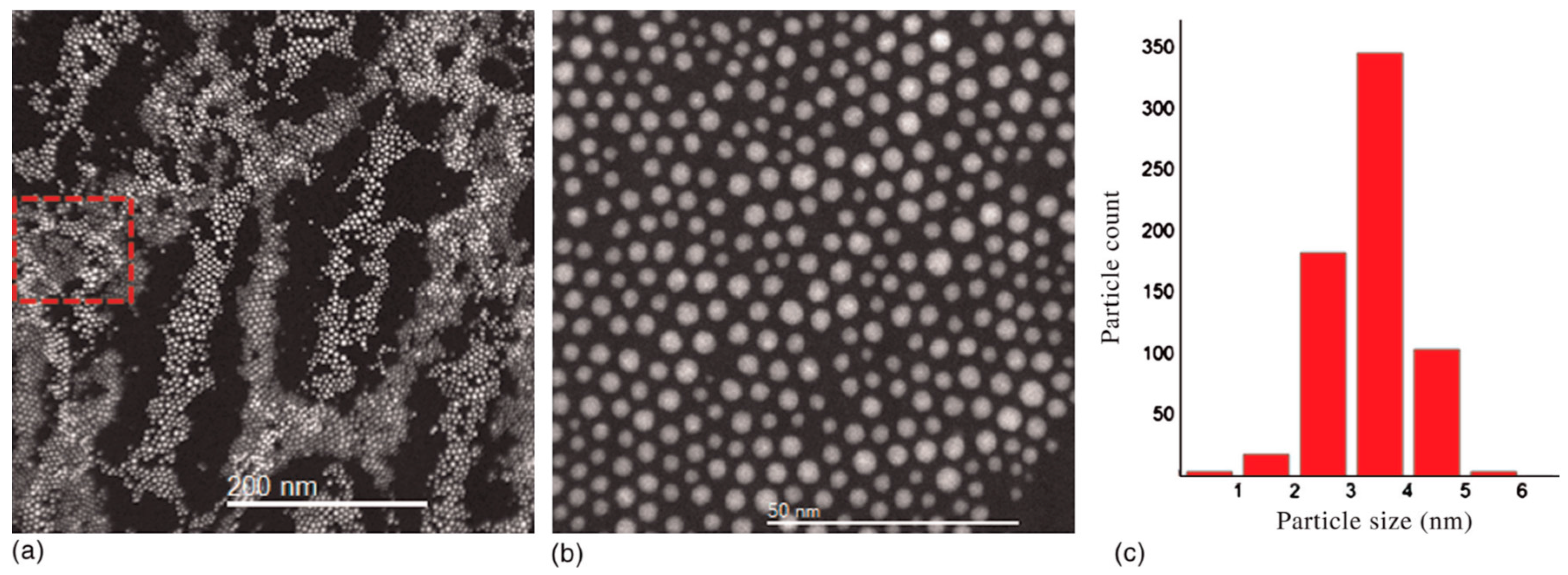
Figure 3.
High resolution TEM images showing the Ag-Fe3O4 nanocomposites, and elemental mapping illustrating the location of the silver and iron oxide. Reprinted with permission from [29]. Copyright Elsevier, 2017.
Figure 3.
High resolution TEM images showing the Ag-Fe3O4 nanocomposites, and elemental mapping illustrating the location of the silver and iron oxide. Reprinted with permission from [29]. Copyright Elsevier, 2017.

Figure 4.
TEM (transmission electron microscopy) images illustrating the size dependence of the supramolecular spiky nanostructures on the amount of smooth Fe3O4@Au seeds initially introduced (a) 4.5 nM; (b) 8.9 nM; (c) 13.2 nM; (d) 17.5 nM. Reprinted with permission from [33]. Copyright WILEY-VCH, 2013.

Figure 5.
TEM images of the Au-Fe3O4 asymmetric nanostars and dumbbells obtained by varying the ratio of dumbbell seeds to gold salt. Reprinted with permission from [39]. Copyright RSC, 2017.
Figure 5.
TEM images of the Au-Fe3O4 asymmetric nanostars and dumbbells obtained by varying the ratio of dumbbell seeds to gold salt. Reprinted with permission from [39]. Copyright RSC, 2017.

Figure 6.
STEM images showing AuCo nanoparticles in amorphous carbon. The histogram illustrates the small size distribution of the nanoparticles. Reprinted with permission from [55]. Copyright MRS, 2013.
Figure 6.
STEM images showing AuCo nanoparticles in amorphous carbon. The histogram illustrates the small size distribution of the nanoparticles. Reprinted with permission from [55]. Copyright MRS, 2013.

Figure 7.
HR-TEM image of silica coated Ag-Fe2O3 dumbbell synthesised by flame spray pyrolysis Reprinted with permission from [56]. Copyright ACS, 2013.
Figure 7.
HR-TEM image of silica coated Ag-Fe2O3 dumbbell synthesised by flame spray pyrolysis Reprinted with permission from [56]. Copyright ACS, 2013.

Figure 8.
(a) A plot illustrating the changes in tumour size in mice following a number of different treatments. The tumours were seen to shrink following treatment with the drug-carrying magnetic plasmonic nanoparticles; (b) Also shown are confocal microscopy images of cells treated with different compounds after a range of incubation times and conditions. Reprinted with permission from [45]. Copyright ACS, 2014.

Figure 9.
Cancer cells labelled with nanoclusters imaged using dark field reflectance. Dark blue-greyish cells are unlabelled. It can be seen that the antigen labelled nanoclusters are only taken up into cells with the complementary antibody. Reprinted with permission from [40]. Copyright WILEY-VCH, 2014.

© 2018 by the authors. Licensee MDPI, Basel, Switzerland. This article is an open access article distributed under the terms and conditions of the Creative Commons Attribution (CC BY) license (http://creativecommons.org/licenses/by/4.0/).
Share and Cite
MDPI and ACS Style
Stafford, S.; Serrano Garcia, R.; Gun’ko, Y.K. Multimodal Magnetic-Plasmonic Nanoparticles for Biomedical Applications. Appl. Sci. 2018, 8, 97. https://doi.org/10.3390/app8010097
AMA Style
Stafford S, Serrano Garcia R, Gun’ko YK. Multimodal Magnetic-Plasmonic Nanoparticles for Biomedical Applications. Applied Sciences. 2018; 8(1):97. https://doi.org/10.3390/app8010097
Chicago/Turabian StyleStafford, Shelley, Raquel Serrano Garcia, and Yurii K. Gun’ko. 2018. "Multimodal Magnetic-Plasmonic Nanoparticles for Biomedical Applications" Applied Sciences 8, no. 1: 97. https://doi.org/10.3390/app8010097
Note that from the first issue of 2016, this journal uses article numbers instead of page numbers. See further details here.