
Synthesis and Characterization of Chitosan-Coated Near-Infrared (NIR) Layered Double Hydroxide-Indocyanine Green Nanocomposites for Potential Applications in Photodynamic Therapy


Abstract
:
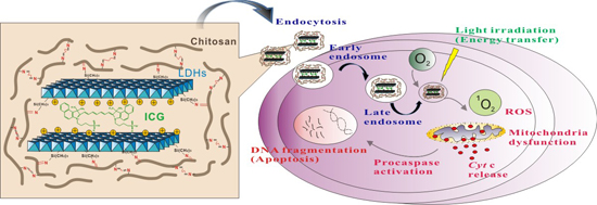
1. Introduction

2. Results and Discussion
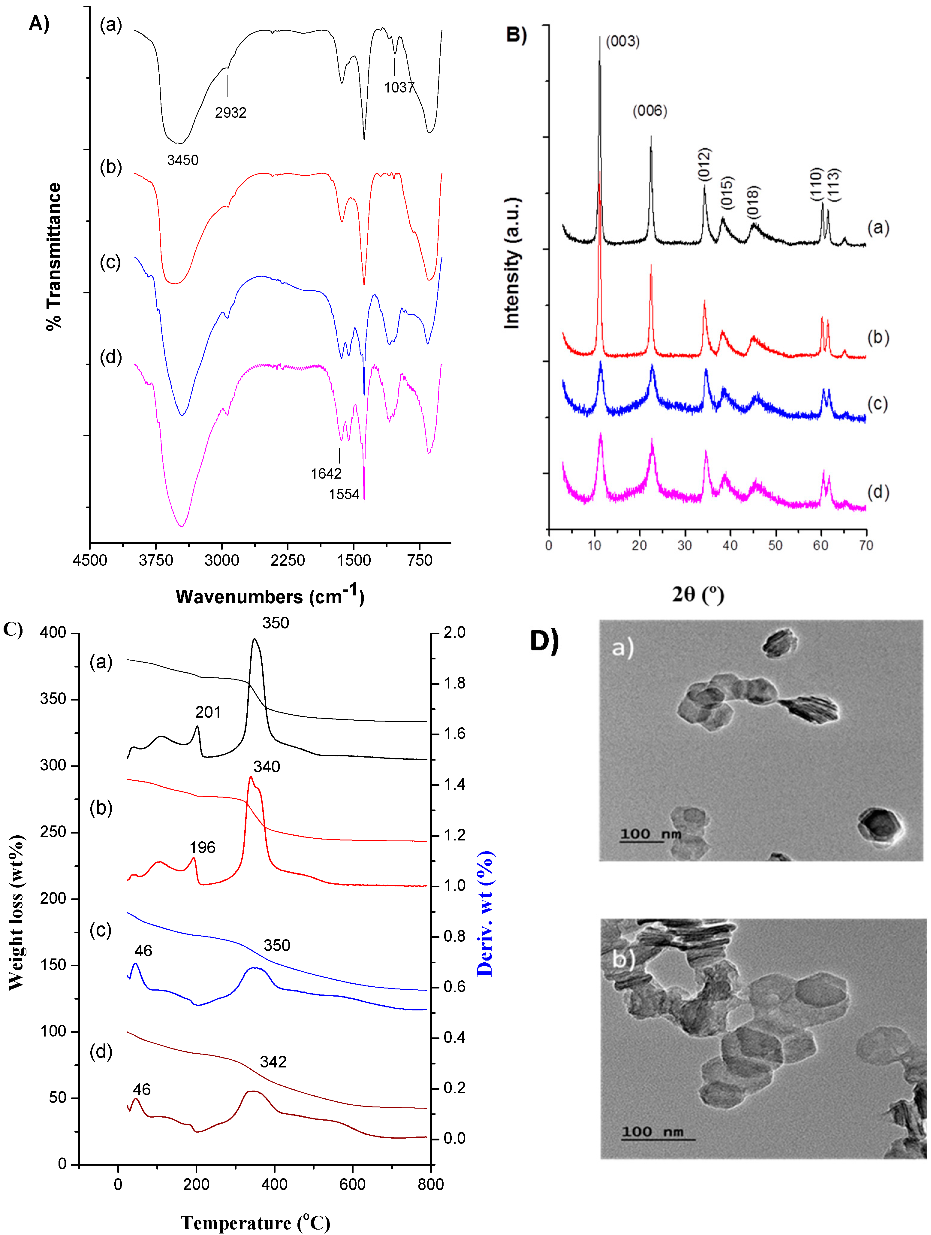
2.1. Physical Characterization of Chitosan-Coated Layered Double Hydroxide (LDH)–NH2–Indocyanine Green (ICG) Matrix

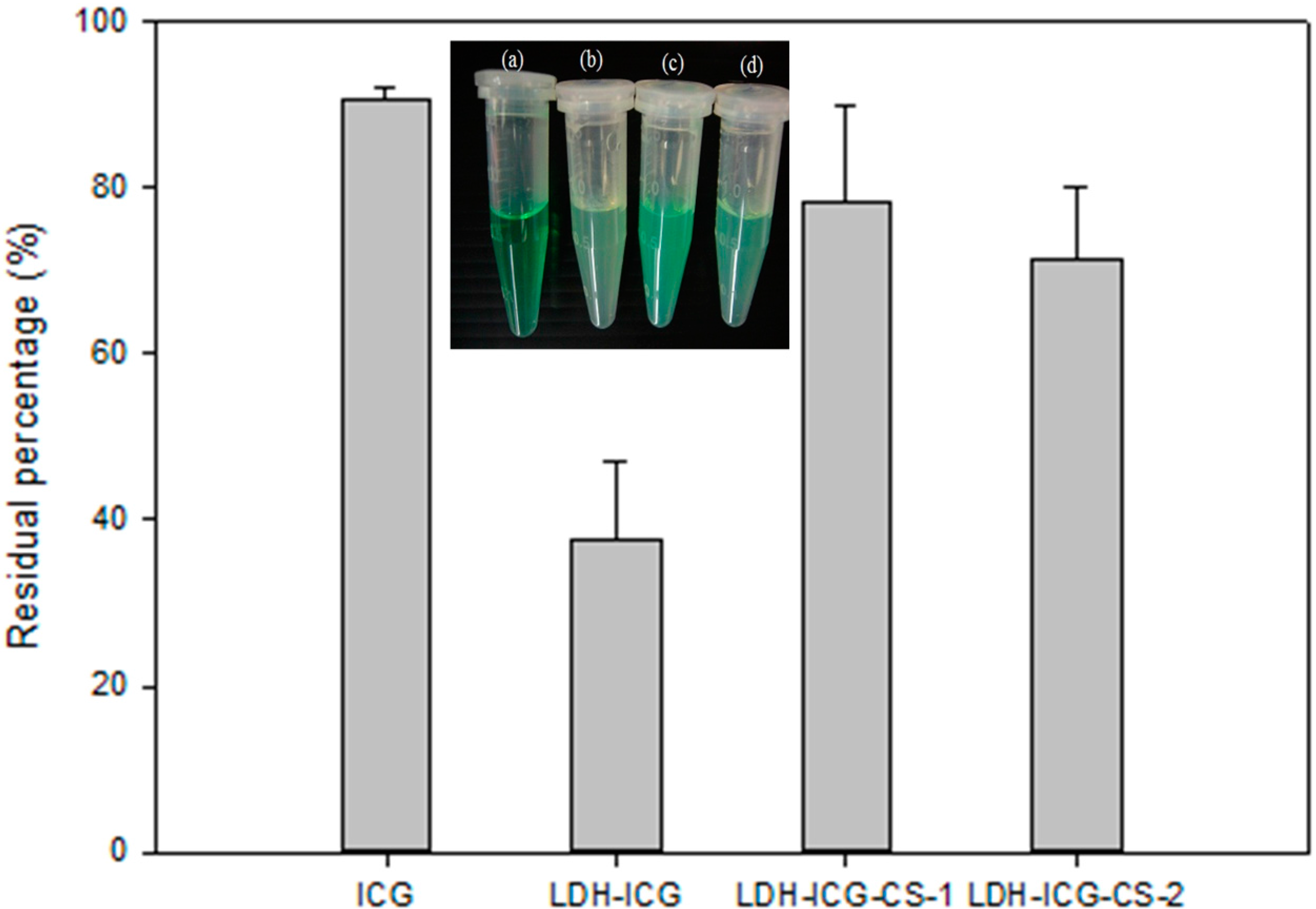
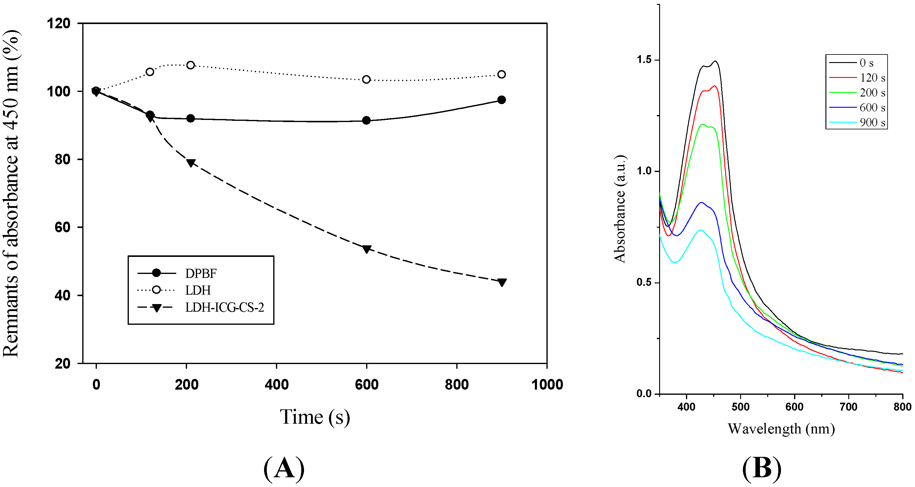
2.2. Photostability of ICG in LDH–Chitosan Matrix

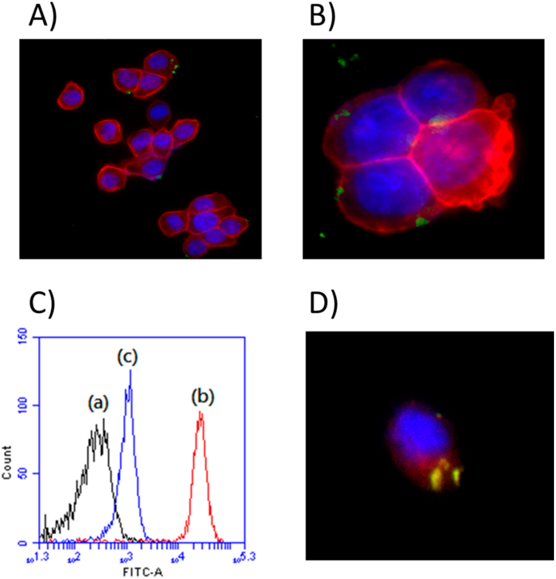
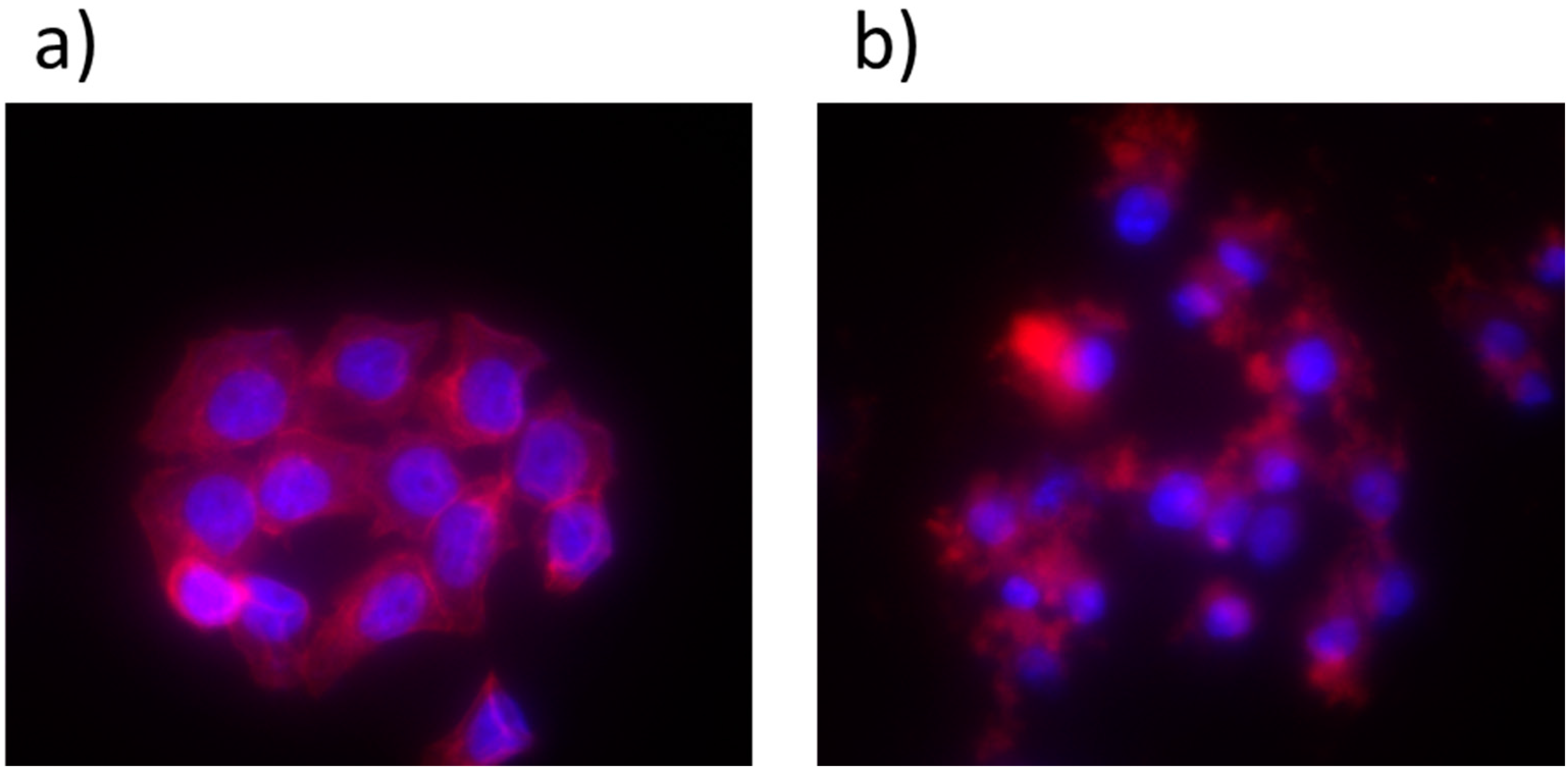
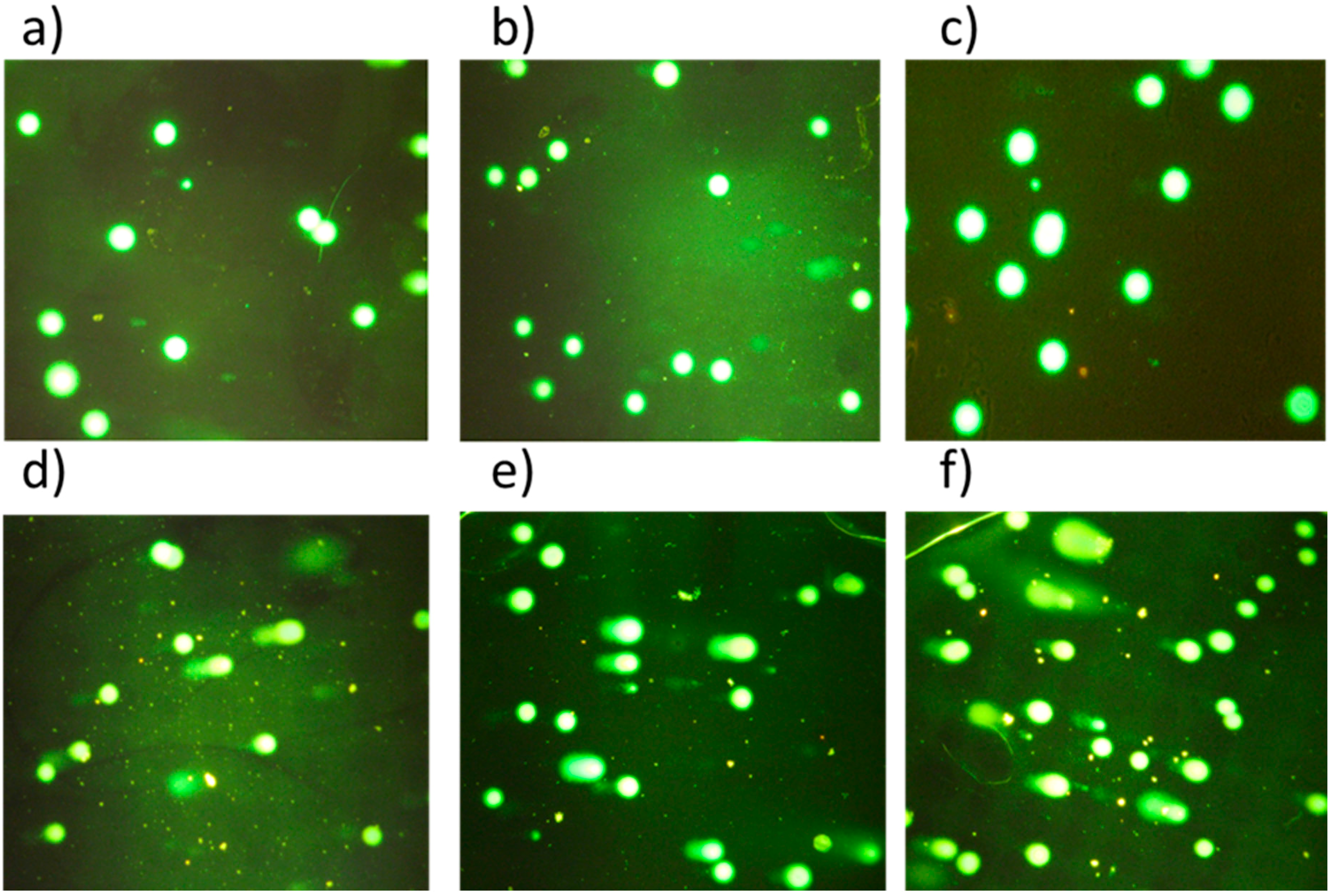
2.3. Cell Uptake and in Vitro Cell Imaging

2.4. Determination of Singlet Oxygen Generation

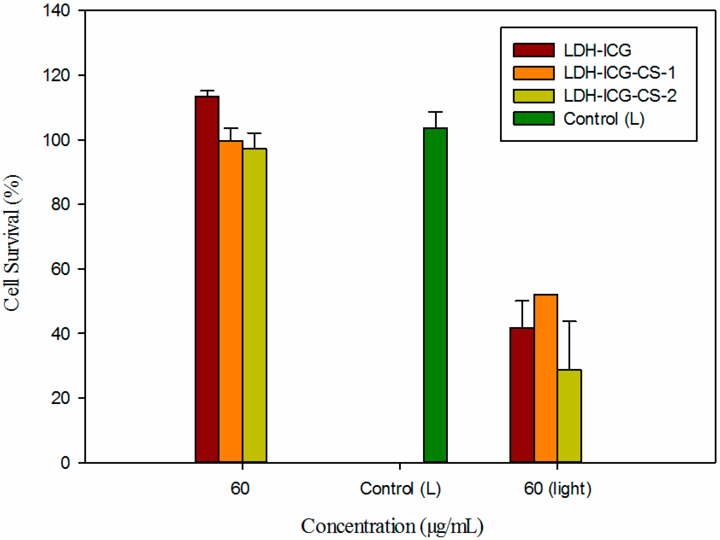
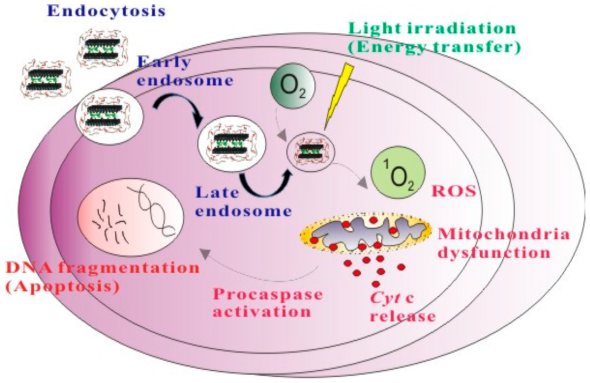
2.5. Phototherapy Assay

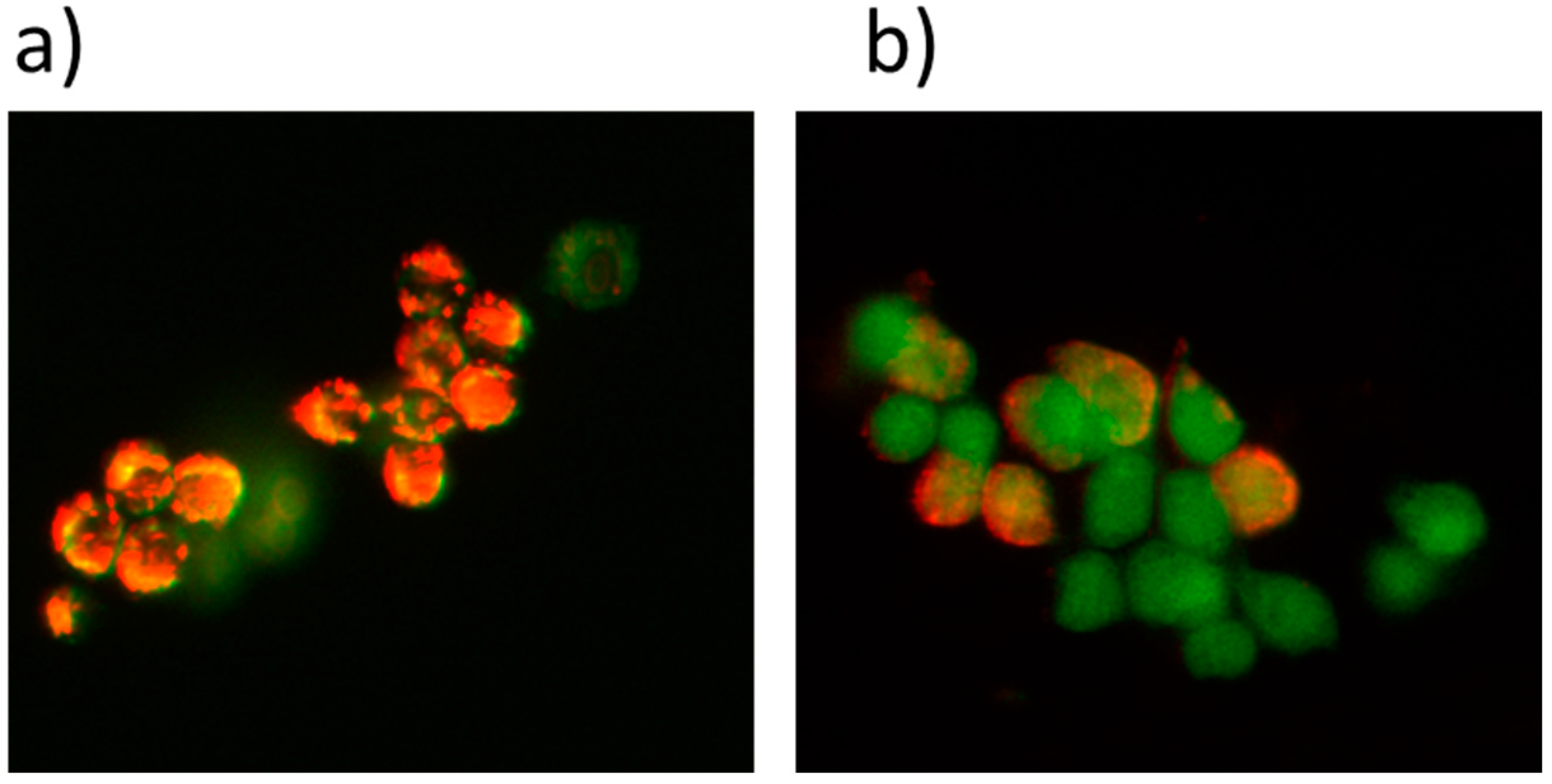
2.6. Apoptosis


3. Experimental Section
3.1. Materials
3.2. Characterization and Instruments
3.3. Synthesis of LDH Nanoparticles
3.4. Synthesis of LDHs–NH2
3.5. Synthesis of LDHs–NH2–ICG
3.6. Synthesis of Chitosan-Coated LDHs–NH2–ICG
3.7. Synthesis of LDH–NH2–FITC–CS (Fluorescein Isothiocyanate–Chitosan)
3.8. Photostability Assay
3.9. Cellular Uptake and Imaging by Confocal Microscopy
3.10. Lysosome Staining
3.11. In Vitro Determination of Singlet Oxygen Production by 1,3-Diphenylisobenzofuran (DPBF)
3.12. Photodynamic Effect Measurements
3.13. Measurements of the Mitochondrial Membrane Potential
3.14. Comet Assay
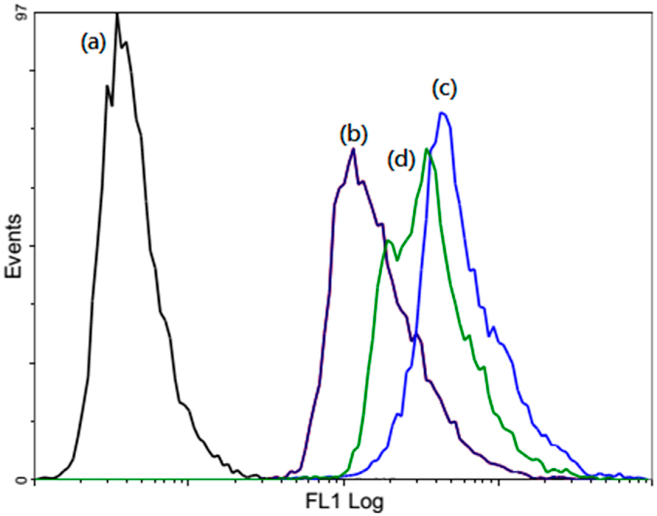
3.15. Flow Cytometric Detections
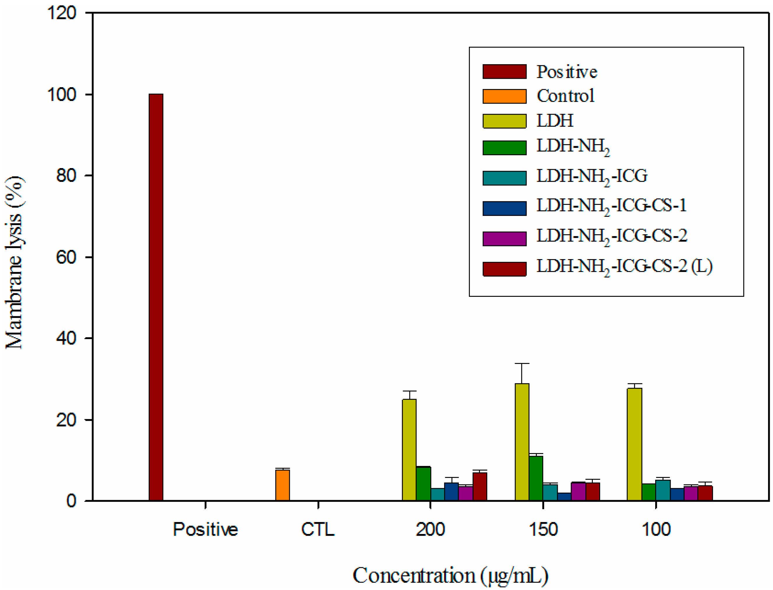
3.16. Lactate Dehydrogenase Assay
4. Conclusions
Supplementary Materials
Acknowledgments
Author Contributions
Conflicts of Interest
References
- Goldenberg, G.; Hamid, O. Nonsurgical treatment options for basal cell carcinoma-focus on advanced disease. J. Drugs Dermatol. 2013, 12, 1369–1378. [Google Scholar] [PubMed]
- Quereux, G.; Brocard, A.; Saint-Jean, M.; Peuvrel, L.; Knol, A.C.; Allix, R.; Khammari, A.; Renaut, J.J.; Dreno, B. Photodynamic therapy with methyl-aminolevulinic acid for paucilesional mycosis fungoides: A prospective open study and review of the literature. J. Am. Acad. Dermatol. 2013, 69, 890–897. [Google Scholar] [CrossRef] [PubMed]
- Ross, K.; Cherpelis, B.; Lien, M.; Fenske, N. Spotlighting the role of photodynamic therapy in cutaneous malignancy: An update and expansion. Dermatol. Surg. 2013, 39, 1733–1744. [Google Scholar] [CrossRef] [PubMed]
- Separovic, D.; Breen, P.; Boppana, N.B.; van Buren, E.; Joseph, N.; Kraveka, J.M.; Rahmaniyan, M.; Li, L.; Gudz, T.I.; Bielawska, A.; et al. Increased killing of SCCVII squamous cell carcinoma cells after the combination of Pc 4 photodynamic therapy and dasatinib is associated with enhanced caspase-3 activity and ceramide synthase 1 upregulation. Int. J. Oncol. 2013, 43, 2064–2072. [Google Scholar] [PubMed]
- Park, J.Y.; Jang, Y.H.; Kim, Y.S.; Sohn, S.; Kim, Y.C. Ultrastructural changes in photorejuvenation induced by photodynamic therapy in a photoaged mouse model. Eur. J. Dermatol. 2013, 23, 471–477. [Google Scholar] [PubMed]
- Almutawa, F.; Thalib, L.; Heckman, D.; Sun, Q.; Hamzavi, I.; Lim, H.W. Efficacy of localized phototherapy and photodynamic therapy for psoriasis: A systematic review and meta-analysis. Photodermatol. Photoimmunol. Photomed. 2015, 31, 5–14. [Google Scholar] [CrossRef] [PubMed]
- Kankala, R.K.; Kuthati, Y.; Liu, C.-L.; Lee, C.-H. Hierarchical coated metal hydroxide nanoconstructs as potential controlled release carriers of photosensitizer for skin melanoma. RSC Adv. 2015, 5, 42666–42680. [Google Scholar] [CrossRef]
- Konan, Y.N.; Gurny, R.; Allemann, E. State of the art in the delivery of photosensitizers for photodynamic therapy. J. Photochem. Photobiol. B 2002, 66, 89–106. [Google Scholar] [CrossRef]
- Van Nostrum, C.F. Delivery of photosensitizers in photodynamic therapy. Adv. Drug Deliv. Rev. 2004, 56, 5–6. [Google Scholar] [CrossRef] [PubMed]
- Kessel, D. Delivery of photosensitizing agents. Adv. Drug Deliv. Rev. 2004, 56, 7–8. [Google Scholar] [CrossRef] [PubMed]
- Anderson, S.J.; Steinman, H.K.; Mazzurco, J.D.; Dixon, A.J. Prolonged adverse events following photodynamic therapy: Regulatory implications. J. Drugs Dermatol. 2014, 13, 62–66. [Google Scholar] [PubMed]
- Chung, I.W.; Eljamel, S. Risk factors for developing oral 5-aminolevulenic acid-induced side effects in patients undergoing fluorescence guided resection. Photodiagnosis Photodyn. Ther. 2013, 10, 362–367. [Google Scholar] [CrossRef] [PubMed]
- Reimer, P.; Balzer, T. Ferucarbotran (Resovist): A new clinically approved res-specific contrast agent for contrast-enhanced mri of the liver: Properties, clinical development, and applications. Eur. Radiol. 2003, 13, 1266–1276. [Google Scholar] [PubMed]
- Rivera Gil, P.; Huhn, D.; del Mercato, L.L.; Sasse, D.; Parak, W.J. Nanopharmacy: Inorganic nanoscale devices as vectors and active compounds. Pharmacol. Res. 2010, 62, 115–125. [Google Scholar]
- Xia, Y. Nanomaterials at work in biomedical research. Nat. Mater. 2008, 7, 758–760. [Google Scholar] [CrossRef] [PubMed]
- Rosenholm, J.M.; Mamaeva, V.; Sahlgren, C.; Linden, M. Nanoparticles in targeted cancer therapy: Mesoporous silica nanoparticles entering preclinical development stage. Nanomedicine (Lond.) 2012, 7, 111–120. [Google Scholar] [CrossRef] [PubMed]
- Bu, L.; Xie, J.; Chen, K.; Huang, J.; Aguilar, Z.P.; Wang, A.; Sun, K.W.; Chua, M.S.; So, S.; Cheng, Z.; et al. Assessment and comparison of magnetic nanoparticles as mri contrast agents in a rodent model of human hepatocellular carcinoma. Contrast Media Mol. Imaging 2012, 7, 363–372. [Google Scholar] [CrossRef] [PubMed]
- Davis, M.E.; Chen, Z.; Shin, D.M. Nanoparticle therapeutics: An emerging treatment modality for cancer. Nat. Rev. Drug Discov. 2008, 7, 771–782. [Google Scholar] [CrossRef] [PubMed]
- Wei, P.-R.; Cheng, S.-H.; Liao, W.-N.; Kao, K.-C.; Weng, C.-F.; Lee, C.-H. Synthesis of chitosan-coated near-infrared layered double hydroxide nanoparticles for in vivo optical imaging. J. Mater. Chem. 2012, 22, 5503–5513. [Google Scholar] [CrossRef]
- Kuthati, Y.; Kankala, R.K.; Lee, C.-H. Layered double hydroxide nanoparticles for biomedical applications: Current status and recent prospects. Appl. Clay Sci. 2015, 112–113, 100–116. [Google Scholar] [CrossRef]
- Wang, Y.; Zhang, D.; Bao, Q.; Wu, J.J.; Wan, Y. Controlled drug release characteristics and enhanced antibacterial effect of graphene oxide-drug intercalated layered double hydroxide hybrid films. J. Mater. Chem. 2012, 22, 23106–23113. [Google Scholar] [CrossRef]
- Kim, T.H.; Lee, J.A.; Choi, S.J.; Oh, J.M. Polymer coated CaAl-layered double hydroxide nanomaterials for potential calcium supplement. Int. J. Mol. Sci. 2014, 15, 22563–22579. [Google Scholar] [CrossRef] [PubMed]
- Zhang, K.; Xu, Z.P.; Lu, J.; Tang, Z.Y.; Zhao, H.J.; Good, D.A.; Wei, M.Q. Potential for layered double hydroxides-based, innovative drug delivery systems. Int. J. Mol. Sci. 2014, 15, 7409–7428. [Google Scholar] [CrossRef] [PubMed]
- Barahuie, F.; Hussein, M.Z.; Fakurazi, S.; Zainal, Z. Development of drug delivery systems based on layered hydroxides for nanomedicine. Int. J. Mol. Sci. 2014, 15, 7750–7786. [Google Scholar] [CrossRef] [PubMed]
- Ariga, K.; Yamauchi, Y.; Rydzek, G.; Ji, Q.; Yonamine, Y.; Wu, K.C.W.; Hill, J.P. Layer-by-layer nanoarchitectonics: Invention, innovation, and evolution. Chem. Lett. 2014, 43, 36–68. [Google Scholar] [CrossRef]
- Kankala, R.K.; Kuthati, Y.; Sie, H.-W.; Shih, H.-Y.; Lue, S.-I.; Kankala, S.; Jeng, C.-C.; Deng, J.-P.; Weng, C.-F.; Liu, C.-L.; et al. Multi-laminated metal hydroxide nanocontainers for oral-specific delivery for bioavailability improvement and treatment of inflammatory paw edema in mice. J. Colloid Interface Sci. 2015, 458, 217–228. [Google Scholar] [CrossRef] [PubMed]
- Miao, Y.-E.; Zhu, H.; Chen, D.; Wang, R.; Tjiu, W.W.; Liu, T. Electrospun fibers of layered double hydroxide/biopolymer nanocomposites as effective drug delivery systems. Mater. Chem. Phys. 2012, 134, 623–630. [Google Scholar] [CrossRef]
- DeLeon, V.H.; Nguyen, T.D.; Nar, M.; D’Souza, N.A.; Golden, T.D. Polymer nanocomposites for improved drug delivery efficiency. Mater. Chem. Phys. 2012, 132, 409–415. [Google Scholar] [CrossRef]
- Zhang, L.; Gu, F.X.; Chan, J.M.; Wang, A.Z.; Langer, R.S.; Farokhzad, O.C. Nanoparticles in medicine: Therapeutic applications and developments. Clin. Pharmacol. Ther. 2008, 83, 761–769. [Google Scholar] [CrossRef] [PubMed]
- Kuo, Y.-M.; Kuthati, Y.; Kankala, R.K.; Wei, P.-R.; Weng, C.-F.; Liu, C.-L.; Sung, P.-J.; Mou, C.-Y.; Lee, C.-H. Layered double hydroxide nanoparticles to enhance organ-specific targeting and the anti-proliferative effect of cisplatin. J. Mater. Chem. B 2015, 3, 3447–3458. [Google Scholar] [CrossRef]
- Choy, J.H.; Jung, J.S.; Oh, J.M.; Park, M.; Jeong, J.; Kang, Y.K.; Han, O.J. Layered double hydroxide as an efficient drug reservoir for folate derivatives. Biomaterials 2004, 25, 3059–3064. [Google Scholar] [CrossRef] [PubMed]
- Choy, J.H.; Kwak, S.Y.; Jeong, Y.J.; Park, J.S. Inorganic layered double hydroxides as nonviral vectors. Angew. Chem. Int. Ed. Engl. 2000, 39, 4042–4045. [Google Scholar]
- Oh, J.M.; Choi, S.J.; Kim, S.T.; Choy, J.H. Cellular uptake mechanism of an inorganic nanovehicle and its drug conjugates: Enhanced efficacy due to clathrin-mediated endocytosis. Bioconjug. Chem. 2006, 17, 1411–1417. [Google Scholar] [CrossRef] [PubMed]
- Oh, J.-M.; Choi, S.-J.; Lee, G.-E.; Kim, J.-E.; Choy, J.-H. Inorganic metal hydroxide nanoparticles for targeted cellular uptake through clathrin-mediated endocytosis. Chem. Asian J. 2009, 4, 67–73. [Google Scholar] [CrossRef] [PubMed]
- Choi, S.J.; Choy, J.H. Layered double hydroxide nanoparticles as target-specific delivery carriers: Uptake mechanism and toxicity. Nanomedicine (Lond.) 2011, 6, 803–814. [Google Scholar] [CrossRef] [PubMed]
- Wong, Y.; Markham, K.; Xu, Z.P.; Chen, M.; Max Lu, G.Q.; Bartlett, P.F.; Cooper, H.M. Efficient delivery of siRNA to cortical neurons using layered double hydroxide nanoparticles. Biomaterials 2010, 31, 8770–8779. [Google Scholar] [CrossRef] [PubMed]
- Xu, Z.P.; Walker, T.L.; Liu, K.L.; Cooper, H.M.; Lu, G.Q.M.; Bartlett, P.F. Layered double hydroxide nanoparticles as cellular delivery vectors of supercoiled plasmid DNA. Int. J. Nanomed. 2007, 2, 163–174. [Google Scholar]
- Ladewig, K.; Niebert, M.; Xu, Z.P.; Gray, P.P.; Lu, G.Q. Controlled preparation of layered double hydroxide nanoparticles and their application as gene delivery vehicles. Appl. Clay Sci. 2010, 48, 280–289. [Google Scholar] [CrossRef]
- Shao, M.; Ning, F.; Zhao, J.; Wei, M.; Evans, D.G.; Duan, X. Preparation of Fe3O4@SiO2@layered double hydroxide core-shell microspheres for magnetic separation of proteins. J. Am. Chem. Soc. 2012, 134, 1071–1077. [Google Scholar] [CrossRef] [PubMed]
- Gu, Z.; Rolfe, B.E.; Xu, Z.P.; Campbell, J.H.; Lu, G.Q.; Thomas, A.C. Antibody-targeted drug delivery to injured arteries using layered double hydroxide nanoparticles. Adv. Healthc. Mater. 2012, 1, 669–673. [Google Scholar] [CrossRef] [PubMed]
- San Román, M.S.; Holgado, M.J.; Salinas, B.; Rives, V. Drug release from layered double hydroxides and from their polylactic acid (PLA) nanocomposites. Appl. Clay Sci. 2013, 71, 1–7. [Google Scholar]
- Kura, A.U.; Ain, N.M.; Hussein, M.Z.; Fakurazi, S.; Hussein-Al-Ali, S.H. Toxicity and metabolism of layered double hydroxide intercalated with levodopa in a Parkinson’s disease model. Int. J. Mol. Sci. 2014, 15, 5916–5927. [Google Scholar] [CrossRef] [PubMed]
- Eili, M.; Shameli, K.; Ibrahim, N.A.; Yunus, W.M. Degradability enhancement of poly(lactic acid) by stearate-Zn3Al LDH nanolayers. Int. J. Mol. Sci. 2012, 13, 7938–7951. [Google Scholar] [CrossRef] [PubMed]
- Kapusetti, G.; Misra, N.; Singh, V.; Kushwaha, R.K.; Maiti, P. Bone cement/layered double hydroxide nanocomposites as potential biomaterials for joint implant. J. Biomed. Mater. Res. A 2012, 100A, 3363–3373. [Google Scholar] [CrossRef] [PubMed]
- Liu, J.; Harrison, R.; Zhou, J.Z.; Liu, T.T.; Yu, C.; Lu, G.Q.; Qiao, S.Z.; Xu, Z.P. Synthesis of nanorattles with layered double hydroxide core and mesoporous silica shell as delivery vehicles. J. Mater. Chem. 2011, 21, 10641–10644. [Google Scholar] [CrossRef]
- Stoica, G.; Castello Serrano, I.; Figuerola, A.; Ugarte, I.; Pacios, R.; Palomares, E. Layered double hydroxides as carriers for quantum dots@silica nanospheres. Nanoscale 2012, 4, 5409–5419. [Google Scholar] [CrossRef] [PubMed]
- El Hadrami, A.; Adam, L.R.; el Hadrami, I.; Daayf, F. Chitosan in plant protection. Mar. Drugs 2010, 8, 968–987. [Google Scholar]
- No, H.K.; Meyers, S.P.; Prinyawiwatkul, W.; Xu, Z. Applications of chitosan for improvement of quality and shelf life of foods: A review. J. Food Sci. 2007, 72, R87–R100. [Google Scholar] [CrossRef] [PubMed]
- Hirano, S.; Hirochi, K.; Hayashi, K.-I.; Mikami, T.; Tachibana, H. Cosmetic and pharmaceutical uses of chitin and chitosan. In Cosmetic and Pharmaceutical Applications of Polymers; Gebelein, C., Cheng, T., Yang, V., Eds.; Springer US: New York, NY, USA, 1991; pp. 95–104. [Google Scholar]
- Patel, M.P.; Patel, R.R.; Patel, J.K. Chitosan mediated targeted drug delivery system: A review. J. Pharm. Pharm. Sci. 2010, 13, 536–557. [Google Scholar] [PubMed]
- Wang, Z.; Ma, R.; Yan, L.; Chen, X.; Zhu, G. Combined chemotherapy and photodynamic therapy using a nanohybrid based on layered double hydroxides to conquer cisplatin resistance. Chem. Commun. 2015, 51, 11587–11590. [Google Scholar] [CrossRef] [PubMed]
- Li, X.-S.; Ke, M.-R.; Huang, W.; Ye, C.-H.; Huang, J.-D. A pH-responsive layered double hydroxide (LDH)–phthalocyanine nanohybrid for efficient photodynamic therapy. Chemistry 2015, 21, 3310–3317. [Google Scholar] [CrossRef] [PubMed]
- Stefanakis, D.; Seimenis, I.; Ghanotakis, D. Synthesis and characterization of gadolinium nanosheets with bound rose bengal: Potential use in photodynamic therapy and MRI. J. Nanopart. Res. 2014, 16, 1–9. [Google Scholar] [CrossRef]
- Liang, R.; Tian, R.; Ma, L.; Zhang, L.; Hu, Y.; Wang, J.; Wei, M.; Yan, D.; Evans, D.G.; Duan, X. A supermolecular photosensitizer with excellent anticancer performance in photodynamic therapy. Adv. Funct. Mater. 2014, 24, 3144–3151. [Google Scholar] [CrossRef]
- Merchan, M.; Ouk, T.S.; Kubat, P.; Lang, K.; Coelho, C.; Verney, V.; Commereuc, S.; Leroux, F.; Sol, V.; Taviot-Gueho, C. Photostability and photobactericidal properties of porphyrin-layered double hydroxide-polyurethane composite films. J. Mater. Chem. B 2013, 1, 2139–2146. [Google Scholar] [CrossRef]
- Lang, K.; Bezdička, P.; Bourdelande, J.L.; Hernando, J.; Jirka, I.; Káfuňková, E.; Kovanda, F.; Kubát, P.; Mosinger, J.; Wagnerová, D.M. Layered double hydroxides with intercalated porphyrins as photofunctional materials: Subtle structural changes modify singlet oxygen production. Chem. Mater. 2007, 19, 3822–3829. [Google Scholar] [CrossRef]
- Kantonis, G.; Trikeriotis, M.; Ghanotakis, D.F. Biocompatible protoporphyrin IX-containing nanohybrids with potential applications in photodynamic therapy. J. Photochem. Photobiol. A Chem. 2007, 185, 62–66. [Google Scholar] [CrossRef]
- Reddi, E.; Zhou, C.; Biolo, R.; Menegaldo, E.; Jori, G. Liposome- or LDL-administered Zn(ii)-phthalocyanine as a photodynamic agent for tumours. I. Pharmacokinetic properties and phototherapeutic efficiency. Br. J. Cancer 1990, 61, 407–411. [Google Scholar] [CrossRef] [PubMed]
- Lee, S.J.; Koo, H.; Jeong, H.; Huh, M.S.; Choi, Y.; Jeong, S.Y.; Byun, Y.; Choi, K.; Kim, K.; Kwon, I.C. Comparative study of photosensitizer loaded and conjugated glycol chitosan nanoparticles for cancer therapy. J. Control. Release 2011, 152, 21–29. [Google Scholar] [CrossRef] [PubMed]
- Sunil Virkar, A.C. Synthesis and characterization of nanocomposites based on polyethylene and Mg-Al layered double hydroxide with intercalated compounds. Adv. Mater. Sci. Appl. 2014, 3, 150–156. [Google Scholar]
- Wang, Z.Y.; Zhao, Y.; Ren, L.; Jin, L.H.; Sun, L.P.; Yin, P.; Zhang, Y.F.; Zhang, Q.Q. Novel gelatin-siloxane nanoparticles decorated by tat peptide as vectors for gene therapy. Nanotechnology 2008, 19, 445103. [Google Scholar] [CrossRef] [PubMed]
- Watson, P.; Jones, A.T.; Stephens, D.J. Intracellular trafficking pathways and drug delivery: Fluorescence imaging of living and fixed cells. Adv. Drug Deliv. Rev. 2005, 57, 43–61. [Google Scholar] [CrossRef] [PubMed]
- Lee, S.J.; Koo, H.; Lee, D.E.; Min, S.; Lee, S.; Chen, X.; Choi, Y.; Leary, J.F.; Park, K.; Jeong, S.Y.; et al. Tumor-homing photosensitizer-conjugated glycol chitosan nanoparticles for synchronous photodynamic imaging and therapy based on cellular on/off system. Biomaterials 2011, 32, 4021–4029. [Google Scholar] [CrossRef] [PubMed]
- Van Nostrum, C.F. Polymeric micelles to deliver photosensitizers for photodynamic therapy. Adv. Drug Deliv. Rev. 2004, 56, 9–16. [Google Scholar]
- Dougherty, T.J.; Gomer, C.J.; Henderson, B.W.; Jori, G.; Kessel, D.; Korbelik, M.; Moan, J.; Peng, Q. Photodynamic therapy. J. Natl. Cancer Inst. 1998, 90, 889–905. [Google Scholar] [CrossRef] [PubMed]
- McNair, F.I.; Marples, B.; West, C.M.; Moore, J.V. A comet assay of DNA damage and repair in K562 cells after photodynamic therapy using haematoporphyrin derivative, methylene blue and meso-tetrahydroxyphenylchlorin. Br. J. Cancer 1997, 75, 1721–1729. [Google Scholar] [CrossRef] [PubMed]
- Haylett, A.K.; Ward, T.H.; Moore, J.V. DNA damage and repair in gorlin syndrome and normal fibroblasts after aminolevulinic acid photodynamic therapy: A comet assay study. Photochem. Photobiol. 2003, 78, 337–341. [Google Scholar] [CrossRef]
- Woods, J.A.; Traynor, N.J.; Brancaleon, L.; Moseley, H. The effect of photofrin on DNA strand breaks and base oxidation in hacat keratinocytes: A comet assay study. Photochem. Photobiol. 2004, 79, 105–113. [Google Scholar] [CrossRef] [PubMed]
- El-Hussein, A.; Harith, M.; Abrahamse, H. Assessment of DNA damage after photodynamic therapy using a metallophthalocyanine photosensitizer. Int. J. Photoenergy 2012, 2012, 281068. [Google Scholar] [CrossRef]
- Miller, J.D.; Baron, E.D.; Scull, H.; Hsia, A.; Berlin, J.C.; McCormick, T.; Colussi, V.; Kenney, M.E.; Cooper, K.D.; Oleinick, N.L. Photodynamic therapy with the phthalocyanine photosensitizer Pc 4: The case experience with preclinical mechanistic and early clinical-translational studies. Toxicol. Appl. Pharmacol. 2007, 224, 290–299. [Google Scholar] [CrossRef] [PubMed]
- Oh, J.-M.; Choi, S.-J.; Lee, G.-E.; Han, S.-H.; Choy, J.-H. Inorganic drug-delivery nanovehicle conjugated with cancer-cell-specific ligand. Adv. Funct. Mater. 2009, 19, 1617–1624. [Google Scholar] [CrossRef]
- Xu, Z.P.; Stevenson, G.S.; Lu, C.Q.; Lu, G.Q.; Bartlett, P.F.; Gray, P.P. Stable suspension of layered double hydroxide nanoparticles in aqueous solution. J. Am. Chem. Soc. 2006, 128, 36–37. [Google Scholar] [CrossRef] [PubMed]
- Ladewig, K.; Niebert, M.; Xu, Z.P.; Gray, P.P.; Lu, G.Q. Efficient sirna delivery to mammalian cells using layered double hydroxide nanoparticles. Biomaterials 2010, 31, 1821–1829. [Google Scholar] [CrossRef] [PubMed]
- Xu, Z.P.; Stevenson, G.; Lu, C.Q.; Lu, G.Q. Dispersion and size control of layered double hydroxide nanoparticles in aqueous solutions. J. Phys. Chem. B 2006, 110, 16923–16929. [Google Scholar] [CrossRef] [PubMed]
- Mathur, A.; Hong, Y.; Kemp, B.K.; Barrientos, A.A.; Erusalimsky, J.D. Evaluation of fluorescent dyes for the detection of mitochondrial membrane potential changes in cultured cardiomyocytes. Cardiovasc. Res. 2000, 46, 126–138. [Google Scholar] [CrossRef]
- Mortera, R.; Vivero-Escoto, J.; Slowing, I.I.; Garrone, E.; Onida, B.; Lin, V.S.Y. Cell-induced intracellular controlled release of membrane impermeable cysteine from a mesoporous silica nanoparticle-based drug delivery system. Chem. Commun. 2009, 3219–3221. [Google Scholar] [CrossRef] [PubMed]
© 2015 by the authors; licensee MDPI, Basel, Switzerland. This article is an open access article distributed under the terms and conditions of the Creative Commons Attribution license (http://creativecommons.org/licenses/by/4.0/).
Share and Cite
Wei, P.-R.; Kuthati, Y.; Kankala, R.K.; Lee, C.-H. Synthesis and Characterization of Chitosan-Coated Near-Infrared (NIR) Layered Double Hydroxide-Indocyanine Green Nanocomposites for Potential Applications in Photodynamic Therapy. Int. J. Mol. Sci. 2015, 16, 20943-20968. https://doi.org/10.3390/ijms160920943
Wei P-R, Kuthati Y, Kankala RK, Lee C-H. Synthesis and Characterization of Chitosan-Coated Near-Infrared (NIR) Layered Double Hydroxide-Indocyanine Green Nanocomposites for Potential Applications in Photodynamic Therapy. International Journal of Molecular Sciences. 2015; 16(9):20943-20968. https://doi.org/10.3390/ijms160920943
Chicago/Turabian StyleWei, Pei-Ru, Yaswanth Kuthati, Ranjith Kumar Kankala, and Chia-Hung Lee. 2015. "Synthesis and Characterization of Chitosan-Coated Near-Infrared (NIR) Layered Double Hydroxide-Indocyanine Green Nanocomposites for Potential Applications in Photodynamic Therapy" International Journal of Molecular Sciences 16, no. 9: 20943-20968. https://doi.org/10.3390/ijms160920943