Article

Phytotoxicity, Bioaccumulation, and Degradation of Nonylphenol in Different Microalgal Species without Bacterial Influences

1
College of Life Science and Resources and Environment, Yichun University, Yichun 336000, China
2
School of Environment and Energy, South China University of Technology, Guangzhou 510006, China
3
Southern Marine Science and Engineering Guangdong Laboratory (Zhuhai), Zhuhai Key Laboratory of Marine Bioresources and Environment, Guangdong Provincial Key Laboratory of Marine Resources and Coastal Engineering, School of Marine Sciences, Sun Yat-Sen University, Guangzhou 510275, China
4
Institute for Marine & Antarctic Studies, University of Tasmania, Private Bag 49, Hobart, TAS 7001, Australia
5
South China Institute of Environmental Science, Ministry of Ecology and Environment, NO.18 Ruihe RD., Guangzhou 510535, China
6
Institute of Hydrobiology, Jinan University, Guangzhou 510632, China
*
Author to whom correspondence should be addressed.
Int. J. Mol. Sci. 2020, 21(4), 1338; https://doi.org/10.3390/ijms21041338
Submission received: 14 December 2019 / Revised: 7 February 2020 / Accepted: 13 February 2020 / Published: 17 February 2020
(This article belongs to the Section Biochemistry)

Abstract

:
Nonylphenol (NP) is a contaminant that has negative impacts on aquatic organisms. To investigate its phytotoxicity, bioaccumulation, and degradation in algae without associated bacteria, six freshwater microalgae—Ankistrodesmus acicularis, Chlorella vulgaris, Chroococcus minutus, Scenedesmus obliquus, Scenedesmus quadricauda, and Selenastrum bibraianum—in bacteria-free cultures were studied. When exposed to 0.5–3.0 mg L−1 NP for 4 days, cell growth and photosynthesis, including maximal photochemistry (Fv/Fm), were suppressed progressively. The antioxidant responses of superoxide dismutase (SOD), catalase (CAT), and peroxidase (POD) showed species differences. While the antioxidant enzymes in C. vulgaris and S. obliquus were more active with the increase of NP (0–3 mg L−1), they dropped in the other four algae at concentrations of 1 and 1.5 mg L−1. Therefore, C. vulgaris and S. obliquus were designated as NP-tolerant species and showed more conspicuous and faster changes of antioxidant reactions compared with the four NP-sensitive species. All six species degraded NP, but A. acicularis was more reactive at low NP concentrations (<1 mg L−1), suggesting its possible application in sewage treatment for its potential for effective NP removal from water bodies in a suitable scope. Therefore, the conclusion is that biodegradation of NP by algae is species specific.

1. Introduction

Nonylphenol (NP) is a common contaminant widely used in industrial, commercial, and household products such as detergents; emulsifiers; wetting, dispersing, and antistatic agents; demulsifiers; and solubilizers [1,2]. It is a microbial biodegradation product of nonylphenol ethoxylates (NPEOs) and is persistent, toxic, and disrupts endocrine function. NP has frequently been detected in rivers, lakes, ocean sediments, and soils. Although it is forbidden by the European Union to use NP or its ethoxylates [3], levels from 10 ng L−1 to over 100 ug L−1 have still been detected in the United States, China, and Japan, especially in polluted areas [4,5,6,7].
The mechanism by which NP is toxic to aquatic animals is not understood. Microalgae have simple life histories and maintain the balance of aquatic ecosystems. They have the potential to take up and degrade contaminants such as herbicides, pesticides, and phenols [8,9]. Our knowledge of the impact of NP on algae is lacking. Of special interest are those algae which are useful for biological research in nutrient enrichment, organic contamination, heavy metals, and various other stresses [10,11,12,13,14,15,16,17]. Therefore, we consider research on the toxicity of and adaption to NP biodegradation in unicellular algae worthwhile.
In nature, microalgae are associated with specific bacteria called the algal microbiome, which plays a critical role in modulating algal populations. Such a microbiome makes it difficult to assess the relationship between microalgae and NP, as the associated bacteria can degrade NP under aerobic conditions [18,19]. Hence, elimination of bacteria from stock algal cultures is important in order to study the toxicity and degradation of NP by microalgae.
Our previous study of removal and biodegradation of NP using microalgae (such as Scenedesmus quadricauda, Ankistrodesmus acicularis, Chlorella vulgaris, and Chroococcus minutus) focused on the growth, removal, and biodegradation rates [20]. Scant data, however, have showed the phytotoxicity effects of NP on various microalgae species. Therefore, a better understanding of physiological irritability variation in NP exposure of microalgae is needed. This study aimed to illustrate the phytotoxicity of NP and its accumulation in six freshwater microalgae obtained from NP-polluted water. We exposed five green algae (Chlorella vulgaris JNU38, Scenedesmus obliquus JNU15, Selenastrum bibraianum JNU28, Ankistrodesmus acicularis JNU14, and Scenedesmus quadricauda JNU39) and one cyanobacterium (Chroococcus minutus JNU17) from bacteria-free cultures to NP. The growth, fluorescence, antioxidant enzyme activity, accumulation of NP, degradation of adsorbed NP, as well as the ability to eliminate NP from the medium were determined. This work establishes the ecotoxicology of unicellular algae without bacterial influence under different NP levels and the adaptive response to organic xenobiotics.

2. Results and Discussion

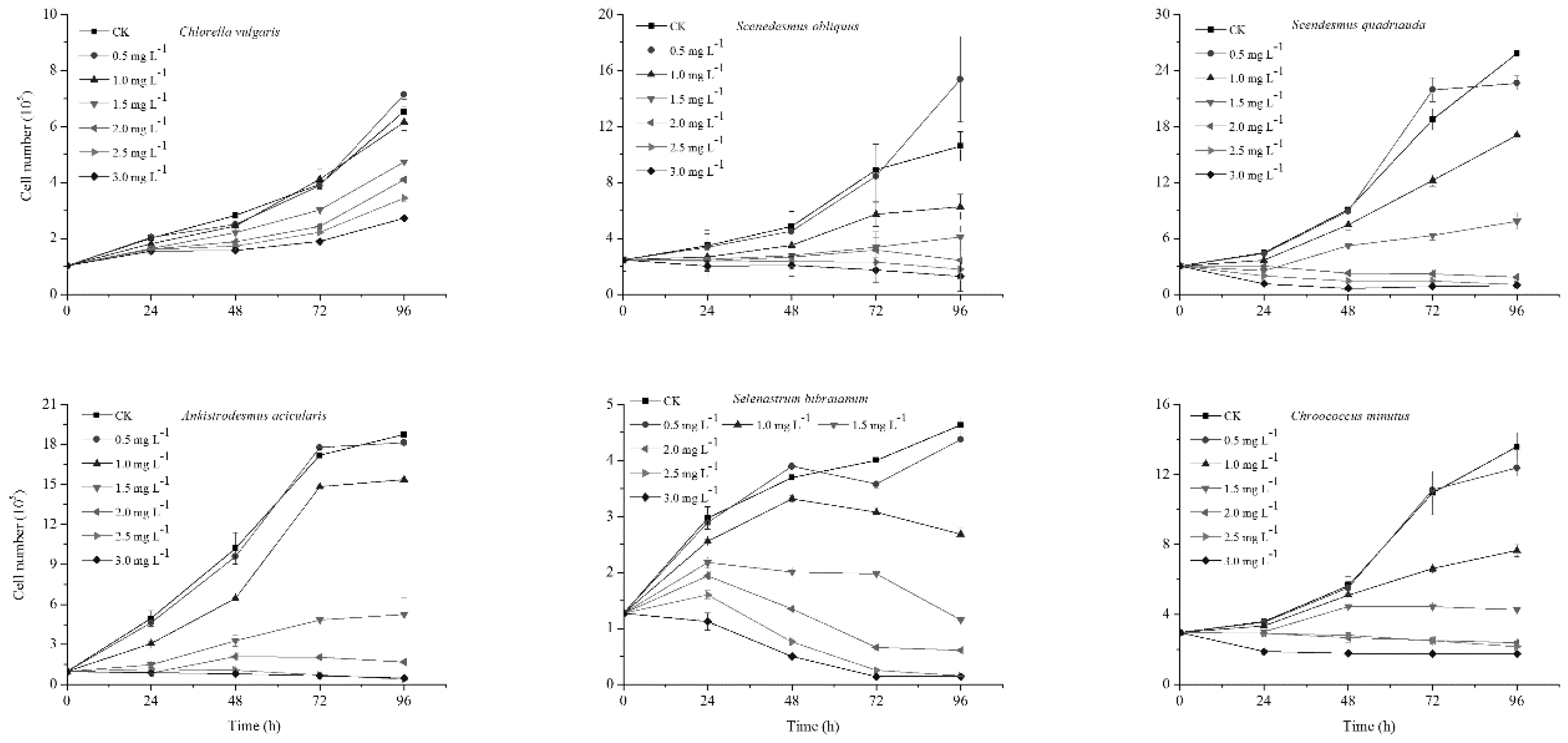
2.1. Affect of NP on Algal Growth

The effects of NP on the growth of C. vulgaris, S. obliquus, S. bibraianum, A. acicularis, S. quadricauda, and C. minutus under different NP concentrations (0.5, 1.0, 1.5, 2.0, 2.5, and 3.0 mg L−1) depended on species, exposure times, and concentrations (Figure 1). Although S. bibraianum and C. minutus showed different drops in growth at 96 h compared with the control, NP at levels between 0 and 0.5 mg L−1 had no influence on growth after 0–72 h exposure. The increase in cell density at low NP concentrations demonstrated the “poison exciting effect (hormesis)” [21]. However, the growth patterns among species varied at NP concentrations from 1.0 to 3.0 mg L−1. At 1.0 mg L−1, cell density in C. vulgaris exhibited a slight decrease at 96 h compared with the control (Figure 1). At 1.0-3.0 mg L−1, the cell densities of S. obliquus, S. bibraianum, A. acicularis, S. quadricauda, and C. minutus dropped at all exposure times, and there were negative correlations between cell density and NP levels. High concentrations of NP reduced algal growth and the toxicity of NP increased with exposure time (Figure 1). For C. vulgaris, S. obliquus, S. bibraianum, A. acicularis, S. quadricauda, and C. minutus, cell density at 3.0 mg L−1 of NP following 4 days of exposure reduced to 41.67%, 12.21%, 3.83%, 2.59%, 3.00%, and 12.85% of the controls, respectively. C. vulgaris showed the highest tolerance to NP, followed by S. obliquus, while the other algae were more sensitive to NP than C. vulgaris and S. obliquus.
Microalgae withstand damage by organic pollutants by many mechanisms. For example, cell walls containing a high amount of carbohydrates and proteins serve as barriers [22]. C. vulgaris produces extra cell wall polysaccharides to cope with high concentrations of pollutants [23]. Further, tolerance to oxidative stress plays a vital role as a defense mechanism [24,25].
A regression equation of NP concentration with the growth inhibition rate was obtained (Table 1). Logistic NP concentrations and inhibitory rate in the present study exhibited a dosage–response relationship. The respective 96 h median inhibitory effect concentration (EC50) values of NP to C. vulgaris, S. obliquus, S. bibraianum, A. acicularis, S. quadricauda, and C. minutus were 1.534, 1.179, 1.177, 1.100, 1.080, and 1.005 mg L−1 (Table 1). The EC50 values of NP to C. vulgaris were much higher than those of other algae. The lowest EC50 values of NP indicated its higher toxicity to C. minutus. Previous studies reported that the EC50 of NP varied from 0.017 to 3.00 mg L−1 to teleosts, and 0.021 to 3.00 mg L−1 to invertebrates [26]. A similar result showed that the 96 h EC50 of NP was 1.01–1.53 mg L−1. The 96 h EC50 of NP for C. minutus was 1.01 mg L−1, illustrating that C. minutus was more sensitive than the other species (Table 2).

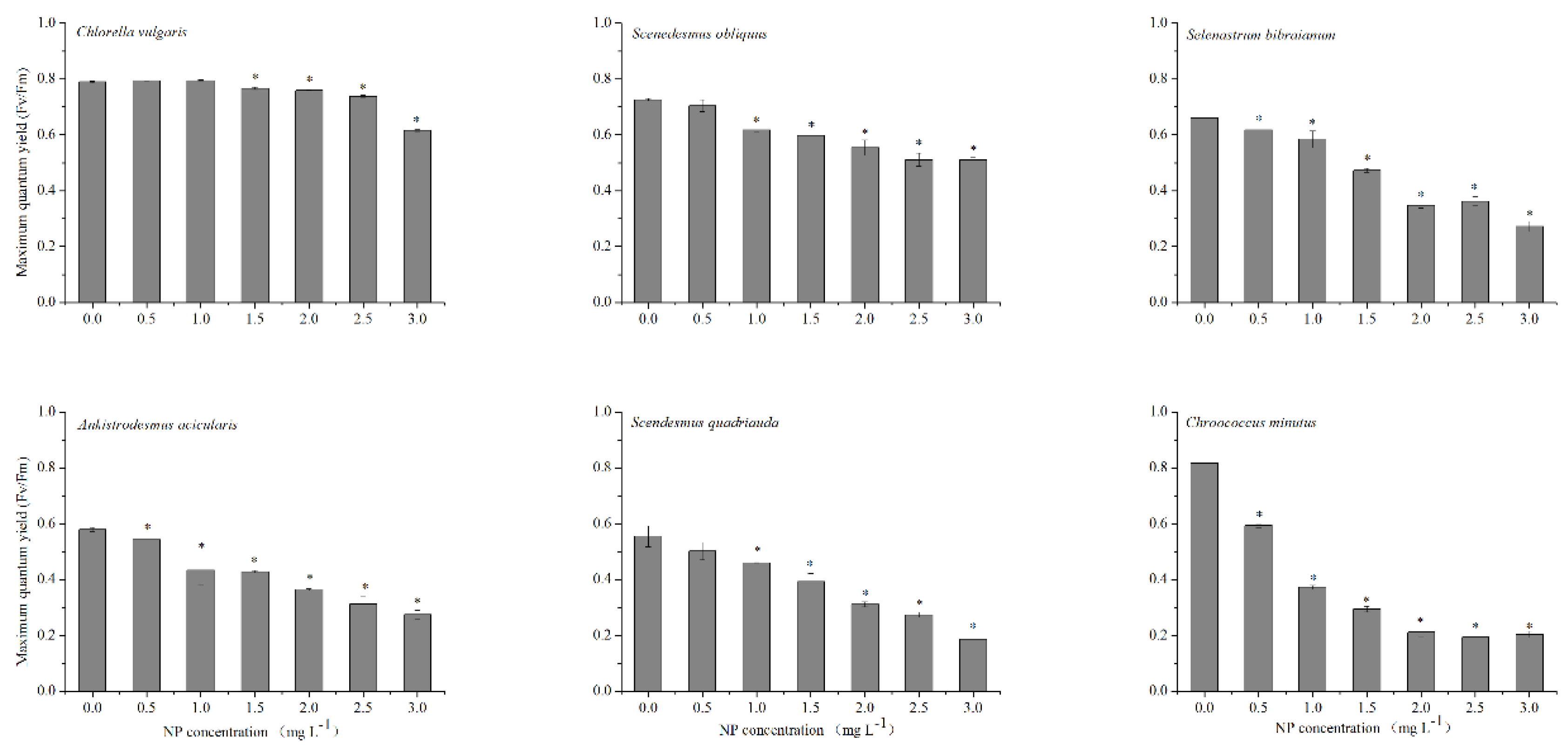
2.2. Influence of NP on Chlorophyll Fluorescence

Maximal photochemistry (Fv/Fm), which originates mainly from the chlorophyll of PSII and illustrates the chlorophyll fluorescence emission of photosynthetic active organisms, has been broadly applied to elucidate environmental stresses [34]. When exposed to high levels of NP (1.5–3.0 mg L−1) after 96 h, Fv/Fm of the six algae were all significantly reduced, while Fv/Fm in C. vulgaris was noticeably higher compared with other algae (p < 0.05) (Figure 2). Fv/Fm of S. obliquus, S. bibraianum, A. acicularis, S. quadricauda, and C. minutus were significantly reduced with the increase of NP concentrations (p < 0.05). However, Fv/Fm in C. vulgaris at low NP concentrations (1.0–1.5 mg L−1) was insignificant (Figure 2). Fv/Fm in C. vulgaris, S. obliquus, S. bibraianum, A. acicularis, S. quadricauda, and C. minutus at 3.0 mg L−1 of NP were 77.9%, 70.3%, 41.2%, 47.4%, 33.6%, and 25.0% of their respective controls. Highly toxic pollutants damage the PSII system, resulting in a decrease of Fv/Fm and inducing a strong inhibition of photosynthetic electron transport, as demonstrated by the decrease of ΦsPSII [12]. Further, NP concentrations higher than 1.5 mg L−1 damaged algal cells, but NP below 1.5 mg L−1 had an undetectable influence on C. vulgaris. In summary, exposure of algae to NP levels higher than 1.5 mg L−1 inactivated PSII reaction centers and thus suppressed electron transport in PSII. By comparison, C. vulgaris exhibited higher tolerance to low NP levels. This corresponded to the estimation of EC50.

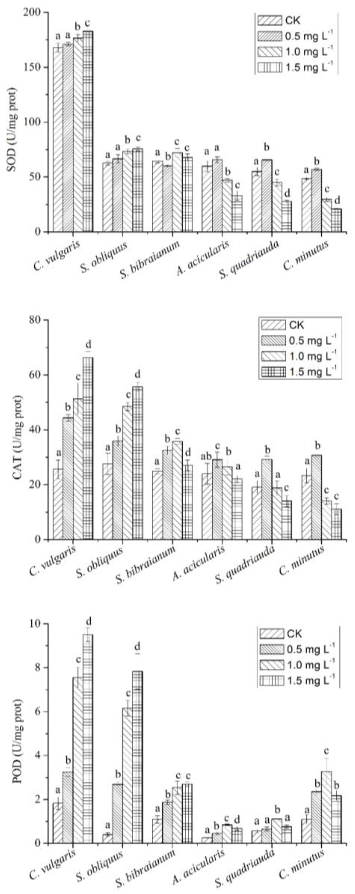
2.3. The Relationship between NP and Antioxidant Enzymes

As demonstrated by previous studies, oxidative stress may be partially responsible for the toxicity of NP due to the promotion of the generation of reactive oxygen species (ROS) and/or inhibition of the antioxidant system [35]. An excessive amount of ROS harms plants, including algae, by reacting to biomolecules at varying degrees and by direct damage to proteins, amino acids, nucleic acids, porphyrins, phenolic substances, and so forth [36]. To defend against the ROS-caused deleterious effects resulting from cellular oxidative stress, antioxidant enzymes play vital roles in the antioxidant system. Therefore, changes in antioxidants induced by NP reflect the toxicity of NP.
Algae respond to oxidative stress by strengthening the antioxidant defense systems, especially by inducing antioxidant enzymes [37]. Superoxide dismutase (SOD), the first line of defense, catalyzes dismutation of O2 to H2O2. The drop of SOD depends on both NP concentration and algal species (Figure 3). SOD in C. vulgaris in the control group was significantly lower in comparison with other groups (p < 0.001). SOD in C. vulgaris was the highest, followed by S. obliquus, S. bibraianum, A. acicularis, S. quadricauda, and C. minutus (Figure 3), which was consistent with EC50 values. For S. bibraianum, C. vulgaris, and S. obliques exposed to 1.5 mg L−1 of NP, the activity of SOD increased by 5%, 9%, and 21%, respectively, compared with their control (p < 0.05) (Figure 3). For A. acicularis, S. quadricauda, and C. minutus, SOD exhibited slight increases with the rise of NP. However, when the NP level was high, SOD was reduced by 45%, 50%, and 57%, respectively, in comparison with their control (p < 0.05) (Figure 3), probably because the protein structure was damaged.
Hydrogen peroxide (H2O2) is a highly toxic by-product in SOD-mediated reactions. Its level, therefore, should be under tight control [38,39]. Catalase (CAT) is one of the key enzymes that scavenge H2O2. The highest CAT activity in this work was observed in C. vulgaris, followed by S. obliquus, S. bibraianum, A. acicularis, and S. quadricauda, and the lowest activity was presented in C. minutus (Figure 2), which was consistent with EC50.
The most significant change in CAT was found in C. vulgaris at 1.5 mg L−1 NP, with a 2.59-fold increase in comparison with the control treatment. CAT in C. vulgaris and S. obliquus showed an increase at all NP treatments. CAT in A. acicularis, S. quadricauda, and C. minutus presented a noticeable increase at low NP but was obviously suppressed at 1.0–1.5 mg L−1 NP (Figure 3). CAT in S. quadricauda and C. minutus decreased 1.36- and 2.09-fold, respectively, at 1.5 mg L−1 of NP compared with the control. CAT in A. acicularis increased when NP was below 0.5 mg L−1, followed by a drop towards the control at 1.5 mg L−1. Peroxidase (POD) showed different patterns in comparison with SOD and CAT, so that the minimum activity was shown in the control group of C. vulgaris, followed by C. minutus, S. bibraianum, S. quadricauda, and S. obliquus (Figure 3).
In this study, the changes in antioxidants indicated that the microalgae were suffering from oxidative stress when exposed to NP (Figure 3). SOD in algae decreased when the NP concentration was high, indicating that NP significantly inhibited SOD production to eliminate H2O2. The increase of CAT and POD indicated that CAT and POD also contributed to the removal of H2O2. In comparison with NP-sensitive species, a recent study showed similarly that C. vulgaris is an NP-tolerant species, as exhibited by a higher and more rapid increase in CAT [35].

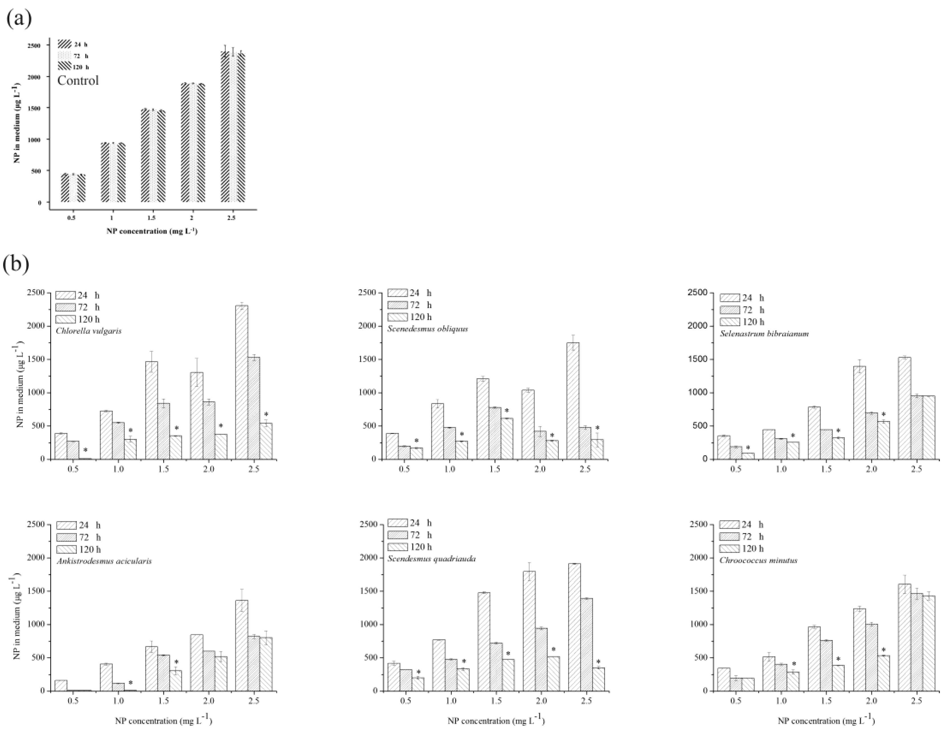
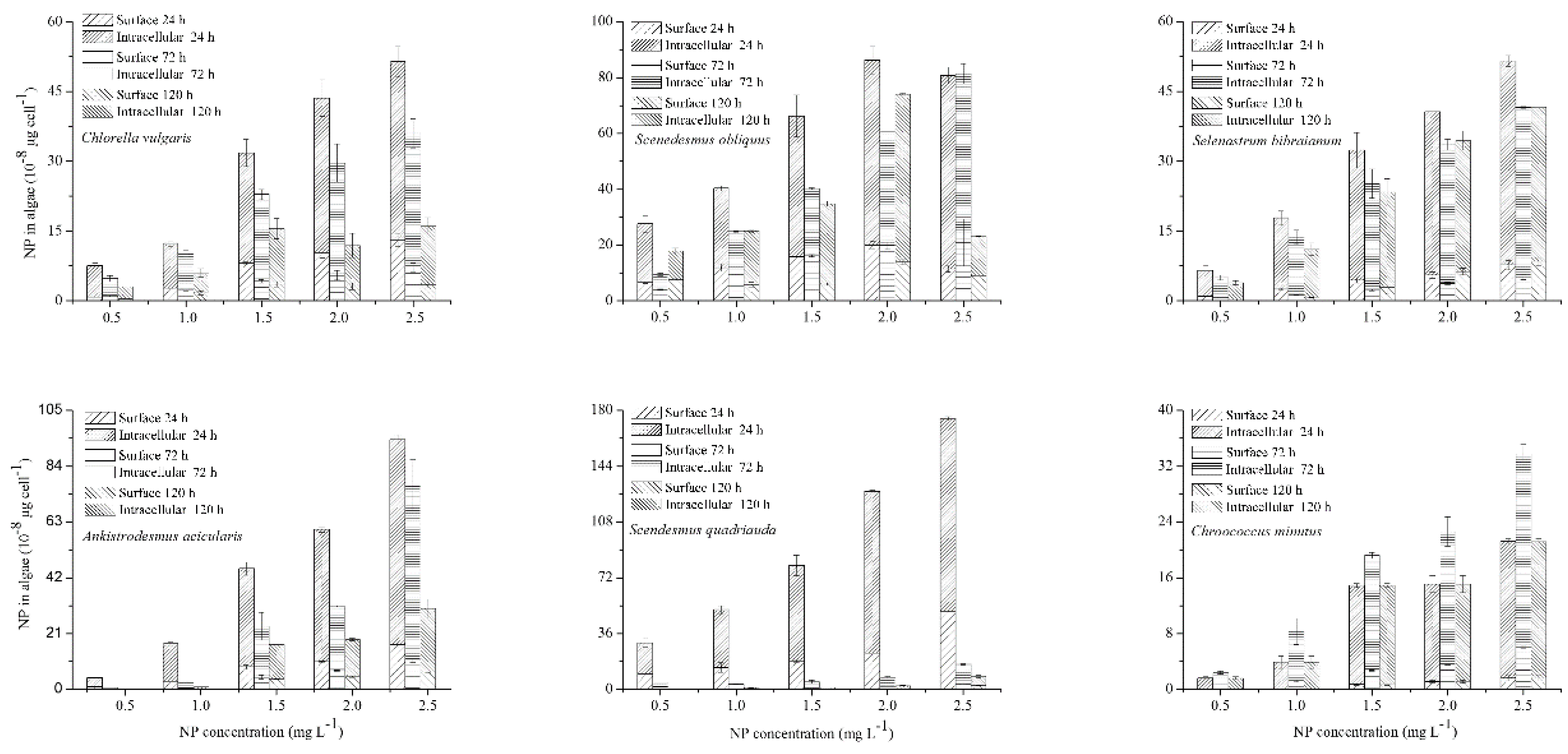
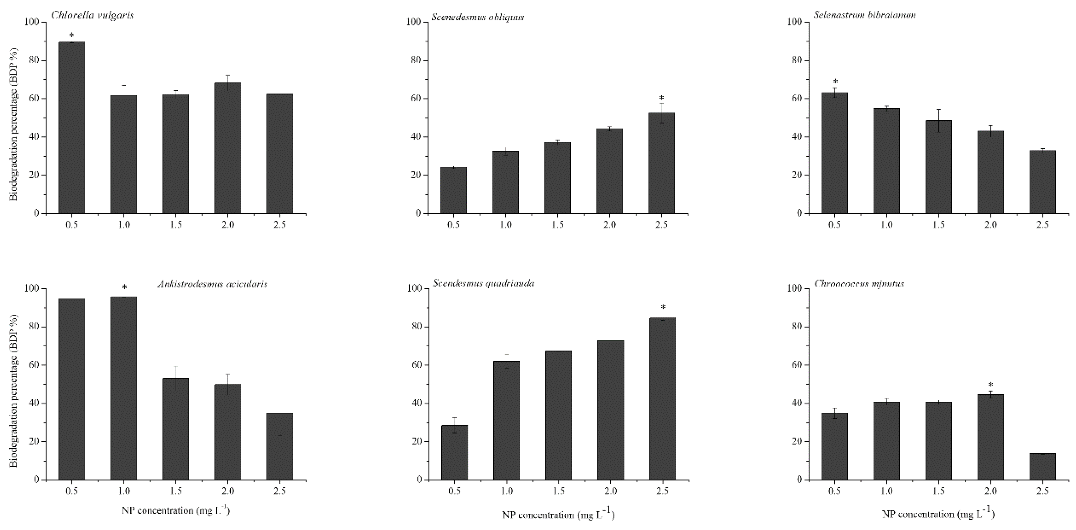
2.4. NP Accumulation and Degradation in Algae

It is known that NP is removed from water solutions in light by abiotic degradation [33]. The residual concentrations of NP in the medium of the control flasks did not show any significant changes during the 120 h experiments (Figure 4a), indicating that the abiotic loss was negligible. On the contrary, the NP level decreased rapidly when algae were available (Figure 4b). To fully evaluate the fate of NP in algae, target compounds were detected under different treatments. NP accumulation increased with the enhancement of the initial NP level (Figure 5). To confirm the accumulation of NP, the residual NP concentration in the medium was evaluated by a time-dependent study demonstrating a gradual decrease of NP over time (Figure 4b). In addition, the medium containing algae had a lower amount of NP compared with the control, suggesting that a certain proportion of NP had accumulated both on the surface and in the interior of the algae.
The quantity of NP removed from the medium was higher than the accumulated amount in algal cells due to the apparent biodegradation of NP. The maximum biodegradation percentages of C. vulgaris, S. obliquus, S. bibraianum, A. acicularis, S. quadricauda, and C. minutus in the present study were 89.5%, 52.5%, 63.1%, 95.6%, 84.6%, and 44.6%, respectively, indicating that the biodegradation of C. vulgaris, A. acicularis, and S. quadricauda was much faster than that of Microcystis aeruginosa, where more than 60% of NP degraded [11,40], and was also faster than other microalgae [40]. The six algae in this study varied in their biological degradation capability. C. vulgaris, S. bibraianum, and A. acicularis decreased with the increase of the initial NP level, and the biodegradation of NP reached the minimum at 2.5 mg L−1 (62.4%, 32.8%, and 34.9%, respectively). Therefore, A. acicularis was the most effective species for NP biodegradation when the NP concentration was below 1.0 mg L−1, indicating that NP biodegradation was associated with algal growth, a finding similar to that of a previous study by Yan et al. on Chlorella pyrenoidosa [41]. Further, in natural water bodies, surface water containing more than 10 μg/L NP is considered highly polluted, water containing 1–10 μg/L is polluted, and surface water containing <1 μg/L of NP has a low pollution level [42]. Even if severe NP pollution occurred in water bodies, the NP concentration was only up to 325 μg L−1 [43]. Obviously, the NP concentration of 1 mg L−1 could cover most of the polluted concentration of water bodies in nature. So, it would appear that A. acicularis could be applied in sewage treatment for its potential to effectively remove NP from water bodies in a suitable scope.
The algae in this study grew well at 0.5-2.5 mg L−1 NP. The concentration of NP decreased significantly during the exponential growth phase (Figure 1 and Figure 4). In contrast, the biodegradation by S. quadricauda and S. obliquus increased with an increase in the initial NP concentration. S. quadricauda was the most effective species for NP biodegradation when the NP concentration was 2.5 mg L−1 (Figure 6). However, the growth was inhibited when NP was higher than 1.0 mg L−1. The metabolism of other phenolic compounds in microalgae displayed similar patterns in comparison with higher plants. Some freshwater microalgae can metabolize bisphenol-A (BPA) to BPA glycosides, which are released into the culture medium [44]. In Tetraselmis marina, the metabolism of p-chlorophenol (p-CP) includes glucosyl transfer followed by malonyl transfer [45]. NP biodegradation in bacteria has been broadly reported, but similar research on microalgae is scarce. Some by-products during biodegradation are disadvantageous to algae growth. For example, 4-n-nonylphenol, a by-product of 4-n-nonylphenol, was degraded by Metarhizium sp. [46] and inhibited growth of the green alga Chlorella sorokiniana at 0.30 mg L−1 [47]. In our study, some by-products during biodegradation blocked S. quadricauda growth.

3. Materials and Methods

3.1. Reagents

Nonylphenol was purchased from Sigma-Aldrich (St. Louis, MO, USA). Methyl alcohol and acetonitrile were chromatographically pure, from Shanghai Anpu Company (Shanghai, China). Other reagents such as those used in culture media were analytical grade, from Guangzhou Chemical Factory (Guangzhou, China).

3.2. Algal Culture and Treatment

Six freshwater microalgae obtained from NP-polluted water at Jinan University, Guangzhou, China were identified using “The freshwater algae of China” (Hu and Wei, 2006). C. vulgaris (JNU38), S. obliquus (JNU15), S. bibraianum (JNU28), A. acicularis (JNU14), S. quadricauda (JNU39), and C. minutus (JNU17) were isolated. The algae were kept individually in conical flasks (2 L) in 1 L of BG11 medium with constant shaking (100 rpm) at 25 ± 2 °C under cool white fluorescent lamps (80 μmol m−2 s−1) at a 12 h light:12 h dark regime. All containers and solutions prior to utilization were autoclaved for 15 min at 121 °C. BG11 medium included the basal culture and trace metal medium [48]. The basal culture medium contained 1.5 g L−1 NaNO3, 40 mg L−1 K2HPO4, 75 mg L−1 MgSO4·7H2O, 36 mg L−1 CaCl2·2H2O, 20 mg L−1 NaHCO3, 6 mg L−1 ferric ammonium citrate, and 6 mg L−1 citric acid. The trace metal solution contained 2.86 mg L−1 H3BO3, 1.81 mg L−1 MnCl2·4H2O, 222 mg L−1 ZnSO4·7H2O, 390 mg L−1 Na2MoO4·2H2O, 79 mg·L−1 CuSO4·5H2O, and 49.4 mg L−1 Co(NO3)2·6H2O.

3.3. Removal of Bacteria from Algal Cultures

After 100 mL algal cultures in mid-exponential phase were filtered through a 10 μm pore size membrane, they were suspended in 50 mL of sterile BG11 medium, followed by 10 min centrifugation at 1000× g. The cells were then washed three times and suspended in sterile medium (50 mL) containing 0.1 M EDTA and 0.005% Tween-80 for 1 h at 20 °C, after which 0.5 mg mL−1 lysozyme (LSZ) and 0.25% sodium dodecyl sulphate (SDS), which had been warmed for 10 min at 20 °C, were added in sequence. Thereafter, cells were centrifuged at 1000× g for 10 min, washed two times to eliminate SDS and LSZ, and resuspended in 50 mL of sterile medium. The antibiotics kanamycin (50 μg mL−1) and penicillin (100 μg mL−1) were added to the algal cultures, which were maintained under a 12h light:12h dark regime at 20 °C for 1 week. Bacterial presence was evaluated, after subculturing three times, by epifluorescence microscopy using 4′,6-diamidino-2-phenylindolestain (DAPI) stain, ensuring a sterile condition [49,50].

3.4. Nonylphenol Treatments

The stock solution of NP was prepared in methanol at a concentration of 1000 mg L−1. Working solutions were set up at concentrations of 0, 0.5, 1.0, 1.5, 2.0, 2.5, and 3.0 mg L−1. Every group was in triplicate. The control group was treated with an equivalent amount of methanol (0.1%). The final concentration of methanol was controlled at 0.40% (v/v) for all experimental media in order to eliminate the effect of methanol on algal cells [51].
All solutions and experimental containers were autoclaved at 121 °C for 15 min. The microalgal cultures in the middle of the log phase of growth were decanted into 100 mL flasks containing 40 mL of medium at 25 ± 2 °C and illuminated with fluorescent lights (90 mol m−2 s−1 photon flux intensity) under a 12:12 h light:dark photoperiod. The algae were transferred to flasks (150 mL) containing 100 mL of BG11 medium, with the initial algal densities of 1–3 × 10⁵ cell m L−1. The experiments lasted for 5 days (120 h) with intermittent shaking.

3.5. Determination of Algal Growth and Inhibitory Effect Concentration

Cell density was indirectly measured by chlorophyll a, and regression equations between cell density and chlorophyll content of the six algae were prepared. A light microscope (Olympus, Japan) was used. Chlorophyll in vivo was evaluated by a TD-700 fluorometer (Turner Design, Fresno, CA, USA) after calibration with a chlorophyll standard. Before measuring, tubes with 10 mL cultures were kept for 20 min in complete darkness at room temperature (RT) followed by constant shaking (100 rpm) three times. Chlorophyll concentration was measured with 420 nm excitation and 680 nm emission spectrum. The content of chlorophyll a was measured at 0, 24, 48, 72, and 96 h.
EC50 was defined as the NP concentrations until the chlorophyll content diminished by half. EC50 at 96 h was calculated by a probability unit and concentration logarithm [52]. The algal growth rate (μ) was calculated according to the following equation:
µ (d − 1) = (Ln Nt − Ln N0)/(t − t0)
where N0 and Nt are the cell density at the beginning (t0) and the end (t) of the selected time interval, respectively.
The percentages (Ir) of algal chlorophyll reduction were calculated according to Equation (2), and the Ir (%) was then used to calculate the EC50 value based on chlorophyll a:
Ir (%) = 100 × (ycys)/ys
where yc and ys are the algal growth rates or chlorophyll a contents in the control medium and test medium, respectively.
According to the probability unit and concentration logarithm, linear regressions were set up and used to obtain EC50 values. Origin software version 8.0 (Microcal Software Inc., USA) was applied.

3.6. Measurement of Fluorescence Transient

A plant efficiency analyzer (PEA; Hansatech Instruments Ltd., UK) was applied for chlorophyll fluorescence transient analysis at 96 h. The algae were placed in complete dark for 20 min at RT. Aqueous-phase attachment of the PEA was applied for measurement. A red light of 3500 µmol photons m−2 s−1 provided by an array of six high-intensity light-emitting diodes was applied to generate transients measured on a time scale from 10 ms to 1 s.
The fluorescence intensity at 50 ms was defined as the initial fluorescence (F0), the peak fluorescence was determined as Fm, and the difference between Fm and F0 (FmF0) was defined as Fv. Fv/Fm was calculated by (FmF0)/Fm [53].

3.7. Assay of Antioxidant Enzyme Activity

After 96 h, algae were harvested following 10 min of centrifugation at 3000× g. The algae were then added to 2 mL of ice-cold extraction buffer including Tris–HCl (50 mM, pH 7.8), EDTA (1 mM), ascorbate (1 mM), as well as polyvinylpyrrolidone (1.5%, w/w). Then, the cocktails were centrifuged at 15,000× g for 20 min at 4 °C. The supernatants were used to estimate the activities of antioxidant enzymes. SOD (EC1.1.5.1.1) was determined by photochemical inhibition of nitro blue tetrazolium (NBT) [12]. CAT (EC 1.11.1.6) was spectrophotometrically estimated by the ammonium molybdate method [12]. POD activity was evaluated at 420 nm [54].

3.8. Quantification of NP in Culture Medium and Algae

The concentrations of NP in the medium were measured at 24, 72, and 120 h, and algae only at 120 h. Liquid–liquid microextraction (DLLME) [50] was applied during measurement. In brief, a sample (5 mL) was mixed with 0.2 mL of chlorobenzene and acetone (1:2), with a milky cloudy mixture generated after gentle shaking, and then subsequently centrifuged at 4500× g (15 min, 4 °C). A microsyringe (50 μL, zero dead volume, cone-tip needle) was used to withdraw the dispersed fine particles of the extraction phase that settled at the bottom of the conical test tube, repeated three times. The sediment fractions were combined for further analysis by high-performance liquid chromatography (HPLC) (Agilent, Santa Clara, CA, USA) [20]. All the extraction was performed at room temperature (23 ± 2 °C).
Before DLLME, cells were separated from the 5 mL cultures withdrawn from the flasks through centrifugation at 4500× g for 15 min at 4 °C. NP concentration in the culture medium could be measured by analyzing the NP distribution in the supernatant using DLLME and HPLC. The cell pellets were washed with 5 mL of 10% methanol with shaking for approximately 60 s, and the wash water was separated and used to analyze the NP adsorbed on the surface [40]. The cell pellet was mixed with anhydrous Na2SO4 and dichloromethane-methanol (1:2 v/v, 3 mL) by sonication for 20 min and centrifuged for 5 min at 3500× g. The extraction was done three times and the solvent fractions were combined for the analysis of NP absorbed by cells [55].

3.9. Determination of Biodegradation Percentage of NP

The biodegradation percentage (BDP) of NP was calculated as
BDP (%) = 100 × (Ci − Cr − Ca − Cd × Wa − Cc × Wa) / Ci
where Ci is the NP initial concentration in the solution (mg L−1), Cr is the NP residual concentration in the solution (mg L−1), Ca is the NP in abiotic elimination (mg L−1), Cd is the concentration (mg g−1) dry weight of NP adsorbed on the cell wall, Cc is the concentration (mg g−1) dry weight of NP accumulated in algal cells, and Wa is the dry mass of algae (g L−1) [49].

3.10. Statistical Analysis

All data in this research are presented as mean ± standard deviation (SD) (n = 3). One-way analysis of variance (ANOVA) was applied to identify the differences among treatments, followed by the least significant difference (LSD) test if the ANOVA result was significant (p < 0.05). The statistical analyses were performed with SPSS 12.0. The linear correlation was performed with Origin 8.0 using the least-squares fitting method.

4. Conclusions

Both the concentration and exposure time of NP were found to affect the responses of microalgae. High levels of NP (≥1.5 mg L−1) were highly toxic to all microalgae in which various antioxidant mechanisms were involved. C. vulgaris and S. obliquus, which are NP-tolerant species, responded rapidly to antioxidation compared with NP-sensitive species, especially when the NP concentration was high. The result that NP showed strong acute toxicity to C. minutus suggests that it could be a promising tool for the study of NP toxicity. All six microalgae species biodegraded NP at a low concentration but in a species-specific manner. Our results indicate that A. acicularis degraded NP when the concentration was below 1.0 mg L−1, while S. quadricauda more actively biodegraded NP when the concentration was above 1.5 mg L−1. However, considering NP pollution in natural water and algal growth, A. acicularis is more suitable to be applied in sewage treatment.

Author Contributions

Conceptualization, X.S. and S.D.; methodology, N.H.; software, S.W.; validation, N.H., W.L. and D.S.; formal analysis, N.H.; writing—original draft preparation, N.H. and X.S.; writing—review and editing, Z.L.; visualization, X.S.; funding acquisition, X.S. and S.D. All authors have read and agreed to the published version of the manuscript.

Funding

This work was funded by the Natural Science Foundation of China (No. 41676099) and the Science and Technology Research Project of Jiangxi Provincial Department of Education (No. GJJ170916).

Conflicts of Interest

All authors declare no conflict of interest.

References

  1. Sood, A.; Uniyal, P.L.; Prasanna, R.; Ahluwalia, A.S. Phytoremediation Potential of Aquatic Macrophyte, Azolla. AMBIO 2012, 41, 122–137. [Google Scholar] [CrossRef] [Green Version]
  2. Soares, A.; Guieysse, B.; Jefferson, B.; Cartmell, E.; Lester, J.N. Nonylphenol in the environment: A critical review on occurrence, fate, toxicity and treatment in wastewaters. Environ. Int. 2008, 34, 1033–1049. [Google Scholar] [CrossRef]
  3. Commission, E. Directive 2003/53/EC of the European Parliament and of the Council of 18 June 2003 amending for the 26th time Council Directive 76/769/EEC relating to restrictions on the marketing and use of certain dangerous substances and preparations (nonylphenol, nonylphenol ethoxylate and cement). In Official Journal of the European Union; Publications Office of the European Union: Luxembourg, 2003; pp. 24–178. [Google Scholar]
  4. Zgoła-Grześkowiak, A. Dispersive liquid–liquid microextraction applied to isolation and concentration of alkylphenols and their short-chained ethoxylates in water samples. J. Chromatogr. A 2010, 1217, 1761–1766. [Google Scholar] [CrossRef]
  5. Mao, Z.; Zheng, X.F.; Zhang, Y.-Q.; Tao, X.-X.; Li, Y.; Wang, W. Occurrence and Biodegradation of Nonylphenol in the Environment. Int. J. Mol. Sci. 2012, 13, 491–505. [Google Scholar] [CrossRef]
  6. Klečka, G.M.; Naylor, C.G.; Staples, C.A.; Losey, B. Occurrence of Nonylphenol Ethoxylates and Their Metabolites in Municipal Wastewater Treatment Plants and Receiving Waters. Water Environ. Res. 2010, 82, 447–454. [Google Scholar] [CrossRef] [PubMed]
  7. Fan, Z.; Hu, J.; An, W.; Yang, M. Detection and Occurrence of Chlorinated Byproducts of Bisphenol A, Nonylphenol, and Estrogens in Drinking Water of China: Comparison to the Parent Compounds. Environ. Sci. Technol. 2013, 47, 10841–10850. [Google Scholar] [CrossRef] [PubMed]
  8. Newsted, J.L. Effect of light, temperature, and pH on the accumulation of phenol by Selenastrum capricornutum, a green alga. Ecotoxicol. Environ. Saf. 2004, 59, 237–243. [Google Scholar] [CrossRef] [PubMed]
  9. Dosnon-Olette, R.; Trotel-Aziz, P.; Couderchet, M.; Eullaffroy, P. Fungicides and herbicide removal in Scenedesmus cell suspensions. Chemosphere 2010, 79, 117–123. [Google Scholar] [CrossRef] [PubMed]
  10. McCormick, P.V.; Cairns, J. Algae as indicators of environmental change. J Appl Phycol 1994, 6, 509–526. [Google Scholar] [CrossRef]
  11. Eullaffroy, P.; Vernet, G. The F684/F735 chlorophyll fluorescence ratio: A potential tool for rapid detection and determination of herbicide phytotoxicity in algae. Water. Res. 2003, 37, 1983–1990. [Google Scholar] [CrossRef]
  12. Corcoll, N.; Bonet, B.; Leira, M.; Guasch, H. Chl-a fluorescence parameters as biomarkers of metal toxicity in fluvial biofilms: An experimental study. Hydrobiologia 2011, 673, 119–136. [Google Scholar] [CrossRef]
  13. Sun, X.; Zhong, Y.; Huang, Z.; Yang, Y. Selenium Accumulation in Unicellular Green Alga Chlorella vulgaris and Its Effects on Antioxidant Enzymes and Content of Photosynthetic Pigments. PLoS ONE 2014, 9, e112270. [Google Scholar] [CrossRef] [PubMed] [Green Version]
  14. Kvíderová, J. Rapid algal toxicity assay using variable chlorophyll fluorescence for Chlorella kessleri (chlorophyta). Environ. Toxicol 2010, 25, 554–563. [Google Scholar] [CrossRef] [PubMed]
  15. Choi, C.J.; Berges, J.A.; Young, E.B. Rapid effects of diverse toxic water pollutants on chlorophyll a fluorescence: Variable responses among freshwater microalgae. Water. Res. 2012, 46, 2615–2626. [Google Scholar] [CrossRef]
  16. Wang, S.; Chen, F.; Mu, S.; Zhang, D.; Pan, X.; Lee, D.-J. Simultaneous analysis of photosystem responses of Microcystis aeruginoga under chromium stress. Ecotox. Environ. Safe 2013, 88, 163–168. [Google Scholar] [CrossRef]
  17. Wang, J.; Xie, P. Antioxidant enzyme activities of Microcystis aeruginosa in response to nonylphenols and degradation of nonylphenols by M. aeruginosa. Environ. Geochem. Health. 2007, 29, 375–383. [Google Scholar] [CrossRef]
  18. Shan, J.; Jiang, B.; Yu, B.; Li, C.; Sun, Y.; Guo, H.; Wu, J.; Klumpp, E.; Schaeffer, A.; Ji, R. Isomer-Specific Degradation of Branched and Linear 4-Nonylphenol Isomers in an Oxic Soil. Environ. Sci. Technol. 2011, 45, 8283–8289. [Google Scholar] [CrossRef]
  19. Xu, P.; Lai, C.; Zeng, G.; Huang, D.; Chen, M.; Song, B.; Peng, X.; Wan, J.; Hu, L.; Duan, A.; et al. Enhanced bioremediation of 4-nonylphenol and cadmium co-contaminated sediment by composting with Phanerochaete chrysosporium inocula. Bioresour. Technol. 2018, 250, 625–634. [Google Scholar] [CrossRef]
  20. He, N.; Sun, X.; Zhong, Y.; Sun, K.; Liu, W.; Duan, S. Removal and Biodegradation of Nonylphenol by Four Freshwater Microalgae. Int. J. Environ. Res. Public Health 2016, 13, 1239. [Google Scholar] [CrossRef] [Green Version]
  21. Shen, H.; Zhou, P. Advance in the studies on effect of environmental organic pollutants on the algae growth. Acta Hydrobiol. Sin. 2002, 26, 529–535. [Google Scholar]
  22. Tsang, C.K.; Lau, P.S.; Tam, N.F.Y.; Wong, Y.S. Biodegradation capacity of tributyltin by two Chlorella species. Environ. Pollut. 1999, 105, 289–297. [Google Scholar] [CrossRef]
  23. Andrade, L.R.; Leal, R.N.; Noseda, M.; Duarte, M.E.R.; Pereira, M.S.; Mourão, P.A.S.; Farina, M.; Amado Filho, G.M. Brown algae overproduce cell wall polysaccharides as a protection mechanism against the heavy metal toxicity. Mar. Pollut. Bull. 2010, 60, 1482–1488. [Google Scholar] [CrossRef] [PubMed]
  24. Osundeko, O.; Davies, H.; Pittman, J.K. Oxidative stress-tolerant microalgae strains are highly efficient for biofuel feedstock production on wastewater. Biomass Bioenergy 2013, 56, 284–294. [Google Scholar] [CrossRef]
  25. Sutherland, D.; Ralph, P. Microalgal bioremediation of emerging contaminants - Opportunities and challenges. Water. Res. 2019, 164, 114921. [Google Scholar] [CrossRef]
  26. Servos, M. Review of the Aquatic Toxicity, Estrogenic Responses and Bioaccumulation of Alkylphenols and Alkylphenol Polyethoxylates. Water Qual. Res. J. Can. 1999, 34, 123–177. [Google Scholar] [CrossRef]
  27. Wang, J.; Xie, P.; Guo, N. Effects of nonylphenol on the growth and microcystin production of Microcystis strains. Environ. Res. 2007, 103, 70–78. [Google Scholar] [CrossRef] [PubMed]
  28. Wang, J.J.; Qian, X.J.; An, M.; Duan, S.S. Effect of combined exposure of diethylphthalate and nonylphenolon on growth of Dunaliella salina. Ecol. Sci. 2012, 31, 370–376. [Google Scholar]
  29. Wu, W.; Qu, J.; Chen, J.; Hu, G. Toxic effects of nonylphenol ethoxylates and its degradation product on aquatic organisms. J Zhanjiang Ocean. Univ./Zhanjiang Haiyang Daxue Xuebao 2003, 23, 39–44. [Google Scholar]
  30. Hense, B.A.; Jüttner, I.; Welzl, G.; Severin, G.F.; Pfister, G.; Behechti, A.; Schramm, K.W. Effects of 4-nonylphenol on phytoplankton and periphyton in aquatic microcosms. Environ Toxicol Chem 2003, 22, 2727. [Google Scholar] [CrossRef] [Green Version]
  31. Guan, C.; SUN, Z.; Min, A.; Duan, S. The ecological toxic effects of Nonylphenol on Phaeocystis globosa. Ecol. Environ. Sci. 2011, 640–645. [Google Scholar]
  32. Liu, X.; Zhao, J.; Dan, L.X.; Shi, X.; Liang, S. Toxic effects of nonylphenol on dominant microalgae species in Jiaozhou Bay. Mar. Environ. Sci. 2012, 31, 667–673. [Google Scholar]
  33. Liu, Y.; Dai, X.; Wei, J. Toxicity of the xenoestrogen nonylphenol and its biodegradation by the alga Cyclotella caspia. J. Environ. Sci. 2013, 25, 1662–1671. [Google Scholar] [CrossRef]
  34. Liu, Y.; Luan, T.-G.; Lu, N.-N.; Lan, C.-Y. Toxicity of Fluoranthene and Its Biodegradation by Cyclotella caspia Alga. J. Integr. Plant. Biol. 2006, 48, 169–180. [Google Scholar] [CrossRef]
  35. Gao, Q.T.; Wong, Y.S.; Tam, N.F.Y. Antioxidant responses of different microalgal species to nonylphenol-induced oxidative stress. J. Appl. Phycol. 2017, 29, 1317–1329. [Google Scholar] [CrossRef]
  36. Mittler, R. Oxidative stress, antioxidants and stress tolerance. Trends Plant Sci. 2002, 7, 405–410. [Google Scholar] [CrossRef]
  37. Bartosz, G. Oxidative stress in plants. Acta. Physiol. Plant. 1997, 19, 47–64. [Google Scholar] [CrossRef]
  38. Šepič, E.; Bricelj, M.; Leskovšek, H. Toxicity of fluoranthene and its biodegradation metabolites to aquatic organisms. Chemosphere 2003, 52, 1125–1133. [Google Scholar] [CrossRef]
  39. Choo, K.; Snoeijs, P.; Pedersén, M. Oxidative stress tolerance in the filamentous green algae Cladophora glomerata and Enteromorpha ahlneriana. J. Exp. Mar. Biol. Ecol. 2004, 298, 111–123. [Google Scholar] [CrossRef]
  40. Zhou, G.; Peng, F.; Zhang, L.; Ying, G. Biosorption of zinc and copper from aqueous solutions by two freshwater green microalgae Chlorella pyrenoidosa and Scenedesmus obliquus. Environ. Sci. Pollut. Res. 2012, 19, 2918–2929. [Google Scholar] [CrossRef]
  41. Yan, H.; Ye, C.; Yin, C. Kinetics of phthalate ester biodegradation by Chlorella pyrenoidosa. Environ. Toxicol. Chem. 1995, 14, 931–938. [Google Scholar] [CrossRef]
  42. Vazquez-Duhalt, R.; Marquez-Rocha, F.; Ponce Rivas, E.; Licea, A.; Viana, M. Teresa Nonylphenol, an integrated vision of a pollutant. Appl. Ecol. Environ. Res. 2005, 4, 1–25. [Google Scholar] [CrossRef]
  43. Gross-Sorokin, M.Y.; Grist, E.P.M.; Cooke, M.; Crane, M. Uptake and Depuration of 4-Nonylphenol by the Benthic Invertebrate Gammarus pulex:  How Important Is Feeding Rate? Environ. Sci. Technol. 2003, 37, 2236–2241. [Google Scholar] [CrossRef] [PubMed]
  44. Nakajima, N.; Teramoto, T.; Kasai, F.; Sano, T.; Tamaoki, M.; Aono, M.; Kubo, A.; Kamada, H.; Azumi, Y.; Saji, H. Glycosylation of bisphenol A by freshwater microalgae. Chemosphere 2007, 69, 934–941. [Google Scholar] [CrossRef] [PubMed]
  45. Petroutsos, D.; Wang, J.; Katapodis, P.; Kekos, D.; Sommerfeld, M.; Hu, Q. Toxicity and metabolism of p-chlorophenol in the marine microalga Tetraselmis marina. Aquat. Toxicol. 2007, 85, 192–201. [Google Scholar] [CrossRef] [PubMed]
  46. Nowak, M.; Soboń, A.; Litwin, A.; Różalska, S. 4-n-nonylphenol degradation by the genus Metarhizium with cytochrome P450 involvement. Chemosphere 2019, 220, 324–334. [Google Scholar] [CrossRef] [PubMed]
  47. Wang, L.; Kang, Y.; Liang, S.; Chen, D.; Zhang, Q.; Zeng, L.; Luo, J.; Jiang, F. Synergistic effect of co-exposure to cadmium (II) and 4-n-nonylphenol on growth inhibition and oxidative stress of Chlorella sorokiniana. Ecotoxicol. Environ. Saf. 2018, 154, 145–153. [Google Scholar] [CrossRef] [PubMed]
  48. Rippka, R.; Deruelles, J.; Waterbury, J.B.; Herdman, M.; Stanier, R.Y. Generic Assignments, Strain Histories and Properties of Pure Cultures of Cyanobacteria. Microbiology 1979, 111, 1–61. [Google Scholar] [CrossRef] [Green Version]
  49. Su, J.Q.; Yang, X.; Zheng, T.; Hong, H. An efficient method to obtain axenic cultures of Alexandrium tamarense—A PSP-producing dinoflagellate. J. Microbiol. Meth. 2007, 69, 425–430. [Google Scholar] [CrossRef]
  50. Rezaee, M.; Assadi, Y.; Milani Hosseini, M.-R.; Aghaee, E.; Ahmadi, F.; Berijani, S. Determination of organic compounds in water using dispersive liquid–liquid microextraction. J. Chromatogr. A 2006, 1116, 1–9. [Google Scholar] [CrossRef]
  51. Zhou, G.J.; Peng, F.Q.; Yang, B.; Ying, G.G. Cellular responses and bioremoval of nonylphenol and octylphenol in the freshwater green microalga Scenedesmus obliquus. Ecotoxicol. Environ. Saf. 2013, 87, 10–16. [Google Scholar] [CrossRef]
  52. Chi, J.; Li, Y.; Gao, J. Interaction between three marine microalgae and two phthalate acid esters. Ecotoxicol. Environ. Saf. 2019, 170, 407–411. [Google Scholar] [CrossRef]
  53. Sharma, D.K.; Fernández, J.O.; Rosenqvist, E.; Ottosen, C.O.; Andersen, S.B. Genotypic response of detached leaves versus intact plants for chlorophyll fluorescence parameters under high temperature stress in wheat. J. plant. physiol. 2014, 171, 576–586. [Google Scholar] [CrossRef] [PubMed]
  54. Salin, M.L.; Day, E.D.; Crapo, J.D. Isolation and characterization of a manganese-containing superoxide dismutase from rat liver. Arch. Biochem. Biophys. 1978, 187, 223–228. [Google Scholar] [CrossRef]
  55. Correa-Reyes, G.; Viana, M.T.; Marquez-Rocha, F.J.; Licea, A.F.; Ponce, E.; Vazquez-Duhalt, R. Nonylphenol algal bioaccumulation and its effect through the trophic chain. Chemosphere 2007, 68, 662–670. [Google Scholar] [CrossRef] [PubMed]
Figure 1. Effect of nonylphenol (NP) on the cell number of microalgae. Algae were treated with NP at 0–3.0 mg L−1 culture for 96 h. Values are the mean ± standard deviation (SD) (n = 3).
Ijms 21 01338 g001
Figure 2. Effect of NP on the maximal PSII activity (Fv/Fm) in microalgae. Algae were treated with NP at 0–3.0 mg L−1 culture for 96 h. Mean and standard deviation of three replicates are shown. Values are the mean ± standard deviation (SD) (n = 3). Asterisks indicate the significant differences between the NP treatments and control (p < 0.05).
Ijms 21 01338 g002
Figure 3. Effects of NP on activities of superoxide dismutase (SOD), catalase (CAT), and peroxidase (POD) in microalgae. Algae were treated with NP at 0.5, 1.0, and 1.5 mg L−1 culture for 96 h, and then the activities were assayed. Mean and standard deviation of three replicates are shown. Means with different letters at each NP concentration for each algal species indicate that they were significantly different at p < 0.05 according to a one-way ANOVA test. NS: not significant.
Ijms 21 01338 g003
Figure 4. Residual NP in the medium: (a) control and (b) six algae. Algae were treated with NP at 0.5–2.5 mg L−1 culture for 120 h. Values are the mean ± standard deviation (SD) (n = 3). Asterisks indicate the significant differences compared to other NP treatments (p < 0.05).
Ijms 21 01338 g004
Figure 5. NP accumulation in microalgae. Algae were treated with NP at 0.5–2.5 mg L−1 culture for 120 h. Values are the mean ± standard deviation (SD) (n = 3).
Ijms 21 01338 g005
Figure 6. Biodegradation of NP by microalgae. Algae were treated with NP at 0.5–2.5 mg L−1 culture for 120 h. Asterisks indicate the significant differences compared to other NP treatments (p < 0.05).
Ijms 21 01338 g006
Table 1. Acute toxicity of NP (mg L−1) on six microalgae species at 96 h culture time.
Microalgal SpeciesRegression EquationR2EC50 (mg L−1)
Chlorella vulgarisy = 0.875x + 0.12570.9751.534
Scenedesmusobliquusy = 1.716x + 0.21820.9041.179
Selenastrumbibraianumy = 1.6141x + 0.2370.9611.177
Ankistrodesmusacicularisy = 1.3231x + 0.3740.9111.100
Scenedesmusquadricauday = 1.3366x + 0.39740.9611.080
Chroococcusminutusy = 1.1814x + 0.49410.9701.005
The concentrations of NP were in the ranges of 0.5–3.0 mg L−1. EC50: the median inhibitory effect concentration (mg L−1). R2: correlation coefficient. p-value significance of linear regression with 95% confidence limits in ANOVA.
Table 2. The 96 h EC50 of NP on different microalgae.
Microalgal SpeciesEC50 (mg L−1)Reference
Microcystisaeruginosa0.67–2.96 [27]
Dunaliellasalina1.47[28]
Scenedesmusobliquus1.0 [29]
Scenedesmussubspicatus0.87–0.98[30]
Phaeocystisglobosa0.42[31]
Skeletonemacostatum0.13[32]
Chaetoceroscurvisetus0.22[32]
Cyclotellacaspia0.18[33]

