Blue-Light-Excited Eu3+/Sm3+ Co-Doped NaLa(MoO4)2 Phosphors: Synthesis, Characterizations and Red Emission Enhancement for WLEDs

1
College of Materials Science and Engineering, Nanjing Tech University, Nanjing 210009, China
2
School of Resources, Environment and Materials, Guangxi University, Nanning 530004, China
*
Authors to whom correspondence should be addressed.
Materials 2018, 11(7), 1090; https://doi.org/10.3390/ma11071090
Submission received: 17 May 2018
/
Revised: 7 June 2018
/
Accepted: 15 June 2018
/
Published: 26 June 2018
Abstract
:The system NaLa(MoO4)2:Eu3+/Sm3+ phosphors were prepared by solid-state reaction followed by heat treatment at 450–600 °C. As shown by X-ray powder diffraction, the phosphors had a body-centered tetragonal structure, high phase purity and high crystallinity. The photoluminescence measurements carried out under excitation at 464 nm indicated the main emission at about 615 nm corresponding to the electric dipole transition 5D0→7F2 of Eu3+, which agreed well with the emission wavelengths of the GaN-based blue LED chips. In NaLa(MoO4)2:Eu3+/Sm3+ phosphors, Sm3+ can efficiently transfer excitation energy to Eu3+, which resulted in a significant improvement of the fluorescence intensity, and the fluorescence intensity of the phosphors calcined at 550 °C maximized at the doping concentrations of Eu3+ and Sm3+ of 15.0 and 2.0 mol %, respectively. The decay curves and CIE (Commission Internationale de L’ Eclairage) coordinates of Sm3+ or/and Eu3+ doped phosphors were analyzed for the investigation of the energy transfer mechanism and color variation trend. Thus, NaLa(MoO4)2:Eu3+,Sm3+ can be classified as a potential red-emitting phosphor for white light-emitting diodes (WLEDs).
1. Introduction
These days, the white light-emitting diodes (WLEDs) regarded as the new-style solid-state photosources achieve much more research enthusiasm than before owing to the energy conservation and environmental protection, long service time, high reliability and function [1,2,3,4]. However, the development of WLEDs is largely limited because there is no excellent red phosphor [5,6]. In the last few years, great attention has been focused on double alkaline rare-earth molybdates in virtue of their unique structures and excellent luminous properties [7,8,9]. NaLa(MoO4)2 pertaining to the double molybdate family is of tetragonal structure with space group I41/a wherein Mo6+ combining with four neighboring O2− forms tetrahedral MoO42− anions to make phosphor matrix with high physical–chemical stability [10,11] and both Na+ and La3+ randomly occupy S4 lattice sites with no symmetrical center, which is propitious to relieve further parity forbidden of the electric dipole transition of RE3+ [12]. Furthermore, the MoO42− group can absorb the energy of the ultraviolet light through Mo-O charge transfer interaction, and then improve rare-earth fluorescence materials’ luminous efficiency and intensity via transferring energy to rare-earth ions [13,14].
Europium ion (Eu3+)-doped compounds are the most promising candidates as the efficient red phosphors for WLEDs due to their advantages of high color purity and the strongest characteristic red light emission peak at about 615 nm attributing to the 5D0→7F2 transition [15,16,17]. Nowadays, Eu3+-doped molybdates are regarded as excellent red phosphors, but possess low luminescence intensity, improvement of which has been tested through many efforts [18]. For example, Zuo et al. [5] found that red emission intensities of Eu3+ were overwhelmingly improved by co-doping Bi3+ in the KLa(MoO4)2 phosphors. Zhou et al. [19] reported that intensity of LiY(MoO4)2:Eu3+,Sm3+ phosphors was heightened when the compounds were prepared by sol–gel process. However, for all we know, no researchers have reported the energy transfer pathway or photoluminescence (PL) properties of the Sm3+/Eu3+ co-doped NaLa(MoO4)2 matrix.
Thus, we synthesized the Eu3+ or/and Sm3+ doped NaLa(MoO4)2 phosphors through a solid-state reaction. The influence of a calcination temperature on a crystal phase as well as luminescent intensity being discussed, and the photoluminescence (PL) spectra, decay behaviors and chromatic properties were analyzed at length.
2. Experimental
2.1. Synthesis
The Eu(NO3)3·6H2O (99.99%), Sm(NO3)3·6H2O (99.9%), La(NO3)3·6H2O (99.0%), NaNO3 (99.0%) and (NH4)6Mo7O24·4H2O (99.0%) used as raw materials were bought from Shanghai Aladdin Bio-Chem Technology Co., Ltd. (Shanghai, China). The Eu3+-doped NaLa(MoO4)2 (abbreviated as NL1−xMO:xEu3+; the mol % x = 5, 10, 15 and 20%) and Eu3+/Sm3+co-doped NaLa(MoO4)2 (abbreviated as NL1−x−yMO:15%Eu3+,ySm3+; the mol % y = 0, 1, 2, 3, 4 and 5%) phosphors were prepared by solid-state reaction. The stoichiometric mixtures of NaNO3, La(NO3)3·6H2O, (NH4)6Mo7O24·4H2O, Eu(NO3)3/Sm(NO3)3 solution (1 M) were ground thoroughly for 1 h in a porcelain mortar to be uniform, and, then, they were oven-dried at 85 °C for 12 h. Finally, appropriate amounts of procedures were calcined in a muffle furnace at 450, 500, 550 or 600 °C for 360 min to form the final products.
2.2. Characterization
The phase composition of the products was determined by the X-ray diffraction (XRD) method (Rigaku-Dmax 2500 X-ray diffractometer ( Japanese science and science company, Tokyo, Japan) with graphite monochromatized Cu Kα radiation, scanning rate was 5°/min, 2θ = 10–75°). The photoluminescence excitation (PLE) and photoluminescence (PL) emission spectra were measured on a HORIBA Fluoromax-4 spectrophotometer (HORIBA Ltd, Irvine, CA, America) equipped with a 150 W Xe-lamp, and the fluorescent decay curves were determined by a Hamamatsu Quantaurus–Tau spectrophotometer (Japanese hamamon photonics Ltd, Hamamatsu, Japan) equipped with a 450 W xenon impulse lamp as the excitation source. The entire tests were put into effect under room temperature conditions.
3. Results and Discussion
3.1. XRD
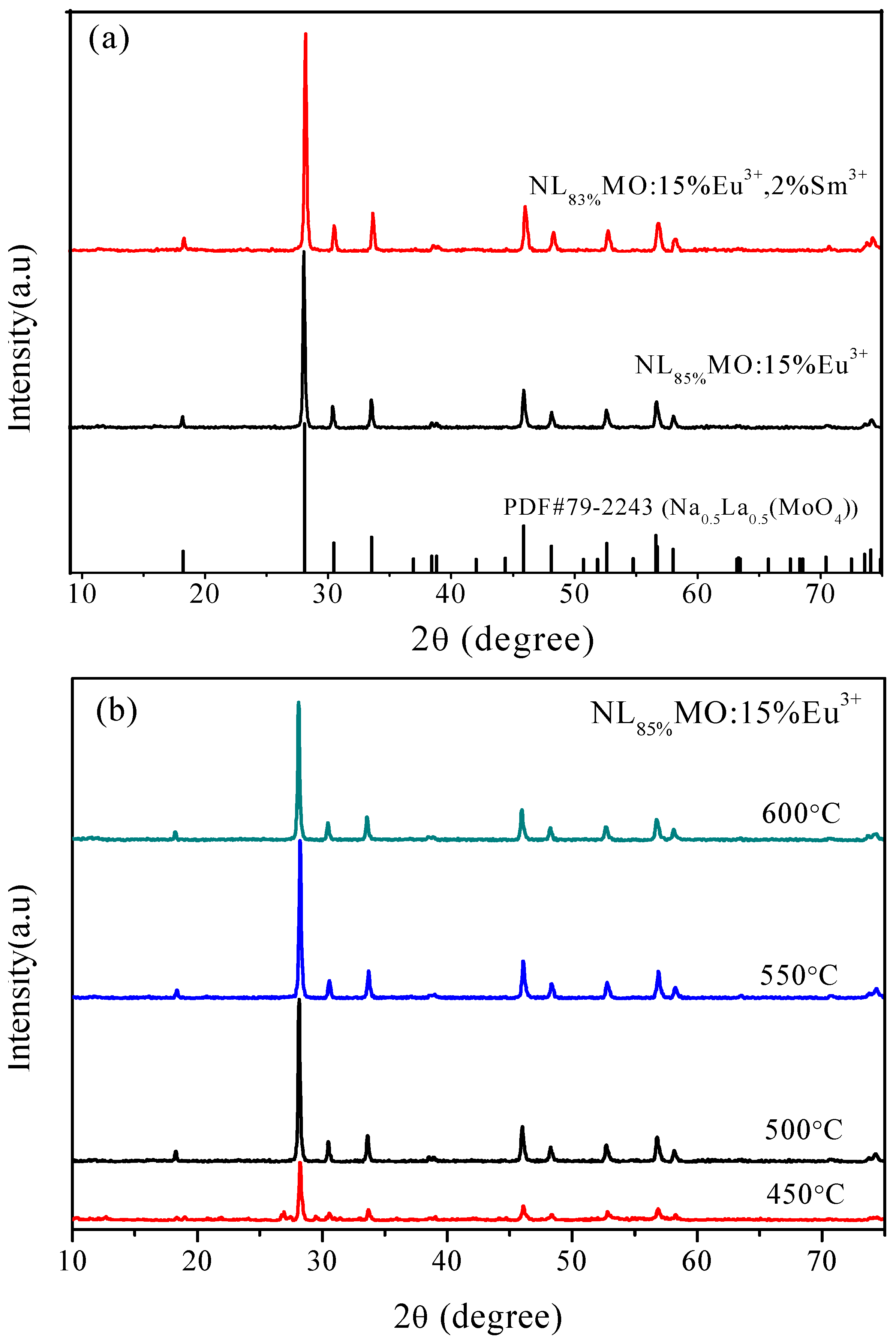
In Figure 1a, the two representative XRD patterns of NL85%MO:15%Eu3+ and NL83%MO:15%Eu3+,2%Sm3+ calcined at 550 °C for 6 h are shown. The diffraction peaks were well attributed to pure tetragonal phase NaLa(MoO4)2 (PDF Card 79-2243, a = b = 0.53424 nm, c = 1.17376 nm), indicating that the doped ions were successfully incorporated into matrix lattice and had not drastically altered the host structure. The presence of foreign phases was not detected. Figure 1b shows the XRD patterns of the products formed at 450, 500, 550 or 600 °C for 6 h. Obviously, the crystalline NLMO:Eu3+ has already formed at 450 °C and the sample calcined at 550 °C is better crystallized and possesses stronger and sharper peaks.
3.2. PL Properties of NLMO: Eu3+ Phosphors
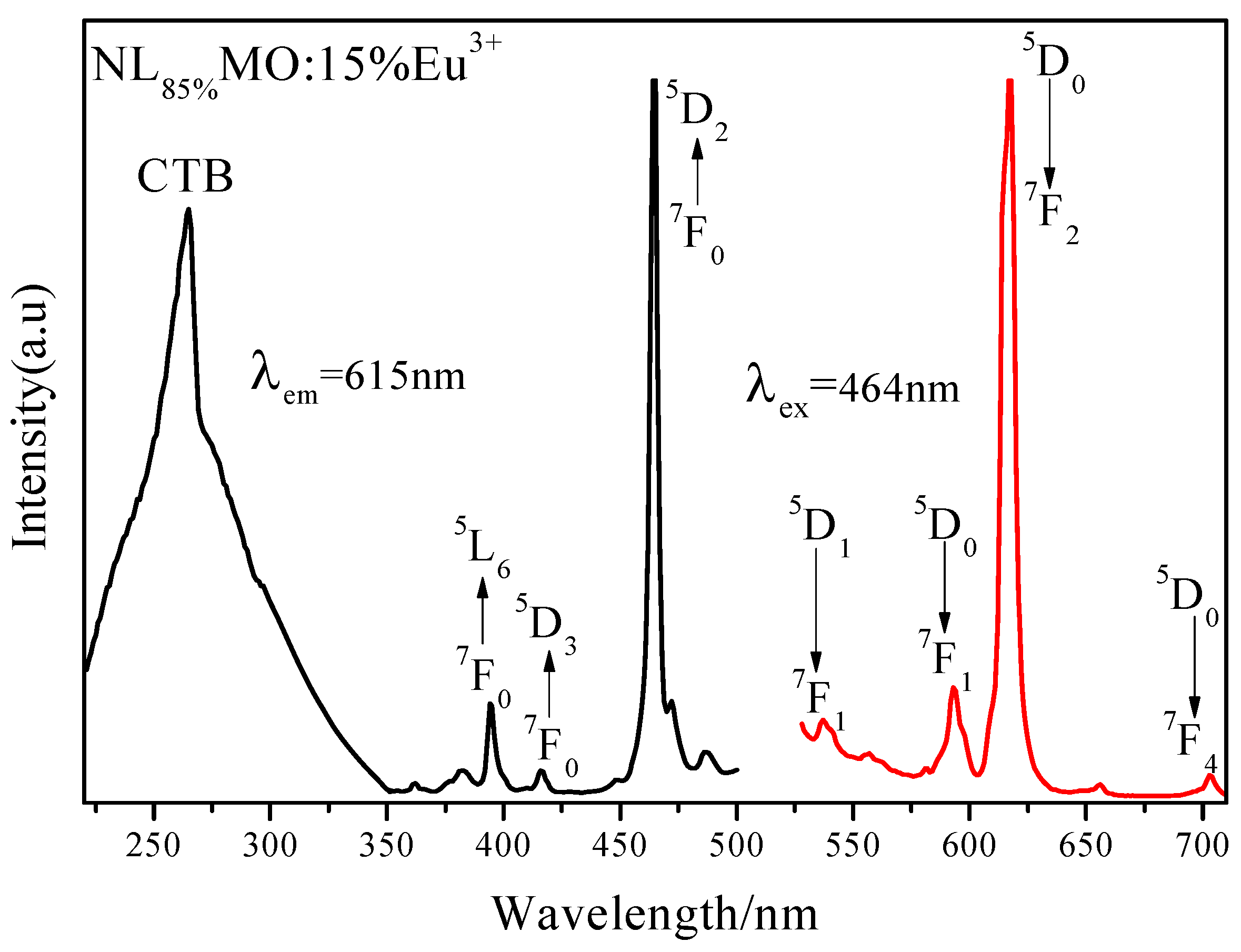
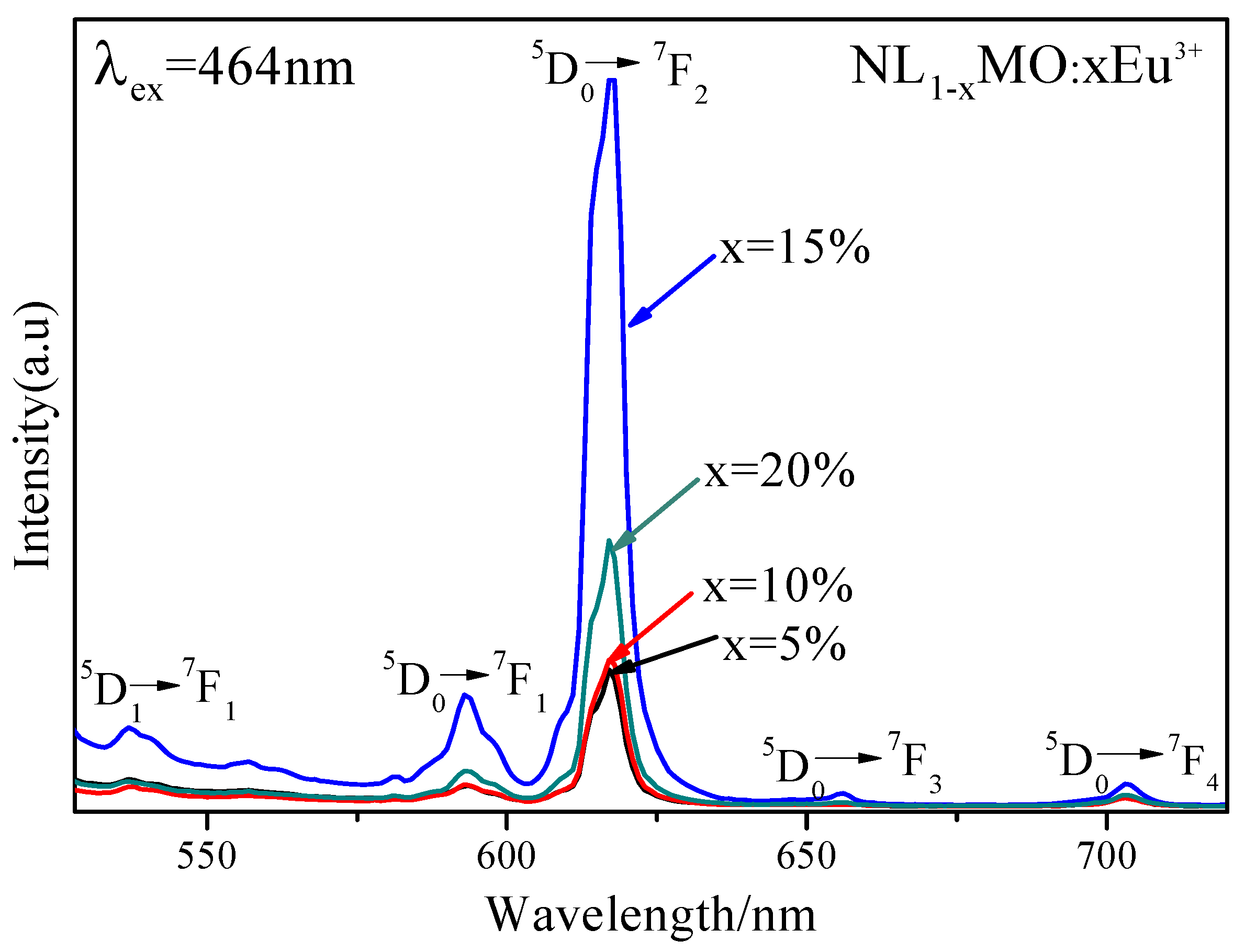
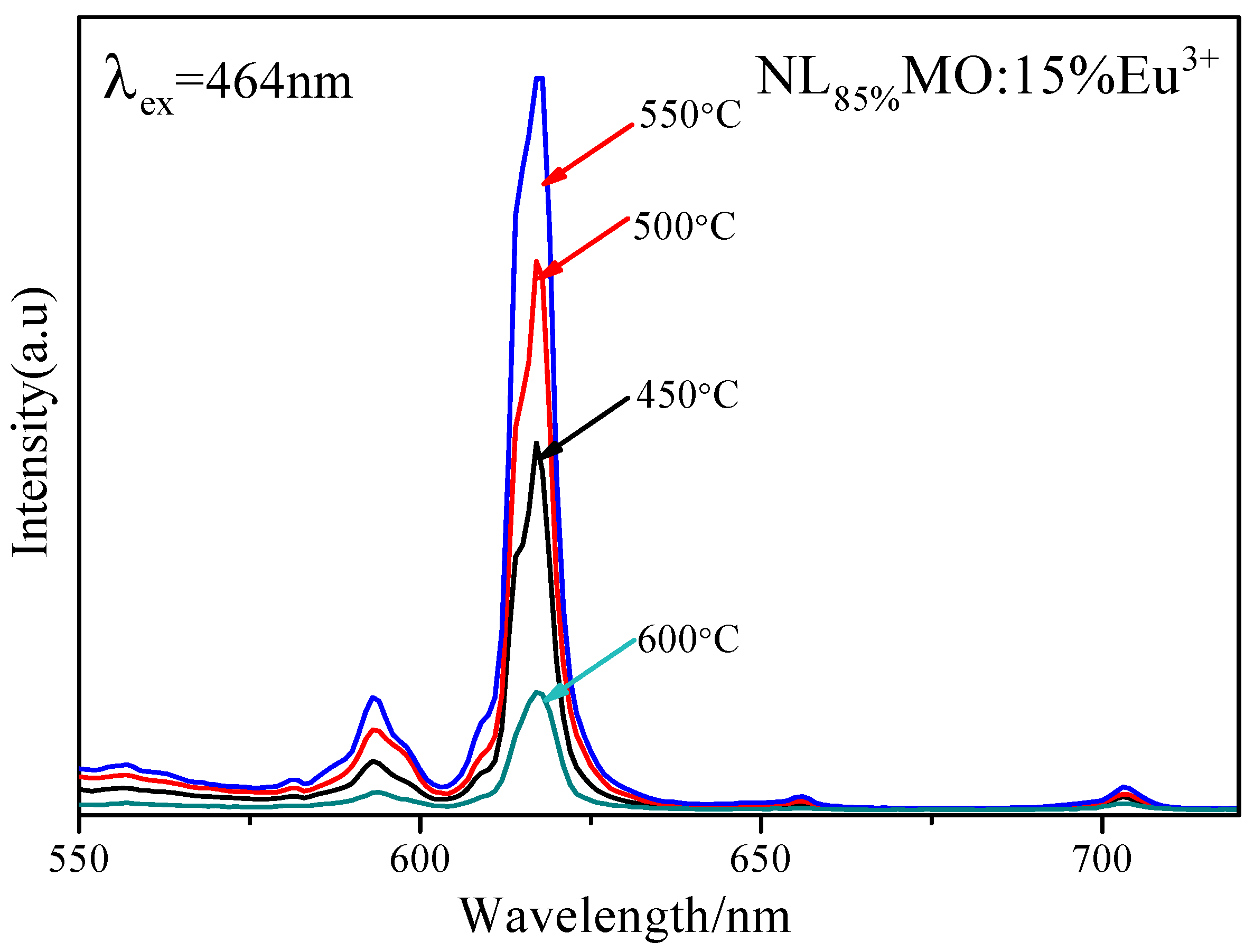
It is generally known that Eu3+ as an excellent activator ion is extensively applied in commercial phosphors because its characteristic emission lines related to the 5D0→7FJ (J = 1–4) transitions are usually distributed in the red spectral region [20]. Clearly, in Figure 2, the PLE spectrum of NL85%MO:15%Eu3+ monitored with the emission at 615 nm correspondent to the 5D0→7F2 transition of Eu3+ is shown. There is a strong broad band ranging from 220 to 350 nm with a maximum at 265 nm as a result of the O-Mo and O-Eu charge transfers, as well as several sharp peaks within 350–500 nm. It is easy to ascribe the sharp peaks at 382, 394, 416 and 464 nm to the intra-configuration 4f–4f transitions of Eu3+ including 7F0→5GJ,5L7, 7F0→5L6, 7F0→5D3 and 7F0→5D2, respectively. Among them, the strongest one at 464 nm is just agreed with the emission wavelengths of the GaN-based blue LED chips. The emission spectrum (PL) obtained under excitation of 464 nm consists of several sharp peaks at 537, 593, 615 and 703 nm due to the transitions of 5D1→7F1 and 5D0→7FJ (J = 1, 2, 4) of Eu3+, respectively. In particular, 5D0→7F1 belongs to magnetic and 5D0→7F2 to electric dipole transitions. According to the Judd–Ofelt theory [21,22], if Eu3+ is located at inversion symmetry, the 5D0→7F1 transition, which is scarcely affected by the crystal field environment of Eu3+, will play a leading role in the PL spectra and the phosphors emit an orange-red light when excited. Otherwise, the 5D0→7F2 transition is stronger and the phosphors emit a red light when excited. Moreover, the 5D0→7F2 transition is dominant in the PL spectra, which clearly indicates that Eu3+ occupies the La3+ site with anti-inversion symmetry and it means high red color purity. Here, the effects of Eu3+ concentration and synthesizing temperature on emission intensity are also shown in Figure 3 and Figure 4. Obviously, the optimal Eu3+ concentration and calcination temperature are 15 mol % and 550 °C, respectively, which were selected as the optimal conditions in the following experiments.
3.3. PL Properties and Energy Transfer Pathway of NLMO:Eu3+/Sm3+ Phosphors
To investigate how Sm3+ transfers its energy to Eu3+ in NLMO, we analyzed the PLE and PL spectra in detail. Differing from the PLE spectrum of NL85%MO:15%Eu3+, that of NL83%MO:15%Eu3+,2%Sm3+ consists of the characteristic excitation peaks of Eu3+ at about 395, 464 and 535 nm and an excitation peak at about 403 nm attributed to the transition of 6H5/2→4F7/2 of Sm3+ (Figure 5).
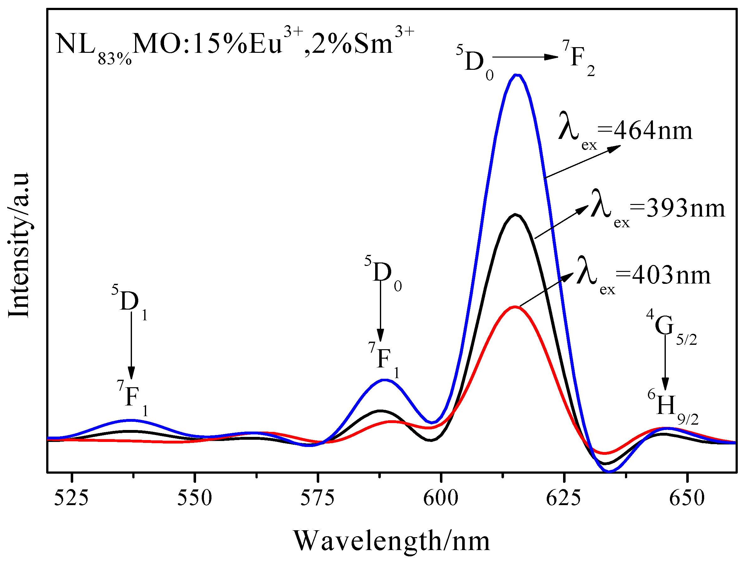
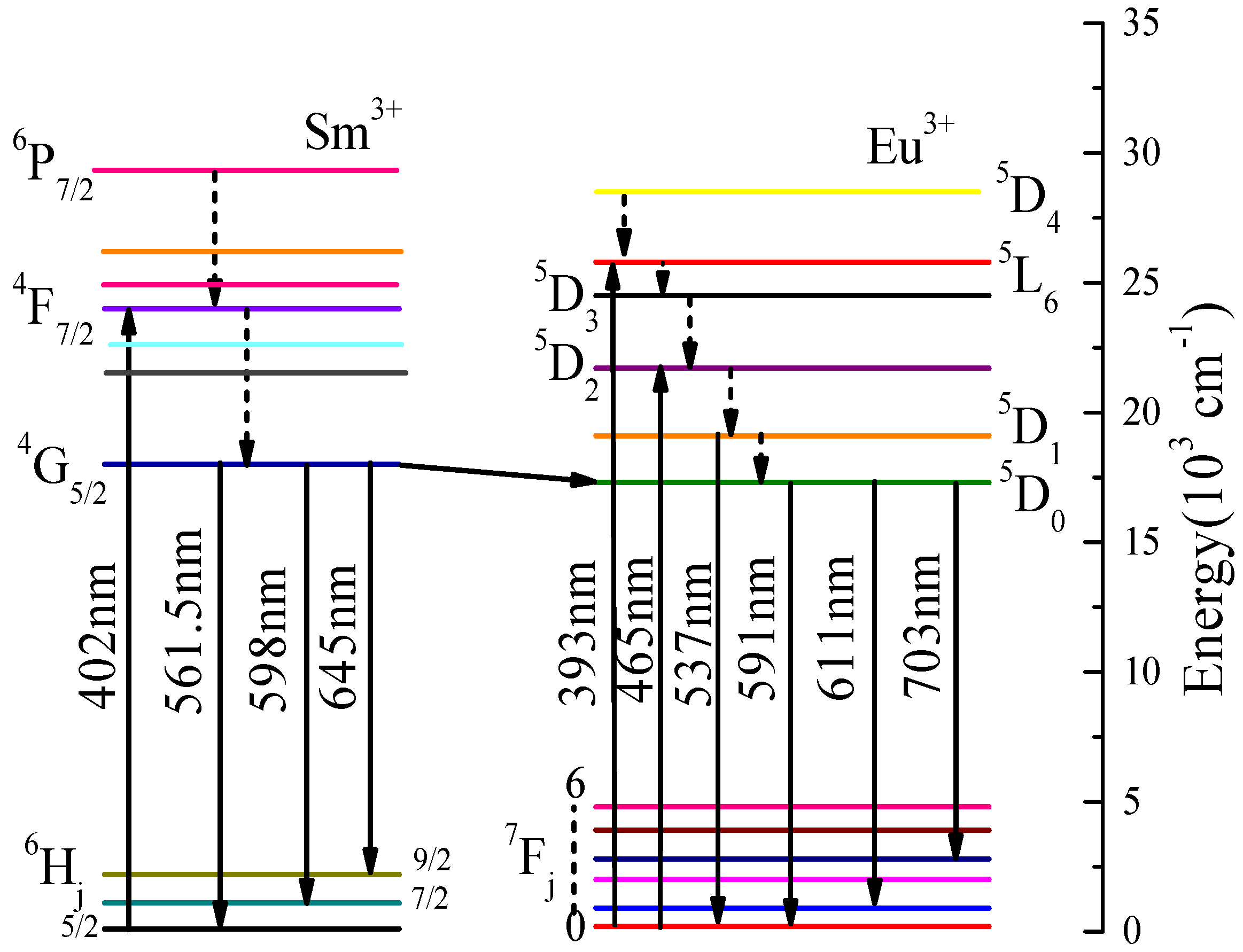
The PL spectrum of NL83%MO:15%Eu3+,2%Sm3+ excited at 403 nm is closely similar to those excited at 393 and 465 nm, while the emission bands at 537 nm correspondent to the 5D1→7F1 transition of Eu3+ and the relative peak intensities are different (Figure 6). The three PL spectra exhibit two significant emission peaks at about 593 and 615 nm related to the transitions of 5D0→7F1 and 5D0→7F2 of Eu3+, respectively, indicating the Sm3+ transferred efficiently its excitation energy to Eu3+. Further comparison shows that there are the emission bands at 537 nm in NLMO:Eu3+ and NLMO:Eu3+/Sm3+ excited at 393 and 464 nm, but not in NLMO:Eu3+/Sm3+ when excited at 405 nm, while there are still other transitions of Eu3+. Therefore, the energy transfer just occurs from the 4G5/2 state of Sm3+ to the 5D0 state rather than the 5D1 state of Eu3+. The probable energy transfer pathway, as determined from the analysis above, is displayed in Figure 7. The electrons of Sm3+, after being excited into 4K11/2 level, rapidly relaxed down to the 4G5/2 state owing to lattice vibration, and then returned to the ground states through the energy transfer to the 5D0 level rather than the 5D1 level of Eu3+ [23,24].
Figure 8a,b exhibits the PLE (λem = 615 nm) and PL(λex = 464 nm) spectra of NL85%−yMO:15%Eu3+,ySm3+ (y = 0, 1, 2, 3, 4 and 5%), respectively. Clearly, with y increasing from 0 to 2%, the characteristic excitation and emission intensities of Eu3+ are enhanced because Sm3+ transfer its energy to Eu3+, and, then, the intensities are weakened because of concentration quenching. Moreover, the fluorescence intensity of NL83%MO:15%Eu3+,2%Sm3+ is 1.47 times that of NL83%MO:15%Eu3+. The results further demonstrate the efficient energy transfer from Sm3+ to Eu3+.
3.4. Decay Curves and Energy Transfer Mechanism of NLMO:Eu3+/Sm3+ Phosphors
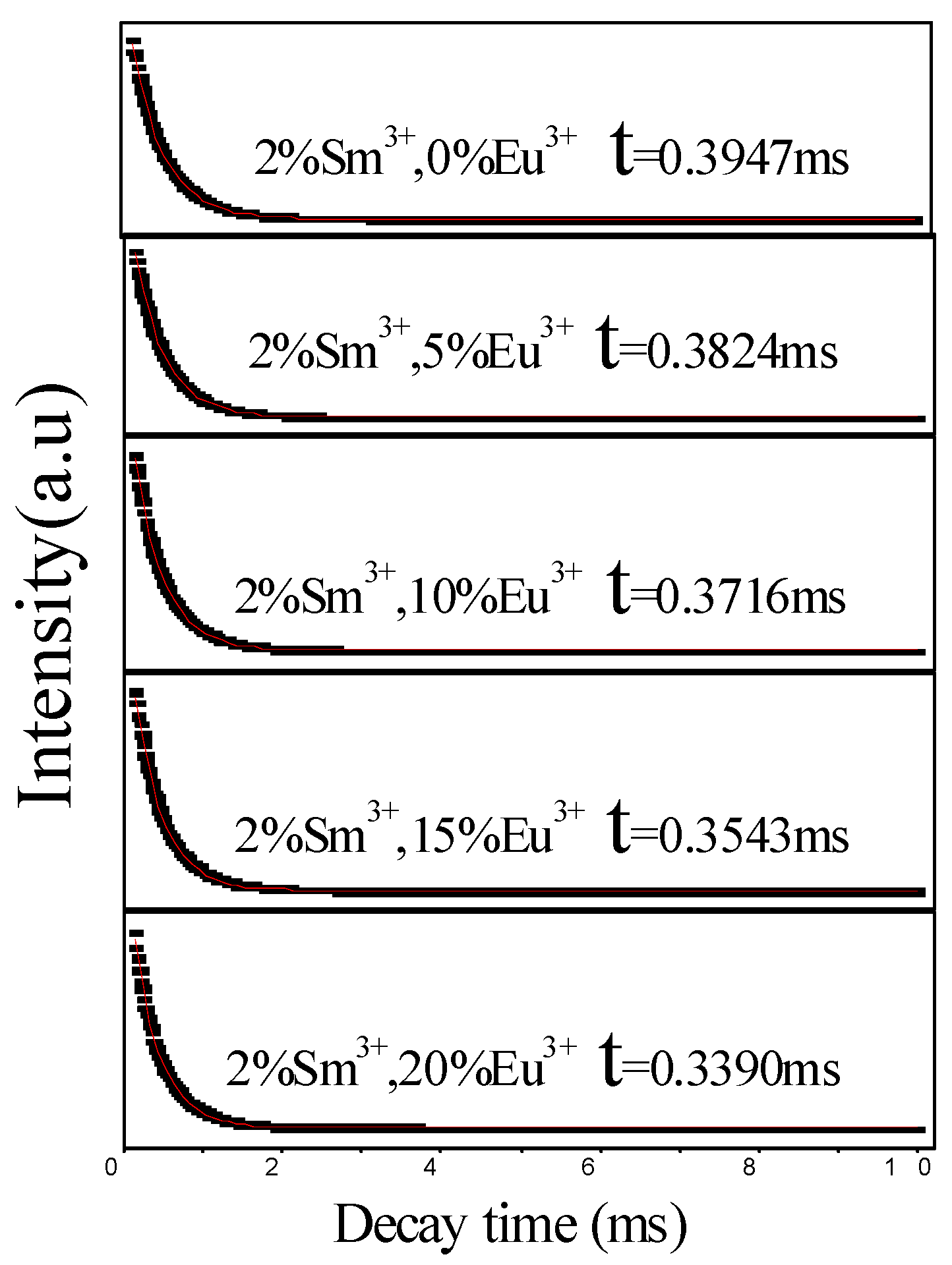
To deeply explore the interaction mechanism of energy transfer from Sm3+ to Eu3+ in the NLMO matrix, we measured the fluorescence lifetime of NL98%−xMO:xEu3+,2%Sm3+ (x = 0, 5, 10, 15 and 20%). The test results and decay curves were shown in Table 1 and Figure 9, respectively.
The fluorescence lifetime of Sm3+ is gradually shortened along with the concentration of Eu3+ increased from 0 to 20%, which is due to the fact that the energy transfer between Sm3+ and Eu3+ shortens the time for excited-electrons to move back to the ground state. To more intuitively understand the energy transfer from Sm3+ to Eu3+, the efficiency (ηET) of energy transfer from Sm3+ to Eu3+ was calculated using the following formula [24,25] and the results are also listed in Table 1:
where τs0 and τs are the lifetime durations of the donor (Sm3+) without and with the acceptor (Eu3+), respectively. All of the above results strongly confirm that Sm3+ transfers its excitation energy to Eu3+ in the NaLa (MoO4)2 matrix.
Energy is transferred through primarily exchange interaction and multipolar interaction, when the distance, R, between the donor ion and the acceptor ion is less than 0.5 nm and more than 0.5 nm, respectively [26,27]. RSm−Eu between Eu3+ and Sm3+ in the NaLa(MoO4)2 matrix can be calculated as follows [28] (Table 1):
where V is the unit cell volume, χc is the total doping mol % concentration of Sm3+ and Eu3+, and N is the number of cation sites. For the NaLa(MoO4)2 crystals, V = 0.33501 nm3 and N = 2. Based on the above data, the RSm−Eu of NL98%−xMO:xEu3+,2%Sm3+ (x = 0%, 5%, 10%, 15% and 20%) can be calculated (Table 1). Obviously, the RSm−Eu values are much greater than 0.5 nm, indicating the exchange interaction is greatly limited. Therefore, Sm3+ transfers its energy to Eu3+ through the multi-pole interaction mechanism. According to the Dexter formula [29] and Reisfeld approximation method [30] of the electric multi-pole interaction mechanism, the following relation can be obtained:
where C is the doping mol % concentration of Sm3+ and Eu3+, and α is relationship index. The values of α correspondent to dipole–dipole, dipole–quadrupole and quadrupole–quadrupole interactions are 6, 8 and 10, respectively. Figure 10a–c shows the Gauss plot of the linear dependency between τs0/τs and Cα/3. Clearly, R2, the goodness of fit, is closer to 1 at α = 10, meaning Sm3+ transfers its energy to Eu3+ in the NaLa(MoO4)2 matrix by the mechanism of quadrupole–quadrupole interaction.
3.5. Chromaticity Coordinate Analysis
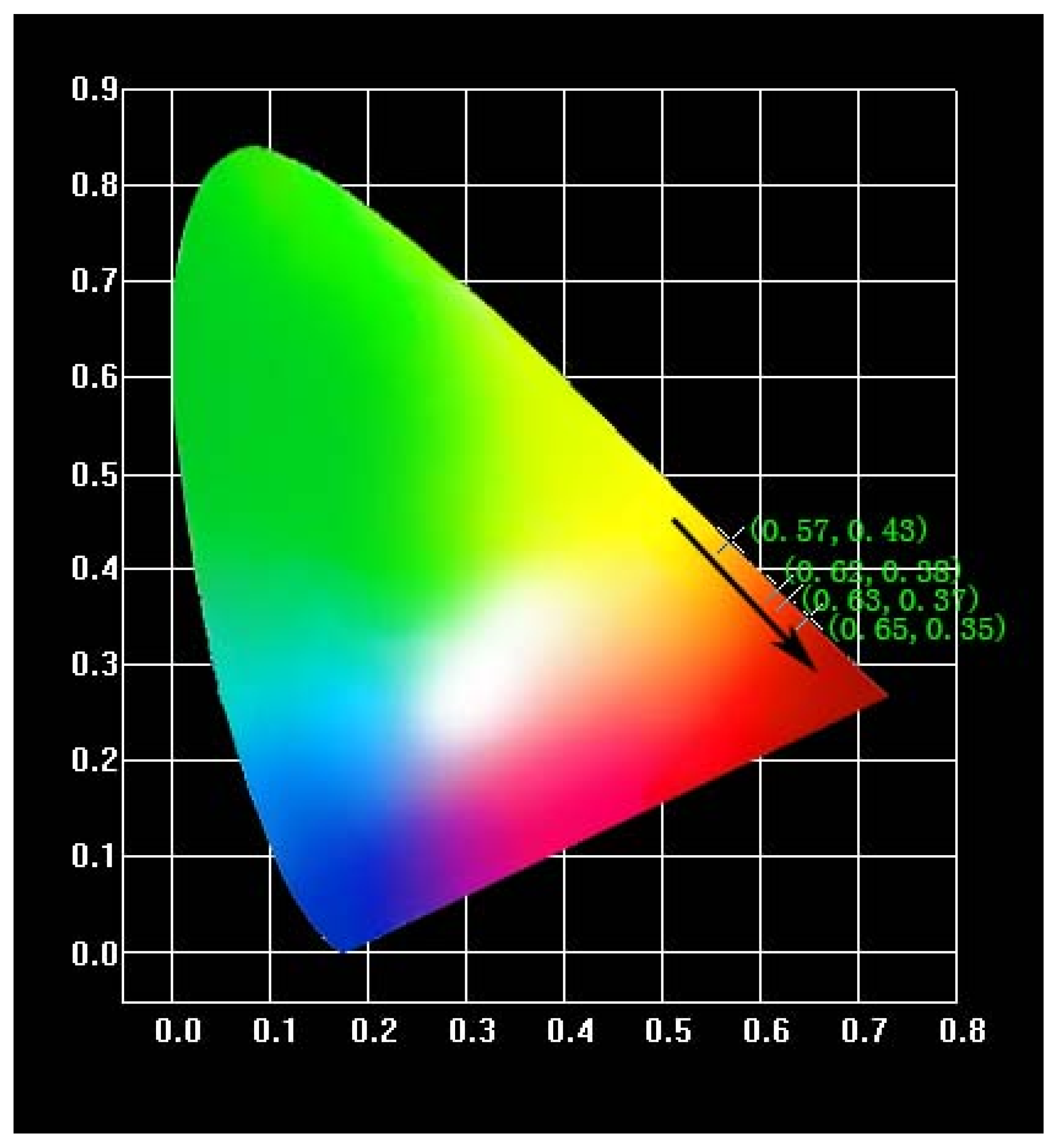
To investigate the luminous color of the phosphors, we depicted the CIE chromaticity diagrams of NL85%-yMO:15%Eu3+,ySm3+ when they are excited at 464 nm (Figure 11). The CIE coordinates are listed in Table 2. As seen, the emission light can be regularly shifted from orange to red with the Sm3+ molar ratio increasing with a maximum at (0.65, 0.35), which is very close to the CIE chromaticity coordinates (0.67, 0.33) of the standard red light, indicating NLMO:Eu3+,Sm3+ has a potential application prospect in blue-light-excited WLEDs.
4. Conclusions
We prepared the NLMO:Eu3+/Sm3+ phosphors via solid-station reaction at 450–600 °C and analyzed their fluorescence spectra. The X-ray diffraction analysis showed that the phosphors possess a body-centered tetragonal structure with high purity and crystallinity. The fluorescence spectrum observation indicates that the phosphor has the strongest excitation and emission peaks at 464 and 615 nm, respectively. The composition possesses a potential for WLEDs because of efficiently emitting red when excited by blue light. In-depth analysis of fluorescence spectra, fluorescence lifetime and inter-ion distance reveals that the energy transfer from the 4G5/2 state of Sm3+ to the 5D0 state of Eu3+ is achieved via the quadrupole–quadrupole mechanism, and it finally broadened the absorption band of Eu3+ and enhanced the luminous intensity.
Author Contributions
G.L. and M.Y. conceived and designed the experiments; Y.W. and J.W. performed the experiments and analyzed the data; Y.W. contributed reagents/materials/analysis tools; G.L. and M.Y. wrote the paper.
Funding
This research received no external funding.
Acknowledgments
This work was supported financially from the Priority Academic Program Development of Jiangsu Higher Education Institutions (PAPD).
Conflicts of Interest
The authors declare no conflict of interest.
References
- Liu, W.R.; Huang, C.H.; Yeh, C.W.; Chiu, Y.C.; Yeh, Y.T.; Liu, R.S. Single-phased white-light-emitting KCaGd(PO4)2:Eu2+,Tb3+,Mn2+ phosphors for LED applications. RSC Adv. 2013, 3, 9023–9028. [Google Scholar] [CrossRef]
- Hou, D.; Liang, H.; Xie, M.; Ding, X.; Zhong, J.; Su, Q.; Tao, Y.; Huang, Y.; Gao, Z. Bright green-emitting, energy transfer and quantum cutting of Ba3Ln(PO4)3:Tb3+(Ln = La, Gd) under VUV-UV excitation. Opt. Express 2011, 19, 11071. [Google Scholar] [CrossRef] [PubMed]
- Long, J.; Luo, Z.; Wang, T.; Pan, L.; Long, Q.; Liao, S.; Huang, Y.; Zhang, H. Enhanced photoluminescence and energy transfer in the novel red emitting phosphors SrZn2(PO4)2:Eu3+,Tb3+,Li+. J. Mater. Sci. Mater. Electron. 2017, 28, 1–4. [Google Scholar] [CrossRef]
- Chen, Y.; Wang, J.; Liu, C.; Kuang, X. A host sensitized reddish-orange Gd2MoO6:Sm3+ phosphor for light emitting diodes. Appl. Phys. Lett. 2011, 98, 191101. [Google Scholar] [CrossRef]
- Zuo, H.; Liu, Y.; Li, J.; Shi, X.; Ma, S.; Zhao, M. Enhancement of red emission in KLa(MoO4)2:Eu3+,Bi3+ phosphor for WLEDs. Ceram. Int. 2015, 41, 14834–14838. [Google Scholar] [CrossRef]
- Jiao, M.; Lv, W.Z.; Lu, W.; Zhao, Q.; Shao, B.Q.; You, H.P. Optical properties and energy transfer of a novel KSrSc2(PO4)3:Ce3+/Eu2+/Tb3+ phosphor for white light emitting diodes. Dalton Trans. 2015, 44, 4080–4087. [Google Scholar] [CrossRef] [PubMed]
- Atuchin, V.V.; Chimitova, O.D.; Gavrilova, T.A. Synthesis, structural and vibrational properties of microcrystalline RbNd(MoO4)2. J. Cryst. Growth 2011, 318, 683–686. [Google Scholar] [CrossRef]
- Atuchin, V.V.; Chimitova, O.D.; Adichtchev, S.V. Synthesis, structural and vibrational properties of microcrystalline β-RbSm(MoO4)2. Mater. Lett. 2013, 106, 26–29. [Google Scholar] [CrossRef]
- Chang, S.L.; Aleksandrovsky, A.S.; Molokeev, M.S. Microwave synthesis and spectroscopic properties of ternary scheelite-type molybdate phosphors NaSrLa(MoO4)3:Er3+, Yb3+. J. Alloy. Compd. 2017, 713, 156–163. [Google Scholar]
- Wang, Y.; Lin, C.; Zheng, H.; Sun, D.; Li, L.; Chen, B. Fluorescent and chromatic properties of visible-emitting phosphor KLa(MoO4)2:Sm3+. J. Alloy. Compd. 2013, 559, 123–128. [Google Scholar] [CrossRef]
- Du, P.; Yu, J.S. Energy transfer mechanism and color controllable luminescence in Dy3+/Eu3+-codoped NaLa(MoO4)2 phosphors. J. Alloy. Compd. 2015, 653, 468–473. [Google Scholar] [CrossRef]
- Li, G.; Lan, S.; Li, L.; Li, M.; Bao, W.; Zou, H.; Xu, X.; Gan, S. Tunable luminescence properties of NaLa(MoO4)2:Ce3+,Tb3+ phosphors for near UV-excited white light-emitting-diodes. J. Alloy. Compd. 2012, 513, 145–149. [Google Scholar] [CrossRef]
- Wu, H.; Chen, H.; Liu, Y.; Lu, Y.; Zhang, D. Highly uniform NaLa(MoO4)2:Eu3+ microspheres: Microwave-assisted hydrothermal synthesis, growth mechanism and enhanced luminescent properties. J. Mater. Sci. Mater. Electron. 2014, 25, 3109–3115. [Google Scholar] [CrossRef]
- Kumar, A.; Kumar, J. Perspective on europium activated fine-grained metal molybdate phosphors for solid state illumination. J. Mater. Chem. 2011, 21, 3788. [Google Scholar] [CrossRef]
- Atuchin, V.V.; Aleksandrovsky, A.S.; Chimitova, O.D. Synthesis and spectroscopic properties of monoclinic α-Eu2(MoO4)3. J. Phys. Chem. C 2014, 28, 15404–15411. [Google Scholar] [CrossRef]
- Shi, P.; Xia, Z.; Molokeev, M.S. Crystal chemistry and luminescence properties of red-emitting CsGd1−xEux(MoO4)2 solid-solution phosphors. Dalton Trans. 2014, 25, 9669–9676. [Google Scholar] [CrossRef] [PubMed]
- Atuchin, V.V.; Subanakov, A.K.; Aleksandrovsky, A.S. Structural and spectroscopic properties of new noncentrosymmetric self-activated borate Rb3EuB6O12 with B5O10 units. Mater. Des. 2018, 140, 488–494. [Google Scholar] [CrossRef]
- Neeraj, S.; Kijima, N.; Cheetham, A.K. Novel red phosphors for solid-state lighting: The system NaM(WO4)2−x(MoO4)x:Eu3+ (M = Gd,Y,Bi). Chem. Phys. Lett. 2004, 387, 2–6. [Google Scholar] [CrossRef]
- Zhou, X.; Wang, G.; Zhou, T.; Zhou, K.; Li, Q.; Wang, Z. Luminescence enhancement in Eu3+, Sm3+ co-doped LiY(MoO4)2 nano-phosphors by sol-gel process. J. Nanosci. Nanotechnol. 2014, 14, 3494. [Google Scholar] [CrossRef] [PubMed]
- Du, P.; Bharat, L.K.; Yu, J.S. Strong red emission in Eu3+/Bi3+ ions codoped CaWO4 phosphors for white light-emitting diode and field-emission display applications. J. Alloy. Compd. 2015, 633, 37–41. [Google Scholar] [CrossRef]
- Judd, B.R. Optical absorption intensities of rare-earth ions. Phys. Rev. 1962, 127, 750–761. [Google Scholar] [CrossRef]
- Ofelt, G.S. Intensities of Crystal Spectra of Rare-Earth Ions. J. Chem. Phys. 1962, 37, 511–520. [Google Scholar] [CrossRef]
- Bi, W.; Meng, Q.; Sun, W. Luminescent properties and energy transfer mechanism of NaGd(MoO4)2:Sm3+, Eu3+ phosphors. Ceram. Int. 2016, 42, 14086–14093. [Google Scholar] [CrossRef]
- Jin, Y.; Hao, Z.D.; Zhang, X.; Luo, Y.S.; Wang, X.J.; Zhang, J.H. Dynamical processes of energy transfer in red emitting phosphor CaMo(W)O4:Sm3+,Eu3+. Opt. Mater. 2011, 33, 1591–1594. [Google Scholar] [CrossRef]
- Rai, S.; Hazarika, S. Fluorescence dynamics of Tb3+ and Tb3+/Ho3+ doped phosphate glasses. Opt. Mater. 2008, 30, 1343–1348. [Google Scholar] [CrossRef]
- Xia, S.; Guan, A.; Chen, P.; Wang, G.; Geng, Y.; Zhou, L. Sol-gel method for preparing a novel red-emitting phosphor YVO4:Sm3+,Eu3+ with ideal red color emission. Superlattice 2016, 97, 319–326. [Google Scholar] [CrossRef]
- Blasse, G. Luminescence Materials; Springer: Berlin, Germany, 1994. [Google Scholar]
- Blasse, G. Materials Science of the Luminescence of Inorganic Solids; Springer US: New York, NY, USA, 1978. [Google Scholar]
- Dexter, D.L. A theory of sensitized luminescence in solids. J. Chem. Phys. 1953, 21, 836–850. [Google Scholar] [CrossRef]
- Reisfeld, R.; Greenberg, E.; Velapoldi, R. Luminescence quantum efficiency of Gd and Tb in borate glasses and the mechanism of energy transfer between them. J. Chem. Phys. 1972, 56, 1698–1705. [Google Scholar] [CrossRef]
Figure 1.
XRD patterns of (a) NL83%MO:15%Eu3+,2%Sm3+ and NL85%MO:15%Eu3+ obtained at 550 °C and (b) NL85%MO:15%Eu3+ formed at different temperatures for 6 h.
Figure 1.
XRD patterns of (a) NL83%MO:15%Eu3+,2%Sm3+ and NL85%MO:15%Eu3+ obtained at 550 °C and (b) NL85%MO:15%Eu3+ formed at different temperatures for 6 h.

Figure 2.
Excitation (λem = 615 nm) and emission (λex = 464 nm) spectra of NL85%MO:15%Eu3+.

Figure 3.
Emission (λex = 464 nm) spectra of NaLa(MoO4)2 with Eu3+ concentration at 5, 10, 15 and 20 mol %.
Figure 3.
Emission (λex = 464 nm) spectra of NaLa(MoO4)2 with Eu3+ concentration at 5, 10, 15 and 20 mol %.

Figure 4.
Emission (λex = 464 nm) spectra of NL85%MO:15%Eu3+ obtained at different calcination temperatures.
Figure 4.
Emission (λex = 464 nm) spectra of NL85%MO:15%Eu3+ obtained at different calcination temperatures.

Figure 5.
PLE and PL spectra of NL85%MO:15%Eu3+ and NL83%MO:15%Eu3+,2%Sm3+.

Figure 6.
PL spectra of NL83%MO:15%Eu3+,2%Sm3+ under the excitation at 393,403 and 465 nm.

Figure 7.
The energy transfer pathway between Eu3+ and Sm3+ in NaLa(MoO4)2.

Figure 8.
(a,b) excitation spectra (λem = 615 nm) and emission spectra (λex = 464 nm) of NL85%-yMO:15%Eu3+,ySm3+ (y = 0, 1, 2, 3, 4 and 5%).
Figure 8.
(a,b) excitation spectra (λem = 615 nm) and emission spectra (λex = 464 nm) of NL85%-yMO:15%Eu3+,ySm3+ (y = 0, 1, 2, 3, 4 and 5%).

Figure 9.
PL decay curves of NL98%-xMO:xEu3+,2%Sm3+.

Figure 10.
Dependence of τs0/τs on Cα/3 with α = (a) 6, (b) 8, (c) 10.

Figure 11.
CIE chromaticity diagram of NL85%−yMO:15%Eu3+,ySm3+ phosphors excited at 464 nm.


Table 1.
Energy transfer efficiency, distance, lifetime of NL98%-xMO:xEu3+,2%Sm3+.
| Xc/mol % | ηET/% | RSm−Eu/nm | τs/ms |
|---|---|---|---|
| 2 | - | 2.5196 | 0.3947 |
| 7 | 3.116 | 1.6595 | 0.3824 |
| 12 | 5.853 | 1.3866 | 0.3716 |
| 17 | 10.236 | 1.2346 | 0.3543 |
| 22 | 14.112 | 1.1329 | 0.3390 |
Table 2.
CIE chromaticity coordinates (x, y) of NL85%-yMO:15%Eu3+,ySm3+ (y = 0, 1, 2, 3, 4 and 5%) excited at 464 nm.
Table 2.
CIE chromaticity coordinates (x, y) of NL85%-yMO:15%Eu3+,ySm3+ (y = 0, 1, 2, 3, 4 and 5%) excited at 464 nm.
| NL85%-yMO:15%Eu3+,ySm3+/% | CIE x | CIE y |
|---|---|---|
| y = 0 | 0.57 | 0.43 |
| 1 | 0.62 | 0.38 |
| 2 | 0.63 | 0.37 |
| 3 | 0.65 | 0.35 |
| 4 | 0.65 | 0.35 |
| 5 | 0.65 | 0.35 |
© 2018 by the authors. Licensee MDPI, Basel, Switzerland. This article is an open access article distributed under the terms and conditions of the Creative Commons Attribution (CC BY) license (http://creativecommons.org/licenses/by/4.0/).
Share and Cite
MDPI and ACS Style
Yan, M.; Liu, G.; Wen, J.; Wang, Y. Blue-Light-Excited Eu3+/Sm3+ Co-Doped NaLa(MoO4)2 Phosphors: Synthesis, Characterizations and Red Emission Enhancement for WLEDs. Materials 2018, 11, 1090. https://doi.org/10.3390/ma11071090
AMA Style
Yan M, Liu G, Wen J, Wang Y. Blue-Light-Excited Eu3+/Sm3+ Co-Doped NaLa(MoO4)2 Phosphors: Synthesis, Characterizations and Red Emission Enhancement for WLEDs. Materials. 2018; 11(7):1090. https://doi.org/10.3390/ma11071090
Chicago/Turabian StyleYan, Man, Guanghui Liu, Jiahao Wen, and Yinlong Wang. 2018. "Blue-Light-Excited Eu3+/Sm3+ Co-Doped NaLa(MoO4)2 Phosphors: Synthesis, Characterizations and Red Emission Enhancement for WLEDs" Materials 11, no. 7: 1090. https://doi.org/10.3390/ma11071090
Note that from the first issue of 2016, this journal uses article numbers instead of page numbers. See further details here.