
Recent Advances in Applications of Droplet Microfluidics

Abstract
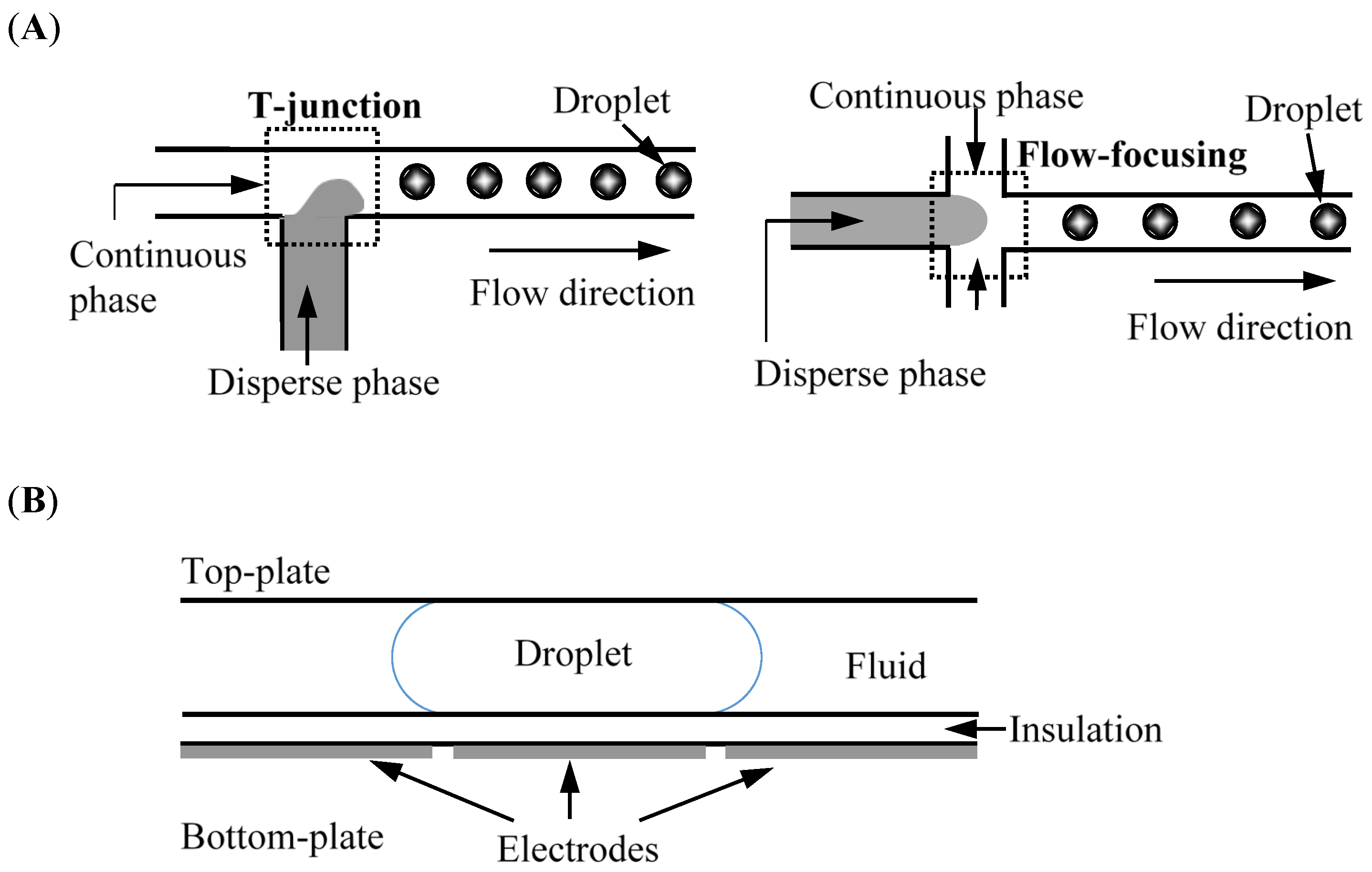
:1. Introduction


2. Molecular Detection

| Topic | Target | Remark | Ref. |
|---|---|---|---|
| A droplet-based fluorescence polarization immunoassay platform for rapid and quantitative analysis of biomarkers | Bovine angiogenin | Accurately determined the angiogenin concentration in cow’s milk, and required a total sample volume of less than 1 nL. | [41] |
| A novel droplet dosing strategy-based versatile microscale biosensor for detection of DNA, protein, and ion | dsDNA, streptavidin, and Hg2+ | The contact-induced droplets dosing based on adsorption and desorption was developed to overcome the channel-fouling problem. | [42] |
| Specific detection of avidin-biotin binding using liquid crystal droplets | Bovine serum albumin, lysozyme, hemoglobin, and chymotrypsinogen | The 5CBPAA-biotin droplets toward avidin were found to have high sensitivity, specificity, and stability. | [43] |
| Glucose sensor using liquid-crystal droplets made by microfluidics | Glucose | The biosensor detected samples under crossed polarizers at concentrations of 0.03 mM and 3-min response times. | [44] |
| Enzyme incorporated microfluidic device for in situ glucose detection in water-in-air microdroplets | Glucose | The fluorescence intensity linearly increased with glucose concentration up to 3 mM, and its detection limit was 6.64 μM. | [45] |
| Integrating bipolar electrochemistry and electrochemiluminescence imaging with microdroplets for chemical analysis | Quinones | Closed bipolar cell sensor could avoid the interference and cross-contamination between analyte solutions and electrochemiluminescence-reporting reagents. | [46] |
| Peptide nucleic acid molecular beacons for the detection of PCR amplicons in droplet-based microfluidic devices | Olea europaea L. and Roundup Ready soybean genes | Efficiently discriminated oligonucleotide sequences carrying single-base mutations at 100 nM. | [47] |
| A highly parallel microfluidic droplet method enabling single-molecule counting for digital enzyme detection | β-Galatosidase | An integrated microfluidic chip offered the feasibility of detecting single-enzyme molecules based on a digital counting method. | [48] |
| Digital microfluidic-enabled single-molecule detection by printing and sealing single magnetic beads in femtoliter droplets | β-Galactosidase | The fluorescent detection had a linear dynamic range of four orders of magnitude ranging from 10 aM to 90 fM. | [49] |
| Protein–protein interaction analysis in single microfluidic droplets using FRET and fluorescence lifetime detection | Bovine serum albumin, avidin, and streptavidin | Could be used for quantitative detection of molecules in direct and competitive assay formats within nM detection limits. | [50] |
3. Imaging
| Topic | Target | Remark | Ref. |
|---|---|---|---|
| Atom-economical in situ synthesis of BaSO4 as imaging contrast agents within poly(N-isopropyl acrylamide) microgels using one-step droplet microfluidics | Microgel | Fourteen-nanometer crystallites of BaSO4 as an X-ray imaging contrast agent were in situ synthesized with interlinking reactions. | [59] |
| Cloud-enabled microscopy and droplet microfluidic platform for specific detection of water | Escherichia coli | Magnetic beads conjugated with fluorescently labeled antibodies could selectively capture and isolate specific bacteria. | [60] |
| Live cell imaging compatible immobilization of Chlamydomonas reinhardtii in microfluidic platform for biodiesel research | Chlamydomonas reinhardtii | Provided real-time monitoring and analysis of lipid accumulation using single cell imaging for rapid optimization of microalgae culture conditions. | [61] |
4. Drug Delivery
5. Diagnostics
6. Cell Biology
| Topic | System | Remark | Ref. |
|---|---|---|---|
| Microfluidic-assisted engineering of polymeric microcapsules with high encapsulation efficiency for protein drug delivery | Polycaprolactone microcapsules encapsulating bovine serum albumin | The high encapsulation efficiency of proteins in the microcapsules reached 84%, and 30% of their content was released within 168 h. | [65] |
| Microfluidic assembly of multistage porous silicon-lipid vesicles for controlled drug release | Thermally hydrocarbonized porous silicon microparticle-lipid vesicle | The drug encapsulation efficiency was 19%, and the whole payload was released after only 6 h at pH 7.4. | [74] |
| Generation of uniform polymer-eccentric and core-centered hollow microcapsules for ultrasound-regulated drug release | Polydimethylsiloxane microcapsules encapsulating Rhodamine 6G and domperidone maleate | The system demonstrated the properties of a floating drug delivery system absorbed in the upper segments of the gastrointestinal tract for a long gastric residence time. | [75] |
| Synthesis of uniform core-shell gelatin-alginate microparticles as intestine-released oral delivery drug carrier | Core-shell gelatin-alginate | The fabricated microparticles could remain intact in gastric juice for more than 3 h, indicating effective protection in an acidic environment. | [76] |
| Controllable microfluidic fabrication of Janus and microcapsule particles for drug delivery applications | Poly(lactic-co-glycolic acid /poly(ε-caprolactone) microcapsule | The microparticles exhibited distinct degradation behavior, implying programmable drug delivery in different manners. | [77] |
| Core-shell structure microcapsules with dual-pH-responsive drug release function | Ampicillin loaded in the chitosan shell and diclofenac loaded in the alginate core | Demonstrated higher drug release efficiency than respective core or shell particles for dual-drug carriers. | [78] |
| Microfluidic-assisted generation of innovative polysaccharide hydrogel microparticles | Pectin-pectin (homo Janus) and pectin-alginate (hetero Janus) encapsulating bovine serum albumin | Facilitated studying the relationships between combined enzymatic hydrolysis and active release for anisotropic microparticles. | [79] |
| Microfluidic synthesis of monodisperse PEGDA microbeads for sustained release of 5-fluorouracil | Poly(ethylene glycol) diacrylate microbeads encapsulating 5-fluorouracil | The drug (0.1 to 0.5% w/w) demonstrated relatively fast elution in the first 12 h and continued to release over the next 156 h to effectively inhibit Huh-7 tumor cells in vitro. | [80] |
| Chitosan/agarose hydrogels: cooperative properties and microfluidic preparation | Chitosan and agarose composite hydrogels containing 5-fluorouracil | The hydrogels released 5-fluorouracil from chitosan/agarose macrogels with dual-pH and temperature properties. | [81] |
| Microfluidic fabrication of monodisperse biocompatible phospholipid vesicles for encapsulation and delivery of hydrophilic drugs or active compounds | Phospholipid vesicles encapsulating doxorubicin hydrochloride | The encapsulation efficiency was approximately 94%, and showed superior sustained release. | [82] |
| Topic | Target | Remark | Ref. |
|---|---|---|---|
| Detecting and tracking nosocomial methicillin-resistant Staphylococcus aureus by using a microfluidic SERS biosensor | Staphylococcus aureus | A reliable detection and epidemiological surveillance of bacterial infections in a bacterial mixture at levels from 5% to 100% was developed. | [31] |
| Ultrarapid generation of femtoliter microfluidic droplets for single-molecule-counting immunoassays | Prostate-specific antigen | The femtodroplet system enabled a single enzyme molecule for prostate cancer to be detected within 10 min and reduced the concentration to 46 fM. | [83] |
| Microfluidic droplet-based liquid-liquid extraction and on-chip IR spectroscopy detection of cocaine in human saliva | Cocaine | Showed a 2−3-fold higher extraction efficiency compared with state-of-the-art H-filters. | [84] |
| A centrifugal microfluidic platform for point-of-care diagnostic applications | Plasma and blood cells | Achieved pumping and valving of fluids and generation of monodisperse droplets on lab-on-a-disk system. | [85] |
| An integrated CMOS quantitative-polymerase-chain-reaction lab-on-chip for point-of-care diagnostics | Staphylococcus aureus | The complementary metal-oxide-semiconductor-integrated circuit had a reliable and sensitive detection of Staphylococcus aureus from 1 to 104 copies per 1.2-nL droplet. | [86] |
| Picoliter droplet microfluidic immunosorbent platform for point-of-care diagnostics | Human anti-tetanus immunoglobulin G | Reduced the reagent volume by four orders of magnitude and the detection time from hours to minutes. | [87] |
| Analysis of single-nucleotide polymorphism in human angiogenin using droplet-based microfluidics | Human angiogenin | The detection of single-nucleotide polymorphism in the droplet could be performed using TaqMan probes on DNA samples amplified offline by using a conventional thermocycler rather an expensive real-time PCR system. | [88] |
| Magnetic bead droplet immunoassay of oligomer amyloid β for the diagnosis of Alzheimer disease using micropillars to enhance the stability of the oil-water interface | Oligomer amyloid β | The platform markedly reduced the assay time to 45 min and the amount of antibody usage to 10–30 ng per assay. | [89] |
| Droplet microfluidic chip based nucleic acid amplification and real-time detection of influenza viruses | Influenza A and C | The detection threshold of the chip-based qRT-PCR for detecting and quantifying viral nucleic acids was approximately five copies per PCR reaction. | [90] |
| Multiplex, quantitative, reverse-transcription PCR detection of influenza viruses using droplet microfluidic technology | Influenza A and B | The qRT-PCR process was found to be less than 10 RNA copies accomplished within 40 min. | [91] |
| Rapid detection of tuberculosis using droplet-based microfluidics | BlaC enzyme | For a 30-μm droplet size, the fluorescent intensity change could be detected after less than 1 h of incubation. | [92] |
| Development of a microfluidic-based optical sensing device for label-free detection of circulating tumor cells through their lactic acid metabolism | Circulating tumor cells | Could detect the targeted cancer cells without interference by the cell species. | [93] |
| Assembly-line manipulation of droplets in microfluidic platform for fluorescence encoding and simultaneous multiplexed DNA detection | Human immunodeficiency virus, variola virus | The result indicated that targets could be simultaneously detected using a time-saving process and without a complex dye-labelling process. | [94] |
| Digital microfluidic platform for the detection of rubella infection and immunity: a proof of concept | Rubella virus | For both rubella viruses IgG and IgM, the performance panel samples demonstrated 100% diagnostic sensitivity and specificity. | [95] |
| Rapid and reproducible analysis of thiocyanate in real human serum and saliva by using a droplet SERS-microfluidic chip | Human serum and saliva | The reaction required less than 15 s in the designed channel, which is at least 40-fold shorter than that for solid metallic substrates. | [96] |
| A novel microbead-based microfluidic device for rapid bacterial identification and antibiotic susceptibility testing | Escherichia coli O157 | The immunocapture efficiency was 85%–92%, higher than 44%–86% of offline immunomagnetic separation. | [97] |
| Rapid detection of bacteriophages in starter culture using water-in-oil-in-water emulsion microdroplets | Escherichia coli BL21 and T7 phages | The lytic phage infection in a bacterial culture could be measured using a simple and inexpensive imaging approach in contrast to flow cytometry and PCR methods. | [98] |
| Rapid enumeration of phage in monodisperse emulsions | T4-LacZ and nonlytic M13 | This quantification was robust and insensitive to environmental fluctuations in contrast to bulk assays. | [99] |
| Highly sensitive and homogeneous detection of membrane protein on a single living cell by using aptameric and nicking enzyme-assisted signal amplification based on microfluidic droplets | Protein tyrosine kinase-7 | Used for constructing a high-throughput platform for detecting a single cell by using aptameric and enzyme-assisted amplification for membrane proteins. | [100] |
| A biocompatible open-surface droplet manipulation platform for detection of multi-nucleotide polymorphism | Multi-nucleotide polymorphism | The entire procedure required only 5 min and the total sample volume consumed in each operation was only 10 μL. | [101] |
| Topography-assisted electromagnetic platform for blood-to-PCR in a droplet | KRAS oncogene | Integrated automatic nucleic acid extraction (only 15 min) with real-time amplification detection of genetic targets. | [102] |
| Single-molecule quantitation and sequencing of rare translocations by using microfluidic-nested digital PCR | Lymphoblasts | Demonstrated quantitative measurement and single-molecule sequencing at extremely low levels (<10−6) in healthy subjects. | [103] |
| Topic | Target | Remark | Ref. |
|---|---|---|---|
| Single-cell analysis and sorting by using droplet-based microfluidics | Mouse hybridoma cells | This protocol displayed the use of two-phase droplet-based microfluidics for high-throughput single-cell analysis and sorting. | [69] |
| Versatile microfluidic droplet array for bioanalysis | HL-60 cells | The novel regional hydrophilic chip demonstrated high-throughput screening in toxic tests of CdSe on cells, and a rapid biosensing approach for carcinoma embryonic antigen was developed. | [104] |
| Mixed hydrogel bead-based tumor spheroid formation and anticancer drug testing | Human cervical carcinoma (HeLa) cells | Multicellular tumor spheroids were formed in the microfluidic droplets, and the viability of cells encapsulated in the mixed hydrogel beads was higher than 90%. | [105] |
| Cell-based drug combination screening with a microfluidic droplet array system | A549 nonsmall lung cancer cells | The sequential operation droplet array technique provided flexible approach for performing cell-based screening, and the reagent consumptions were decreased by two to three orders of magnitude compared with traditional multiwell plates. | [106] |
| Digital microfluidics for time-resolved cytotoxicity studies on single nonadherent yeast cells | Saccharomyces cerevisiae strain BY4741 | Could isolate single nonadherent cells and monitor their dynamic responses at a defined position over time for implementation of high-throughput cytotoxicity assays. | [107] |
| Droplet-based microfluidic platform for high-throughput, multiparameter screening of photosensitizer activity | Escherichia coli | Could detect both live and dead cells online to score cell viability and enable simultaneous measurement of many experiments including those on dark toxicity, photosensitizer concentration, light dose, and oxygenation levels. | [108] |
| Changing growth behavior of heavy-metal tolerant bacteria: media optimization using droplet-based microfluidics | Bacillus sporothermodurans and Streptomyces tendae | The nitrogen source between the light scattering and the fluorescence signal may be used for the production of fluorescent secondary metabolites. | [109] |
| Generation of monodisperse cell-sized microdroplets using a centrifuge-based axisymmetric coflowing microfluidic device | Yeast cells | After the encapsulation process, 87% of the yeast cells were alive in the monodisperse microdroplets. | [110] |
| Real-time image processing for label-free enrichment of Actinobacteria cultivated in picoliter droplets | Actinobacteria | Implemented high-throughput cultivation of soil-derived Actinobacteria and developed trigger imaging for picoliter droplet sorting. | [111] |
| Digital microfluidic processing of mammalian embryos for vitrification | Mammalian embryos | The benefits of this digital microfluidic device over conventional manual operation include cryoprotectant concentration gradient generation, automated operation, and feasibility of loading and retrieval of cells. | [112] |
| Single-cell forensic short tandem repeat typing within microfluidic droplets | Human lymphoid cells | Individual cells were efficiently encapsulated in nanoliter agarose droplets, serving as the reactors for PCR assays. | [113] |
| Microfluidic encapsulation of cells in alginate particles via an improved internal gelation approach | Antibody-secreting hybridoma cells (9E10 cell) and mouse breast cancer cells (M6C cell) | Two mammalian cell types were encapsulated with a viability of higher than 84% and grew well inside the microparticles. | [114] |
| A droplet-based heterogeneous immunoassay for screening single cells secreting antigen-specific antibodies | Alginate microbeads encapsulating antibody-secreting cells | Screened anti-TNF-alpha antibody-secreting cells from a mixture of cells in alginate microbeads as cell culture chambers. | [115] |
| Ultrahigh-throughput detection of single-cell β-galactosidase activity in droplets using microoptical lens array | Escherichia coli | This analytical throughput by a parallelized fluorescent detection compatible with droplet reinjection was larger than those obtained using flow cytometry. | [116] |
| New glycosidase substrates for droplet-based microfluidic screening | Cellobiohydrolase activity on model bacterial strains (Escherichia coli and Bacillus subtilis) | The fluorogenic substrates could be utilized to assay glycosidase activities in a broad pH range (4–11) and with incubation times of more than 24 h in droplet-based microfluidic systems. | [117] |
7. Other Applications
7.1. Particle Shaping
7.2. Food
8. Conclusions
Acknowledgments
Author Contributions
Conflicts of Interest
References
- Puigmartí-Luis, J. Microfluidic platforms: A mainstream technology for the preparation of crystals. Chem. Soc. Rev. 2014, 43, 2253–2271. [Google Scholar] [CrossRef] [PubMed]
- Zhu, Y.; Fang, Q. Analytical detection techniques for droplet microfluidics—A review. Anal. Chim. Acta 2013, 787, 24–35. [Google Scholar] [CrossRef] [PubMed]
- Samiei, E.; Hoorfar, M. Systematic analysis of geometrical based unequal droplet splitting in digital microfluidics. J. Micromech. Microeng. 2015, 25, 055008. [Google Scholar] [CrossRef]
- Oliveira, K.A.; Silva, P.B.M.; de Souza, F.R.; Martins, F.T.; Coltro, W.K.T. Kinetic study of glucose oxidase on microfluidic toner-based analytical devices for clinical diagnostics with image-based detection. Anal. Methods 2014, 6, 4995–5000. [Google Scholar] [CrossRef]
- Jones, C.N.; Hoang, A.N.; Dimisko, L.; Hamza, B.; Martel, J.; Irimia, D. Microfluidic platform for measuring neutrophil chemotaxis from unprocessed whole blood. J. Vis. Exp. 2014. [Google Scholar] [CrossRef] [PubMed]
- Bernacka-Wojcik, I.; Lopes, P.; Vaz, A.C.; Veigas, B.; Wojcik, P.J.; Simões, P.; Barata, D.; Fortunato, E.; Baptista, P.V.; Águas, H.; et al. Bio-microfluidic platform for gold nanoprobe based DNA detection-application to mycobacterium tuberculosis. Biosens. Bioelectron. 2013, 48, 87–93. [Google Scholar] [CrossRef] [PubMed]
- Peng, Z.; Young, B.; Baird, A.E.; Soper, S.A. Single-pair fluorescence resonance energy transfer analysis of mRNA transcripts for highly sensitive gene expression profiling in near real time. Anal. Chem. 2013, 85, 7851–7858. [Google Scholar] [CrossRef] [PubMed]
- Vladisavljevic, G.T.; Khalid, N.; Neves, M.A.; Kuroiwa, T.; Nakajima, M.; Uemura, K.; Ichikawa, S.; Kobayashi, I. Industrial lab-on-a-chip: Design, applications and scale-up for drug discovery and delivery. Adv. Drug Deliv. Rev. 2013, 65, 1626–1663. [Google Scholar] [CrossRef] [PubMed] [Green Version]
- Dressler, O.J.; Maceiczyk, R.M.; Chang, S.I.; DeMello, A.J. Droplet-based microfluidics: Enabling impact on drug discovery. J. Biomol. Screen. 2014, 19, 483–496. [Google Scholar] [CrossRef] [PubMed]
- Sun, X.; Tang, K.; Smith, R.D.; Kelly, R.T. Controlled dispensing and mixing of pico- to nanoliter volumes using on-demand droplet-based microfluidics. Microfluid. Nanofluid. 2013, 15, 117–126. [Google Scholar] [CrossRef] [PubMed]
- Lin, Y.S.; Yang, C.H.; Wu, C.T.; Grumezescu, A.M.; Wang, C.Y.; Hsieh, W.C.; Chen, S.Y.; Huang, K.S. A microfluidic chip using phenol formaldehyde resin for uniform-sized polycaprolactone and chitosan microparticle generation. Molecules 2013, 18, 6521–6531. [Google Scholar] [CrossRef] [PubMed]
- Lin, Y.S.; Yang, C.H.; Hsu, Y.Y.; Hsieh, C.L. Microfluidic synthesis of tail-shaped alginate microparticles using slow sedimentation. Electrophoresis 2013, 34, 425–431. [Google Scholar] [CrossRef]
- Huang, K.S.; Lin, Y.S.; Chang, W.R.; Wang, Y.L.; Yang, C.H. A facile fabrication of alginate microbubbles using a gas foaming reaction. Molecules 2013, 18, 9594–9602. [Google Scholar] [CrossRef]
- Yang, C.H.; Wang, W.T.; Grumezescu, A.M.; Huang, K.S.; Lin, Y.S. One-step synthesis of platinum nanoparticles loaded in alginate bubbles. Nanoscale Res. Lett. 2014, 9, 277. [Google Scholar] [CrossRef] [PubMed]
- Yang, C.H.; Huang, K.S.; Grumezescu, A.M.; Wang, C.Y.; Tzeng, S.C.; Chen, S.Y.; Lin, Y.H.; Lin, Y.S. Synthesis of uniform poly(d,l-lactide) and poly(d,l-lactide-co-glycolide) microspheres using a microfluidic chip for comparison. Electrophoresis 2014, 35, 316–322. [Google Scholar] [CrossRef]
- Gañán-Calvo, A.M.; Montanero, J.M.; Martín-Banderas, L.; Flores-Mosquera, M. Building functional materials for health care and pharmacy from microfluidic principles and flow focusing. Adv. Drug Deliv. Rev. 2013, 65, 1447–1469. [Google Scholar] [CrossRef] [PubMed]
- Sarvothaman, M.K.; Kim, K.S.; Seale, B.; Brodersen, P.M.; Walker, G.C.; Wheeler, A.R. Dynamic fluoroalkyl polyethylene glycol co-polymers: A new strategy for reducing protein adhesion in lab-on-a-chip devices. Adv. Funct. Mater. 2015, 25, 506–515. [Google Scholar] [CrossRef]
- Seiffert, S.; Friess, F.; Lendlein, A.; Wischke, C. Faster droplet production by delayed surfactant-addition in two-phase microfluidics to form thermo-sensitive microgels. J. Colloid Interface Sci. 2015, 452, 38–42. [Google Scholar] [CrossRef] [PubMed]
- Pirbodaghi, T.; Vigolo, D.; Akbari, S.; DeMello, A. Investigating the fluid dynamics of rapid processes within microfluidic devices using bright-field microscopy. Lab Chip 2015, 15, 2140–2144. [Google Scholar] [CrossRef] [PubMed]
- Musterd, M.; Van Steijn, V.; Kleijn, C.R.; Kreutzer, M.T. Calculating the volume of elongated bubbles and droplets in microchannels from a top view image. RSC Adv. 2015, 5, 16042–16049. [Google Scholar] [CrossRef]
- Zantow, M.; Dendere, R.; Douglas, T.S. Image-based analysis of droplets in microfluidics. Eng. Med. Biol. Soc. 2013, 1776–1779. [Google Scholar]
- Luo, Y.; Chakrabarty, K. Design of pin-constrained general-purpose digital microfluidic biochips. Comput. Aided Des. Integr. Circuits Syst. 2013, 32, 1307–1320. [Google Scholar] [CrossRef]
- Jan-Willi, J.; Marian, W.; Gerri, K.; Jonathon, H.; Thomas, S.; Ilia, P.; Spatz, J.P. Key factors for stable retention of fluorophores and labeled biomolecules in droplet-based microfluidics. Anal. Chem. 2015, 87, 2063–2067. [Google Scholar]
- Pan, I.; Mukherjee, R.; Rahaman, H.; Samanta, T.; Dasgupta, P. Optimization algorithms for the design of digital microfluidic biochips: A survey. Comput. Electr. Eng. 2013, 39, 112–121. [Google Scholar] [CrossRef]
- Lai, K.T.; Yang, Y.T.; Lee, C.Y. An intelligent digital microfluidic processor for biomedical detection. J. Sign Process Syst. 2015, 78, 85–93. [Google Scholar] [CrossRef]
- Huang, K.D.; Liu, C.H.; Lin, H.S. Reactant and waste minimization in multitarget sample preparation on digital microfluidic biochips. IEEE Trans. Comput. Aided Des. Integr. Circuits Syst. 2013, 32, 1484–1494. [Google Scholar] [CrossRef]
- Chen, Y.H.; Hsu, C.L.; Tsai, L.C.; Huang, T.W.; Ho, T.Y. A reliability-oriented placement algorithm for reconfigurable digital microfluidic biochips using 3-D deferred decision making technique. IEEE Trans. Comput. Aided Des. Integr. Circuits Syst. 2013, 32, 1151–1162. [Google Scholar] [CrossRef]
- Maftei, E.; Pop, P.; Madsen, J. Module-based synthesis of digital microfluidic biochips with droplet-aware operation execution. ACM J. Emerg. Technol. Comput. Syst. 2013, 9, 21. [Google Scholar] [CrossRef]
- Luo, Y.; Chakrabarty, K.; Ho, T.Y. Error recovery in cyberphysical digital microfluidic biochips. IEEE Trans. Comput. Aided Des. Integr. Circuits Syst. 2013, 32, 59–72. [Google Scholar] [CrossRef]
- Isgor, P.K.; Marcali, M.; Keser, M.; Elbuken, C. Microfluidic droplet content detection using integrated capacitive sensors. Sens. Actuator B Chem. 2015, 210, 669–675. [Google Scholar] [CrossRef]
- Lu, X.; Samuelson, D.R.; Xu, Y.; Zhang, H.; Wang, S.; Rasco, B.A.; Xu, J.; Konkel, M.E. Detecting and tracking nosocomial methicillin-resistant staphylococcus aureus using a microfluidic SERS biosensor. Anal. Chem. 2013, 85, 2320–2327. [Google Scholar] [CrossRef] [PubMed]
- Kawano, Y.; Otsuka, C.; Sanzo, J.; Higgins, C.; Nirei, T.; Schilling, T.; Ishikawa, T. Expanding imaging capabilities for microfluidics: Applicability of darkfield internal reflection illumination (DIRI) to observations in microfluidics. PLoS ONE 2015, 10. [Google Scholar] [CrossRef] [PubMed]
- Kim, M.; Pan, M.; Gai, Y.; Pang, S.; Han, C.; Yang, C.; Tang, S.K. Optofluidic ultrahigh-throughput detection of fluorescent drops. Lab Chip 2015, 15, 1417–1423. [Google Scholar] [CrossRef] [PubMed]
- Muluneh, M.; Kim, B.; Buchsbaum, G.; Issadore, D. Miniaturized, multiplexed readout of droplet-based microfluidic assays using time-domain modulation. Lab Chip 2014, 14, 4638–4646. [Google Scholar] [CrossRef] [PubMed]
- Zhao, Y.; Chen, D.; Yue, H.; French, J.B.; Rufo, J.; Benkovic, S.J.; Huang, T.J. Lab-on-a-chip technologies for single-molecule studies. Lab Chip 2013, 13, 2183–2198. [Google Scholar] [CrossRef] [PubMed]
- Cretich, M.; Daaboul, G.G.; Sola, L.; Unlu, M.S.; Chiari, M. Digital detection of biomarkers assisted by nanoparticles: Application to diagnostics. Trends Biotechnol. 2015, 33, 343–351. [Google Scholar] [CrossRef] [PubMed]
- Streets, A.M.; Huang, Y. Microfluidics for biological measurements with single-molecule resolution. Curr. Opin. Biotechnol. 2014, 25, 69–77. [Google Scholar] [CrossRef] [PubMed]
- Kang, D.K.; Ali, M.M.; Zhang, K.; Pone, E.J.; Zhao, W. Droplet microfluidics for single-molecule and single-cell analysis in cancer research, diagnosis and therapy. TrAC Trends Anal. Chem. 2014, 58, 145–153. [Google Scholar] [CrossRef]
- Oedit, A.; Vulto, P.; Ramautar, R.; Lindenburg, P.W.; Hankemeier, T. Lab-on-a-chip hyphenation with mass spectrometry: Strategies for bioanalytical applications. Curr. Opin. Biotechnol. 2015, 31, 79–85. [Google Scholar] [CrossRef] [PubMed]
- Zeng, Y.; Wang, T. Quantitative microfluidic biomolecular analysis for systems biology and medicine. Anal. Bioanal. Chem. 2013, 405, 5743–5758. [Google Scholar] [CrossRef] [PubMed]
- Choi, J.W.; Kim, G.J.; Lee, S.; Kim, J.; deMello, A.J.; Chang, S.I. A droplet-based fluorescence polarization immunoassay (DFPIS) platform for rapid and quantitative analysis of biomarkers. Biosens. Bioelectron. 2015, 67, 497–502. [Google Scholar] [CrossRef] [PubMed]
- Chen, J.; Liu, Y.; Ye, T.; Xiang, X.; Ji, X.; He, Z. A novel droplet dosing strategy-based versatile microscale biosensor for detection of DNA, protein and ion. Sens. Actuator B Chem. 2015, 215, 206–214. [Google Scholar] [CrossRef]
- Khan, M.; Park, S.Y. Specific detection of avidin-biotin binding using liquid crystal droplets. Colloid Surf. B Biointerfaces 2015, 127, 241–246. [Google Scholar] [CrossRef] [PubMed]
- Kim, J.; Khan, M.; Park, S.Y. Glucose sensor using liquid-crystal droplets made by microfluidics. ACS Appl. Mater. Interfaces 2013, 5, 13135–13139. [Google Scholar] [CrossRef] [PubMed]
- Piao, Y.; Han, D.J.; Azad, M.R.; Park, M.; Seo, T.S. Enzyme incorporated microfluidic device for in situ glucose detection in water-in-air microdroplets. Biosens. Bioelectron. 2015, 65, 220–225. [Google Scholar] [CrossRef] [PubMed]
- Wu, S.Z.; Zhou, Z.Y.; Xu, L.R.; Su, B.; Fang, Q. Integrating bipolar electrochemistry and electrochemiluminescence imaging with microdroplets for chemical analysis. Biosens. Bioelectron. 2014, 53, 148–153. [Google Scholar] [CrossRef] [PubMed]
- Zanoli, L.M.; Licciardello, M.; D’Agata, R.; Lantano, C.; Calabretta, A.; Corradini, R.; Marchelli, R.; Spoto, G. Peptide nucleic acid molecular beacons for the detection of pcr amplicons in droplet-based microfluidic devices. Anal. Bioanal. Chem. 2013, 405, 615–624. [Google Scholar] [CrossRef] [PubMed]
- Guan, Z.; Zou, Y.; Zhang, M.J.; Lv, J.; Shen, H.; Yang, P.; Zhang, H.; Zhu, Z.; Yang, C.J. A highly parallel microfluidic droplet method enabling single-molecule counting for digital enzyme detection. Biomicrofluidics 2014, 8, 014110. [Google Scholar] [CrossRef] [PubMed]
- Witters, D.; Knez, K.; Ceyssens, F.; Puers, R.; Lammertyn, J. Digital microfluidics-enabled single-molecule detection by printing and sealing single magnetic beads in femtoliter droplets. Lab Chip 2013, 13, 2047–2054. [Google Scholar] [CrossRef] [PubMed]
- Benz, C.; Retzbach, H.; Nagl, S.; Belder, D. Protein-protein interaction analysis in single microfluidic droplets using fret and fluorescence lifetime detection. Lab Chip 2013, 13, 2808–2814. [Google Scholar] [CrossRef] [PubMed]
- Rademeyer, P.; Carugo, D.; Lee, J.Y.; Stride, E. Microfluidic system for high throughput characterisation of echogenic particles. Lab Chip 2015, 15, 417–428. [Google Scholar] [CrossRef] [PubMed]
- Duncanson, W.J.; Arriaga, L.R.; Ung, W.L.; Kopechek, J.A.; Porter, T.M.; Weitz, D.A. Microfluidic fabrication of perfluorohexane-shelled double emulsions for controlled loading and acoustic-triggered release of hydrophilic agents. Langmuir 2014, 30, 13765–13770. [Google Scholar] [CrossRef] [PubMed]
- Sheeran, P.S.; Rojas, J.D.; Puett, C.; Hjelmquist, J.; Arena, C.B.; Dayton, P.A. Contrast-enhanced ultrasound imaging and in vivo circulatory kinetics with low-boiling-point nanoscale phase-change perfluorocarbon agents. Ultrasound Med. Biol. 2015, 41, 814–831. [Google Scholar] [CrossRef] [PubMed]
- Duncanson, W.J.; Kodger, T.E.; Babaee, S.; Gonzalez, G.; Weitz, D.A.; Bertoldi, K. Microfluidic fabrication and micromechanics of permeable and impermeable elastomeric microbubbles. Langmuir 2015, 31, 3489–3493. [Google Scholar] [CrossRef] [PubMed]
- Dhanaliwala, A.H.; Chen, J.L.; Wang, S.; Hossack, J.A. Liquid flooded flow-focusing microfluidic device for in situ generation of monodisperse microbubbles. Microfluid. Nanofluid. 2013, 14, 457–467. [Google Scholar] [CrossRef] [PubMed]
- Neto, B.A.; Carvalho, P.H.; Correa, J.R. Benzothiadiazole derivatives as fluorescence imaging probes: Beyond classical scaffolds. Acc. Chem. Res. 2015, 48, 1560–1569. [Google Scholar] [CrossRef] [PubMed]
- Yu, X.; Cheng, G.; Zhou, M.D.; Zheng, S.Y. On-demand one-step synthesis of monodisperse functional polymeric microspheres with droplet microfluidics. Langmuir 2015, 31, 3982–3992. [Google Scholar] [CrossRef] [PubMed]
- Wang, W.; Zhang, M.J.; Chu, L.Y. Functional polymeric microparticles engineered from controllable microfluidic emulsions. Acc. Chem. Res. 2014, 47, 373–384. [Google Scholar] [CrossRef] [PubMed]
- Wang, Q.; Zhang, D.; Yang, X.; Xu, H.; Shen, A.Q.; Yang, Y. Atom-economical in situ synthesis of BaSO4 as imaging contrast agents within poly(n-isopropylacrylamide) microgels using one-step droplet microfluidics. Green Chem. 2013, 15, 2222–2229. [Google Scholar] [CrossRef]
- Golberg, A.; Linshiz, G.; Kravets, I.; Stawski, N.; Hillson, N.J.; Yarmush, M.L.; Marks, R.S.; Konry, T. Cloud-enabled microscopy and droplet microfluidic platform for specific detection of escherichia coli in water. PLoS ONE 2014, 9. [Google Scholar] [CrossRef] [PubMed]
- Park, J.W.; Na, S.C.; Nguyen, T.Q.; Paik, S.M.; Kang, M.; Hong, D.; Choi, I.S.; Lee, J.H.; Jeon, N.L. Live cell imaging compatible immobilization of chlamydomonas reinhardtii in microfluidic platform for biodiesel research. Biotechnol. Bioeng. 2015, 112, 494–501. [Google Scholar] [CrossRef] [PubMed]
- Seiffert, S. Small but smart: Sensitive microgel capsules. Angew. Chem. Int. Edit. 2013, 52, 11468. [Google Scholar] [CrossRef] [PubMed]
- Kaler, K.; Prakash, R. Droplet microfluidics for chip-based diagnostics. Sensors 2014, 14, 23283–23306. [Google Scholar] [CrossRef] [PubMed]
- Iwanaga, S.; Saito, N.; Sanae, H.; Nakamura, M. Facile fabrication of uniform size-controlled microparticles and potentiality for tandem drug delivery system of micro/nanoparticles. Colloid Surf. B Biointerfaces 2013, 109, 301–306. [Google Scholar] [CrossRef] [PubMed]
- Pessi, J.; Santos, H.A.; Miroshnyk, I.; Yliruusi, J.; Weitz, D.A.; Mirza, S. Microfluidics-assisted engineering of polymeric microcapsules with high encapsulation efficiency for protein drug delivery. Int. J. Pharm. 2014, 472, 82–87. [Google Scholar] [CrossRef] [PubMed]
- Zhao, C.X. Multiphase flow microfluidics for the production of single or multiple emulsions for drug delivery. Adv. Drug Deliv. Rev. 2013, 65, 1420–1446. [Google Scholar] [CrossRef] [PubMed]
- Tasoglu, S.; Gurkan, U.A.; Wang, S.Q.; Demirci, U. Manipulating biological agents and cells in micro-scale volumes for applications in medicine. Chem. Soc. Rev. 2013, 42, 5788–5808. [Google Scholar] [CrossRef] [PubMed]
- Rosenfeld, L.; Lin, T.; Derda, R.; Tang, S.K.Y. Review and analysis of performance metrics of droplet microfluidics systems. Microfluid. Nanofluid. 2014, 16, 921–939. [Google Scholar] [CrossRef]
- Mazutis, L.; Gilbert, J.; Ung, W.L.; Weitz, D.A.; Griffiths, A.D.; Heyman, J.A. Single-cell analysis and sorting using droplet-based microfluidics. Nat. Protoc. 2013, 8, 870–891. [Google Scholar] [CrossRef] [PubMed]
- Rakszewska, A.; Tel, J.; Chokkalingam, V.; Huck, W.T.S. One drop at a time: Toward droplet microfluidics as a versatile tool for single-cell analysis. NPG Asia Mater. 2014, 6, e133. [Google Scholar] [CrossRef]
- Schlicht, B.; Zagnoni, M. Droplet-interface-bilayer assays in microfluidic passive networks. Sci. Rep. 2015, 5, 9951. [Google Scholar] [CrossRef] [PubMed]
- Cao, J.; Köhler, J.M. Droplet-based microfluidics for microtoxicological studies. Eng. Life Sci. 2015, 15, 306–317. [Google Scholar] [CrossRef]
- Au, S.H.; Fobel, R.; Desai, S.P.; Voldman, J.; Wheeler, A.R. Cellular bias on the microscale: Probing the effects of digital microfluidic actuation on mammalian cell health, fitness and phenotype. Integr. Biol. 2013, 5, 1014–1025. [Google Scholar] [CrossRef] [PubMed]
- Herranz-Blanco, B.; Arriaga, L.R.; Mäkilä, E.; Correia, A.; Shrestha, N.; Mirza, S.; Weitz, D.A.; Salonen, J.; Hirvonen, J.; Santos, H.A. Microfluidic assembly of multistage porous silicon-lipid vesicles for controlled drug release. Lab Chip 2014, 14, 1083–1086. [Google Scholar] [CrossRef] [PubMed]
- Huang, J.; Li, W.; Li, Y.; Luo, C.; Zeng, Y.; Xu, Y.; Zhou, J. Generation of uniform polymer eccentric and core-centered hollow microcapsules for ultrasound-regulated drug release. J. Mater. Chem. B 2014, 2, 6848–6854. [Google Scholar] [CrossRef]
- Huang, K.S.; Yang, C.H.; Kung, C.P.; Grumezescu, A.M.; Ker, M.D.; Lin, Y.S.; Wang, C.Y. Synthesis of uniform core-shell gelatin-alginate microparticles as intestine-released oral delivery drug carrier. Electrophoresis 2014, 35, 336. [Google Scholar] [CrossRef] [PubMed]
- Li, W.; Dong, H.; Tang, G.; Ma, T.; Cao, X. Controllable microfluidic fabrication of janus and microcapsule particles for drug delivery applications. RSC Adv. 2015, 5, 23181–23188. [Google Scholar] [CrossRef]
- Yang, C.H.; Wang, C.Y.; Grumezescu, A.M.; Wang, A.H.J.; Hsiao, C.J.; Chen, Z.Y.; Huang, K.S. Core-shell structure microcapsules with dual pH-responsive drug release function. Electrophoresis 2014, 35, 2680. [Google Scholar] [CrossRef] [PubMed]
- Marquis, M.; Davy, J.; Cathala, B.; Fang, A.; Renard, D. Microfluidics assisted generation of innovative polysaccharide hydrogel microparticles. Carbohydr. Polym. 2015, 116, 189–199. [Google Scholar] [CrossRef] [PubMed]
- Xue, P.; Wu, Y.; Menon, N.V.; Kang, Y. Microfluidic synthesis of monodisperse pegda microbeads for sustained release of 5-fluorouracil. Microfluid. Nanofluid. 2014, 18, 333–342. [Google Scholar] [CrossRef]
- Zamora-Mora, V.; Velasco, D.; Hernández, R.; Mijangos, C.; Kumacheva, E. Chitosan/agarose hydrogels: Cooperative properties and microfluidic preparation. Carbohydr. Polym. 2014, 111, 348–355. [Google Scholar] [CrossRef] [PubMed]
- Kong, F.; Zhang, X.; Hai, M. Microfluidics fabrication of monodisperse biocompatible phospholipid vesicles for encapsulation and delivery of hydrophilic drug or active compound. Langmuir 2014, 30, 3905–3912. [Google Scholar] [CrossRef] [PubMed]
- Shim, J.U.; Ranasinghe, R.T.; Smith, C.A.; Ibrahim, S.M.; Hollfelder, F.; Huck, W.T.S.; Klenerman, D.; Abell, C. Ultrarapid generation of femtoliter microfluidic droplets for single-molecule-counting immunoassays. ACS Nano 2013, 7, 5955–5964. [Google Scholar] [CrossRef] [PubMed]
- Wagli, P.; Chang, Y.C.; Hans, K.; Homsy, A.; Hvozdara, L.; Herzig, H.P.; Sigrist, M.; De Rooij, N.F. Microfluidic droplet-based liquid-liquid extraction and on-chip IR spectroscopy detection of cocaine in human saliva. Anal. Chem. 2013, 85, 7558–7565. [Google Scholar] [CrossRef] [PubMed]
- Hugo, S.; Land, K.; Madou, M.; Kido, H. A centrifugal microfluidic platform for point-of-care diagnostic applications. S. Afr. J. Sci. 2014, 110, 1–7. [Google Scholar] [CrossRef]
- Norian, H.; Field, R.M.; Kymissis, I.; Shepard, K.L. An integrated cmos quantitative-polymerase-chain-reaction lab-on-chip for point-of-care diagnostics. Lab Chip 2014, 14, 4076–4084. [Google Scholar]
- Golberg, A.; Yarmush, M.L.; Konry, T. Picoliter droplet microfluidic immunosorbent platform for point-of-care diagnostics of tetanus. Microchim. Acta 2013, 180, 855–860. [Google Scholar] [CrossRef]
- Bardiya, N.; Choi, J.-W.; Chang, S.-I. Analysis of single nucleotide polymorphism in human angiogenin using droplet-based microfluidics. BioChip J. 2014, 8, 15–21. [Google Scholar] [CrossRef]
- Kim, J.A.; Kim, M.; Kang, S.M.; Lim, K.T.; Kim, T.S.; Kang, J.Y. Magnetic bead droplet immunoassay of oligomer amyloid beta for the diagnosis of alzheimer’s disease using micro-pillars to enhance the stability of the oil-water interface. Biosens. Bioelectron. 2015, 67, 724–732. [Google Scholar] [CrossRef] [PubMed]
- Prakash, R.; Pabbaraju, K.; Wong, S.; Wong, A.; Tellier, R.; Kaler, K. Droplet microfluidic chip based nucleic acid amplification and real-time detection of influenza viruses. J. Electrochem. Soc. 2014, 161, B3083–B3093. [Google Scholar] [CrossRef]
- Prakash, R.; Pabbaraju, K.; Wong, S.; Wong, A.; Tellier, R.; Kaler, K. Multiplex, quantitative, reverse transcription pcr detection of influenza viruses using droplet microfluidic technology. Micromachines 2015, 6, 63–79. [Google Scholar] [CrossRef]
- Rosenfeld, L.; Cheng, Y.F.; Rao, J.H.; Tang, S.K.Y. Rapid detection of tuberculosis using droplet-based microfluidics. Proc. SPIE 8976 Microfluid. BioMEMS Med. Microsyst. XII 2014. [Google Scholar] [CrossRef]
- Chiu, T.K.; Lei, K.F.; Hsieh, C.H.; Hsiao, H.B.; Wang, H.M.; Wu, M.H. Development of a microfluidic-based optical sensing device for label-free detection of circulating tumor cells (CTCs) through their lactic acid metabolism. Sensors 2015, 15, 6789–6806. [Google Scholar] [CrossRef] [PubMed]
- Chen, J.; Zhou, G.; Liu, Y.; Ye, T.; Xiang, X.; Ji, X.; He, Z. Assembly-line manipulation of droplets in microfluidic platform for fluorescence encoding and simultaneous multiplexed DNA detection. Talanta 2015, 134, 271–277. [Google Scholar] [CrossRef] [PubMed]
- Ng, A.H.; Lee, M.; Choi, K.; Fischer, A.T.; Robinson, J.M.; Wheeler, A.R. Digital microfluidic platform for the detection of rubella infection and immunity: A proof of concept. Clin. Chem. 2015, 61, 420–429. [Google Scholar] [CrossRef] [PubMed]
- Wu, L.; Wang, Z.; Zong, S.; Cui, Y. Rapid and reproducible analysis of thiocyanate in real human serum and saliva using a droplet sers-microfluidic chip. Biosens. Bioelectron. 2014, 62, 13–18. [Google Scholar] [CrossRef] [PubMed]
- He, J.; Mu, X.; Guo, Z.; Hao, H.; Zhang, C.; Zhao, Z.; Wang, Q. A novel microbead-based microfluidic device for rapid bacterial identification and antibiotic susceptibility testing. Eur. J. Clin. Microbiol. Infect. Dis. 2014, 33, 2223–2230. [Google Scholar] [CrossRef] [PubMed]
- Wang, M.S.; Nitin, N. Rapid detection of bacteriophages in starter culture using water-in-oil-in-water emulsion microdroplets. Appl. Microbiol. Biotechnol. 2014, 98, 8347–8355. [Google Scholar] [CrossRef] [PubMed]
- Tjhung, K.F.; Burnham, S.; Anany, H.; Griffiths, M.W.; Derda, R. Rapid enumeration of phage in monodisperse emulsions. Anal. Chem. 2014, 86, 5642–5648. [Google Scholar] [CrossRef] [PubMed]
- Li, L.; Wang, Q.; Feng, J.; Tong, L.; Tang, B. Highly sensitive and homogeneous detection of membrane protein on a single living cell by aptamer and nicking enzyme assisted signal amplification based on microfluidic droplets. Anal. Chem. 2014, 86, 5101–5107. [Google Scholar] [CrossRef] [PubMed]
- Huang, C.J.; Fang, W.F.; Ke, M.S.; Chou, H.Y.; Yang, J.T. A biocompatible open-surface droplet manipulation platform for detection of multi-nucleotide polymorphism. Lab Chip 2014, 14, 2057–2062. [Google Scholar] [CrossRef] [PubMed]
- Chiou, C.H.; Shin, D.J.; Zhang, Y.; Wang, T.H. Topography-assisted electromagnetic platform for blood-to-pcr in a droplet. Biosens. Bioelectron. 2013, 50, 91–99. [Google Scholar] [CrossRef] [PubMed]
- Shuga, J.; Zeng, Y.; Novak, R.; Lan, Q.; Tang, X.; Rothman, N.; Vermeulen, R.; Li, L.; Hubbard, A.; Zhang, L.; et al. Single molecule quantitation and sequencing of rare translocations using microfluidic nested digital pcr. Nucleic Acids. Res. 2013. [Google Scholar] [CrossRef] [PubMed]
- Hu, S.W.; Xu, B.Y.; Ye, W.K.; Xia, X.H.; Chen, H.Y.; Xu, J.J. Versatile microfluidic droplets array for bioanalysis. ACS Appl. Mater. Interfaces 2015, 7, 935–940. [Google Scholar] [CrossRef] [PubMed]
- Wang, Y.; Wang, J. Mixed hydrogel bead-based tumor spheroid formation and anticancer drug testing. Analyst 2014, 139, 2449–2458. [Google Scholar] [CrossRef] [PubMed]
- Du, G.S.; Pan, J.Z.; Zhao, S.P.; Zhu, Y.; Den Toonder, J.M.; Fang, Q. Cell-based drug combination screening with a microfluidic droplet array system. Anal. Chem. 2013, 85, 6740–6747. [Google Scholar] [CrossRef] [PubMed]
- Kumar, P.T.; Vriens, K.; Cornaglia, M.; Gijs, M.; Kokalj, T.; Thevissen, K.; Geeraerd, A.; Cammue, B.P.; Puers, R.; Lammertyn, J. Digital microfluidics for time-resolved cytotoxicity studies on single non-adherent yeast cells. Lab Chip 2015, 15, 1852–1860. [Google Scholar] [CrossRef] [PubMed]
- Cho, S.; Kang, D.K.; Sim, S.; Geier, F.; Kim, J.Y.; Niu, X.; Edel, J.B.; Chang, S.I.; Wootton, R.C.; Elvira, K.S.; et al. Droplet-based microfluidic platform for high-throughput, multi-parameter screening of photosensitizer activity. Anal. Chem. 2013, 85, 8866–8872. [Google Scholar] [CrossRef] [PubMed]
- Wetzel, K.; Cao, J.; Kothe, E.; Köhler, J.M. Changing growth behavior of heavy-metal tolerant bacteria: Media optimization using droplet-based microfluidics. Eng. Life Sci. 2015, 15, 327–332. [Google Scholar] [CrossRef]
- Yamashita, H.; Morita, M.; Sugiura, H.; Fujiwara, K.; Onoe, H.; Takinoue, M. Generation of monodisperse cell-sized microdroplets using a centrifuge-based axisymmetric co-flowing microfluidic device. J. Biosci. Bioeng. 2015, 119, 492–495. [Google Scholar] [CrossRef] [PubMed]
- Zang, E.; Brandes, S.; Tovar, M.; Martin, K.; Mech, F.; Horbert, P.; Henkel, T.; Figge, M.T.; Roth, M. Real-time image processing for label-free enrichment of actinobacteria cultivated in picoliterliter droplets. Lab Chip 2013, 13, 3707–3713. [Google Scholar] [CrossRef] [PubMed]
- Pyne, D.G.; Liu, J.; Abdelgawad, M.; Sun, Y. Digital microfluidic processing of mammalian embryos for vitrification. PLoS ONE 2014, 9. [Google Scholar] [CrossRef] [PubMed]
- Geng, T.; Novak, R.; Mathies, R.A. Single-cell forensic short tandem repeat typing within microfluidic droplets. Anal. Chem. 2014, 86, 703–712. [Google Scholar] [CrossRef] [PubMed]
- Akbari, S.; Pirbodaghi, T. Microfluidic encapsulation of cells in alginate particles via an improved internal gelation approach. Microfluid. Nanofluid. 2014, 16, 773–777. [Google Scholar] [CrossRef]
- Akbari, S.; Pirbodaghi, T. A droplet-based heterogeneous immunoassay for screening single cells secreting antigen-specific antibodies. Lab Chip 2014, 14, 3275–3280. [Google Scholar] [CrossRef] [PubMed]
- Lim, J.; Vrignon, J.; Gruner, P.; Karamitros, C.S.; Konrad, M.; Baret, J.C. Ultra-high throughput detection of single cell β-galactosidase activity in droplets using micro-optical lens array. Appl. Phys. Lett. 2013, 103, 203704. [Google Scholar] [CrossRef]
- Najah, M.; Mayot, E.; Mahendra-Wijaya, I.P.; Griffiths, A.D.; Ladame, S.; Drevelle, A. New glycosidase substrates for droplet-based microfluidic screening. Anal. Chem. 2013, 85, 9807–9814. [Google Scholar] [CrossRef] [PubMed]
- Polenz, I.; Brosseau, Q.; Baret, J.C. Monitoring reactive microencapsulation dynamics using microfluidics. Soft Matter 2015, 11, 2916–2923. [Google Scholar] [CrossRef] [PubMed]
- Shim, T.S.; Kim, S.-H.; Yang, S.-M. Elaborate design strategies toward novel microcarriers for controlled encapsulation and release. Part. Part. Syst. Charact. 2013, 30, 9–45. [Google Scholar] [CrossRef]
- Seiffert, S. Microgel capsules tailored by droplet-based microfluidics. Chem. Phys. Chem. 2013, 14, 295–304. [Google Scholar] [CrossRef] [PubMed]
- Tang, G.N.; Li, W.X.; Cao, X.D.; Dong, H. In situ microfluidic fabrication of multi-shape inorganic/organic hybrid particles with controllable surface texture and porous internal structure. RSC Adv. 2015, 5, 12872–12878. [Google Scholar] [CrossRef]
- Yan, H.; Kim, C. Formation of monodisperse silica microparticles with various shapes and surface morphologies using double emulsion templates. Colloid Surf. A Physicochem. Eng. Asp. 2014, 443, 88–95. [Google Scholar] [CrossRef]
- Maan, A.A.; Nazir, A.; Khan, M.K.I.; Boom, R.; Schroën, K. Microfluidic emulsification in food processing. J. Food Eng. 2015, 147, 1–7. [Google Scholar] [CrossRef]
- Shewan, H.M.; Stokes, J.R. Review of techniques to manufacture micro-hydrogel particles for the food industry and their applications. J. Food Eng. 2013, 119, 781–792. [Google Scholar] [CrossRef]
© 2015 by the authors; licensee MDPI, Basel, Switzerland. This article is an open access article distributed under the terms and conditions of the Creative Commons Attribution license (http://creativecommons.org/licenses/by/4.0/).
Share and Cite
Chou, W.-L.; Lee, P.-Y.; Yang, C.-L.; Huang, W.-Y.; Lin, Y.-S. Recent Advances in Applications of Droplet Microfluidics. Micromachines 2015, 6, 1249-1271. https://doi.org/10.3390/mi6091249
Chou W-L, Lee P-Y, Yang C-L, Huang W-Y, Lin Y-S. Recent Advances in Applications of Droplet Microfluidics. Micromachines. 2015; 6(9):1249-1271. https://doi.org/10.3390/mi6091249
Chicago/Turabian StyleChou, Wei-Lung, Pee-Yew Lee, Cing-Long Yang, Wen-Ying Huang, and Yung-Sheng Lin. 2015. "Recent Advances in Applications of Droplet Microfluidics" Micromachines 6, no. 9: 1249-1271. https://doi.org/10.3390/mi6091249