Metal Oxide Nanoparticles Against Bacterial Biofilms: Perspectives and Limitations


1
Microbiology Lab of SCAMT Institute, ITMO University, Lomonosova st. 9, 191002 St. Petersburg, Russia
2
Department of Epidemiology, Pasteur Institute, 197101 St. Petersburg, Russia
*
Author to whom correspondence should be addressed.
Microorganisms 2020, 8(10), 1545; https://doi.org/10.3390/microorganisms8101545
Submission received: 12 September 2020
/
Revised: 25 September 2020
/
Accepted: 2 October 2020
/
Published: 7 October 2020
(This article belongs to the Section Biofilm)
Abstract
:At present, there is an urgent need in medicine and industry to develop new approaches to eliminate bacterial biofilms. Considering the low efficiency of classical approaches to biofilm eradication and the growing problem of antibiotic resistance, the introduction of nanomaterials may be a promising solution. Outstanding antimicrobial properties have been demonstrated by nanoparticles (NPs) of metal oxides and their nanocomposites. The review presents a comparative analysis of antibiofilm properties of various metal oxide NPs (primarily, CuO, Fe3O4, TiO2, ZnO, MgO, and Al2O3 NPs) and nanocomposites, as well as mechanisms of their effect on plankton bacteria cells and biofilms. The potential mutagenicity of metal oxide NPs and safety problems of their wide application are also discussed.
1. Introduction
Biofilm is a wide-range life form of bacteria that consists of the association of microorganisms cultures and extracellular polymer matrix (EPM), a complex biochemical mixture of polysaccharides, proteins, glycopeptides, nucleic acids, and lipids [1,2]. This slimy three-dimensional biopolymer is heterogeneous in different layers and contains structures similar to transport and water channels [3]. In biofilm formation, there are three main stages: adhesion, colonization, and maturation [4,5]. From the mature biofilm, the plankton microorganisms are isolated or dispersed into the environment. Thus, biofilm is a complex three-dimensional biological structure with a higher microbial life organization, in many respects, similar to a multicellular organism [6].
The primary function of biofilm is to protect the microorganism inside it from unfavorable physical, chemical, and biological factors in the environment, such as temperature, drying, ultraviolet radiation, biocides, humoral, and cellular factors of immunity [4]. Therefore, biofilms are stable, stress-resistant structures, which are difficult to destroy. They can cause many problems in various areas: water treatment (biofilm can disrupt the microbial community of active silt) [7]; food production (biofilm formation by pathogenic microorganisms and spoilage microorganisms) [8]; implantology (biofilms are capable of infecting medical devices: intravenous catheters, vascular prostheses, heart valve prostheses, urinary catheters, joint prostheses, pacemakers, and contact lenses) [9]; treatment of chronic diseases (according to the Center for Disease Control, biofilms cause 65% of chronic infections, and the National Institute of Health increases this index to 80%) [10,11]. All these problems have emerged since standard antibacterial therapy is practically ineffective against biofilms.
In most cases, the concentrations of antibiotics needed to kill the floating (planktonic) forms of bacteria can be 1000 times less than the required concentration for the complete removal of biofilms [10]. This tolerance to antibiotics causes the transition of infections caused by biofilms to a chronic form [7,12,13]. Initially, it was suggested that a unique biofilm structure could be a physical barrier to the inward penetration of antibiotics. However, more recent studies have shown that the diffusion of antibiotics is not hampered by EPM [14,15]. A further hypothesis is that after binding to polysaccharides, proteins, and DNA present in the biofilm, antibiotics may no longer be biologically active or cannot reach a necessary concentration for effective bacterial destruction [16].
Additionally, there is a gradient of oxygen concentration and nutrients inside the biofilm, which makes antibiotics, whose action is associated with metabolic disorders, much less effective against bacteria due to slowing or stopping metabolism [17]. Besides the high biofilm resistance to antibiotics, there has been an increased amount of information about biofilms formed by multidrug-resistant strains over the past decade. The latter further enhances the problem of combating biofilms in medicine and other areas.
The problems described above cause an urgent need to develop new ways of inhibiting biofilm growth and elimination. One of the potentially successful strategies may be transitioning from standard therapy to high-tech solutions based on nanomaterials.
Nanoparticles (NPs) are considered a promising tool for the treatment of bacterial biofilms. It is due to antibiotic resistance mechanisms not being effective against NPs [18]. Among many NPs, the most promised and widely studied are metal oxide nanoparticles, such as TiO2, Fe3O4, ZnO, CuO [19], and some mixed metal oxides [20,21,22]. It has been shown that many metal oxide NPs exhibit biological properties much better than the NPs of the parent metals. That is why the metal oxide NPs provoked the highest interest in the scientific community [23,24].
In this review, we collected and systematized all of the information received in recent years on the effectiveness of using metal oxide NPs against biofilms. A comparative analysis of CuO, Fe3O4, TiO2, ZnO, MgO, and Al2O3 NPs and nanocomposites of oxides was conducted. Potential mutagenicity and biosafety problems of these nanomaterials are also discussed.
2. The Mechanism of NPs Interaction with Biofilms
EPM of biofilms is heterogeneous from the point of physicochemical properties of a structure containing many polymer molecules carrying a charge [25]. Therefore, biofilm can be considered a three-dimensional filter capable of capturing organic molecules, ions, and NPs.
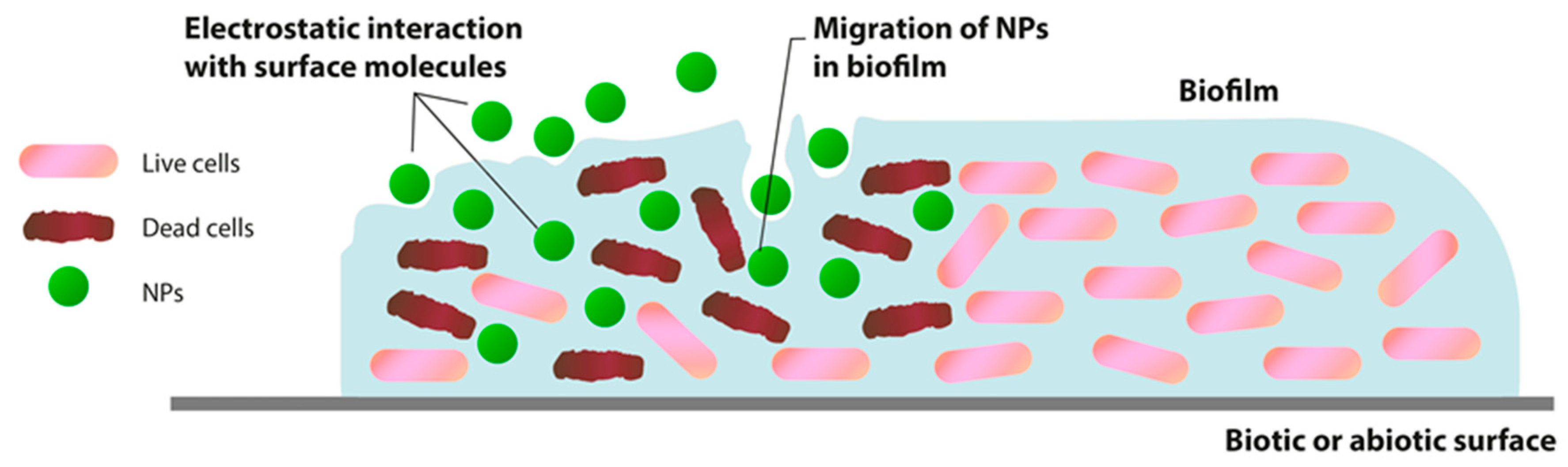
The interaction between NPs and biofilm can be considered as a three-stage process: (1) transfer of NPs in the vicinity of the biofilm; (2) attachment to the biofilm surface; and (3) migration in biofilms (Figure 1). The implementation of each stage is conducted by many factors: the physicochemical characteristics of NPs, EPM, and the environment.
Once the NPs reach the biofilm boundary, the physicochemical characteristics of the EPM determine the initial attachment of NPs to the biofilm surface and their subsequent movement in the matrix. The initial addition of NPs to the outermost surface of biofilms can be affected by various physicochemical interactions.
Primarily, the interaction between NPs and biofilm is determined by their electrostatic characteristics. These features depend on the zeta potential of NPs and the charge of the biofilm matrix [26,27,28,29]. The majority of bacteria have a polyionic biofilm matrix due to the presence of uronic acid or metal-bound pyruvate with the functions of carboxylic acid and residual phosphate or rarely sulfate [30,31]. This negatively charged matrix can interact with positively charged metal ions and organic compounds through electrostatic forces [32,33].
Successfully associated NPs with EPM on the biofilm surface can penetrate deep into the biofilm at different rates. The penetration of NPs and movement within the biofilm is considered to be primarily due to diffusion [34]. In this case, the NPs’ diffusion inside the biofilm may depend on the size of its pores [28], water channels presence [35], the charge of NPs and EPM [34], hydrophobicity of the environment [36], the chemical gradient within the matrix. The EPM pore spaces containing water can have different ion concentrations. Ions and organic molecules diffuse and penetrate the biofilm, move, and are distributed through these pore spaces. This gives a plausible possibility that the interval between EPM pores will be especially crucial in this process. However, this variability on the scale of nanometers is not sufficiently characterized and understandable [37].
Thus, the penetration and migration of NPs inside the biofilm will be determined mainly by the charge and size of the particles and the composition and structure of EPM. However, many details of this interaction have yet to be determined.
3. Effects of Metal Oxide NPs on Plankton Cells and Biofilm
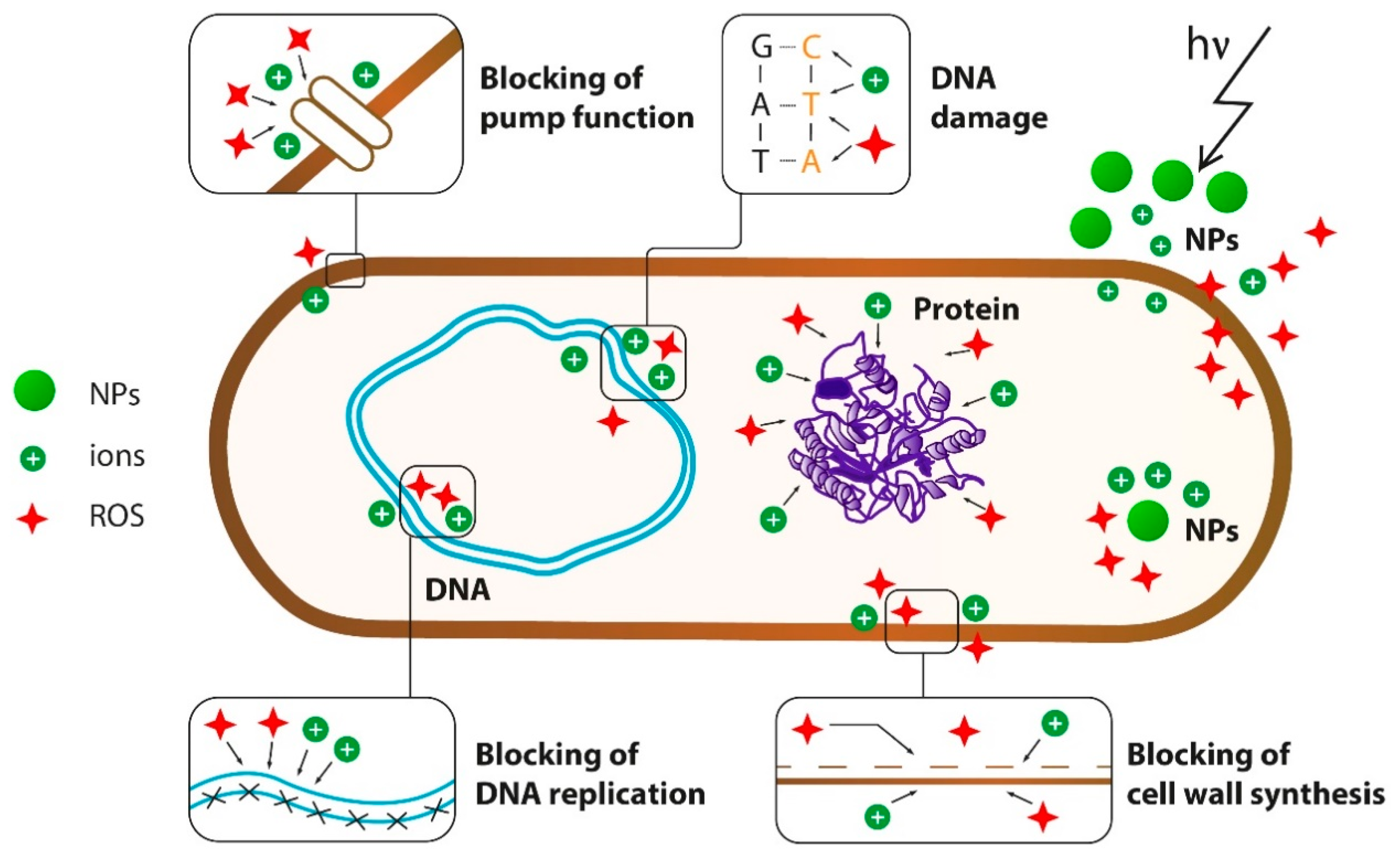
Three main mechanisms of antibacterial effects of NPs are well known: (1) mechanical damage to the cell wall as a result of electrostatic interaction, (2) oxidative stress as a result of the generation of reactive oxygen species (ROS), and (3) disruption of proteins functions and cell structures as a result of metal cations release [18] (Figure 2).
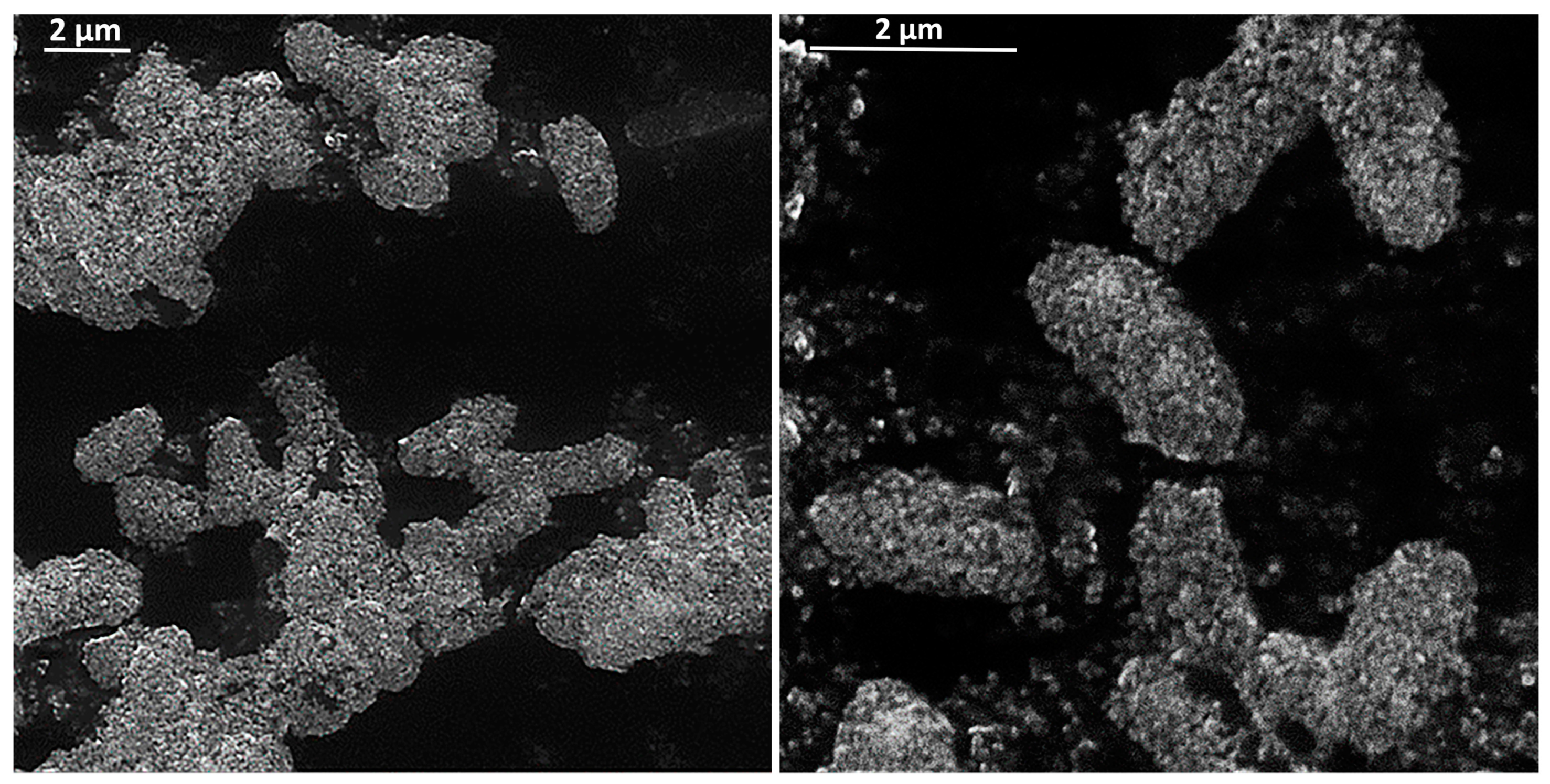
For negatively charged bacteria, adhesion on metal oxide NPs increases due to a positively charged surface of NPs [38]. Figure 3 shows how actively NPs interact with the cell wall on the example of magnetite. In this way, metal oxide NPs binds to the cell wall through electrostatic and Van der Waals interactions, in particular, to cell membrane proteins that disrupt bacteria functions. MgO NPs also show strong interactions not only with the cell surface but also with spores [39,40].
The primary mechanism of bacterial cell destruction under the NPs’ influence is currently considered to be the induction of oxidative stress through the generation of ROS under electromagnetic irradiation of NPs [41]. The generation of ROS induces oxidative stress in the cell, as a result of which it dies. In most cases, ROS production is also directly related to the release of cations. In particular, Fe3O4 NPs release Fe2+ ions, which cause the generation of ROS after a reaction with hydrogen peroxide (Fenton reaction) [42]. Copper ions can also disrupt biochemical processes and significantly damage nucleic acids [43]. It is assumed that after the specific binding of copper to DNA, repeated cyclic redox reactions generate several OH− radicals near the binding site, causing multiple damages to the nucleic acids, but in some microorganisms, copper oxidative damage to the genetic material may occur through the Fenton mechanism [44]
Calcium and magnesium oxides can generate superoxide radical O2, whereas zinc oxide NPs produce H2O2 and hydroxyl radical OH− under ultraviolet and visible light, but not O2 [45,46]. The molecules of OH− and O2 cannot penetrate the cell membrane due to their charges or reactivity [47], and probably remain on the cell surface, whereas H2O2 can penetrate bacterial cells, thereby inducing cell death [48,49]. Meanwhile, copper oxide NPs can produce all four types of reactive oxygen. Thus, CuO NPs are toxic enough for bacteria and have shown significant effects against biofilms.
TiO2 NPs under irradiation can generate electron-hole pairs with photoexcitation, initiate cascade oxidation–reduction reactions on the surface of TiO2, and, consequently, produce ROS for further reactions [50,51]. Thus, TiO2 NPs inhibit bacterial growth by lipid peroxidation in the membranes, DNA damage, nucleotide and amino acid oxidation, or destruction of protein-catalytic sites by photocatalysis [52].
Despite the above, there is evidence that ROS generation does not always directly cause cell death. In particular, the analysis of gene expression has shown that ZnO NPs inhibits the expression of oxidative stress genes, despite the ROS generation. The antibacterial effect may be due to biomimetic action and other mechanisms [53].
The release of metal cations can also have a devastating effect on cells, regardless of ROS products. Different bacterial species have varying sensitivities to metal ions. Cations can interact with sulfhydryl groups in enzymes, with amine and carboxyl groups on microbial cells [54,55,56]. In particular, Zn2+ ions influence peptides by changing their conformation [57]. Cations can cause the mismetallation of proteins in a cell. In particular, copper ions can mismetallate proteins that require a molybdenum or iron cofactor because of their affinity for thiol ligands [58,59]. This process may affect cell metabolism due to improper assembly of biosynthetic enzymes [60]. All this leads to disruption in the functioning of cellular components and cell death in the end.
The NPs discussed in this review exhibit antibacterial properties to varying degrees by implementing the mechanisms mentioned above. Conditionally, NPs can be arranged in the following order of their decreasing antibacterial and antibiofilm properties: CuO–ZnO–MgO–TiO2–Fe3O4–Al2O3. Physical and antibiofilm properties of some NPs oxides are presented in Table 1.
3.1. CuO
The minimum inhibitory concentration (MIC) for CuO NPs against a wide range of Gram-negative and Gram-positive bacteria is 15 µg/mL to 100 µg/mL on average. [63,64,98]. In particular, MIC of CuO NPs for E. coli is 22 µg/mL, and for S. aureus is 15 µg/mL [63,64].
CuO NPs inhibit the formation of biofilms and promote the eradication of already formed biofilms. It is mainly due to the toxicity of copper ions for plankton and biofilm cells. For example, CuO NPs efficiently reduced the biofilm formation by MRSA and E. coli. Almost all MRSA and E. coli biofilm cells died within four days of exposure to CuO NPs [99]. CuO NPs at a 50 µg/mL concentration significantly inhibited the growth of total oral bacteria, extracellular polysaccharide (EPS) production, and biofilm formation on the glass, acrylic dentures, and cultured human epithelial cells as models [100]. It has also been shown that CuO NPs’ inhibitory concentration on Ralstonia solanacearum biofilms is 125 and 250 µg/mL after an incubation period of 24 and 72 h [101]. Thus, it is evident that CuO NPs can be potentially effective against biofilms, as shown in different groups of microorganisms.
3.2. ZnO
For ZnO NPs, the effective antibacterial concentration increases to an average of 20–500 µg/mL [102,103,104,105,106,107,108]. For example, there was 50 µg/mL against E. coli [105]. However, the antibacterial properties of ZnO NPs can be enhanced by additional physical exposure. In particular, cell penetration by NPs can be amplified by ultrasound. The combination of ZnO NPs with ultrasound enhances the antibacterial effect on S. aureus by 76% due to generating more hydrogen peroxide [109]. Besides, ultrasonic treatment can physically promote the dissociation of cell membranes, thereby increasing the penetration of ZnO NPs into cells [18].
ZnO NPs can be considered a potential agent for the inhibition of microbial biofilms. The results of the inhibition of S. pneumoniae biofilm showed that the sub-MIC doses (3, 6, and 12 µg/mL) of ZnO NPs exhibited significant antibiofilm activity. The main mechanism preventing the formation of S. pneumonia biofilms in the presence of zinc oxide NPs is that it reduces the adhesion of cells to the surface [11]. Studies of biofilms on dentures revealed the ZnO NPs’ effectiveness in controlling the formation of biofilms Rothia dentocariosa and R. mucilaginosa. Zinc ion generation inhibits the enzymatic activity of the DapE protein involved in the synthesis of peptidoglycans, which leads to the failure of biofilm formation in the initial stage [79]. The recent studies on the effect on biofilms formed by uropathogenic strains of E. coli have shown that MIC concentration of NPs fully inhibited biofilm formation in 20% of isolates, and 30% of isolates reduce the optical density of biofilm formation from a moderate to weak level [80]. In this way, ZnO NPs also have excellent potential for antibacterial materials’ development.
3.3. MgO
MgO NPs show activity against Gram-positive and Gram-negative bacteria, spores, and viruses [110,111,112,113] at sufficiently high particle concentrations (on average 100–1200 µg/mL). In particular, MIC of MgO NPs for E. coli were determined to be 1 mg/mL [114].
Recent studies have shown the efficiency of MgO NPs in the action against biofilms of E. coli (250 µg/mL), K. pneumoniae (125 µg/mL), and S. aureus (500 µg/mL). Bacterial adhesion to the plastic surface decreased markedly after 12 h incubation of E. coli, S. aureus, and K. pneumonia with MgO, thus preventing the biofilm formation. The effect of MgO on mature biofilms was also detected. Biofilm biomass was significantly reduced when treating biofilms with a subinhibitor concentration of 0.5 MIC. [83]. In another recent study, it was reported that 10 µg/mL significantly complicates the formation of S. aureus biofilms [115]. MgO NPs had a strong inhibitory effect on the formation of biofilms E. coli and S. aureus at a size of 8 nm [85]. MgO NPs reduced the biofilm growth of R. solanacearu, and the biofilm formation gradually decreased with the bulk MgO treatments. The 200 and 250 µg/mL treatments of MgO NPs exhibited high inhibitory effects on the R. solanacearum biofilm formation. The biofilm formation was reduced by 61% and 71% after 24 h and by 67% and 72% after 72 h, respectively [84]
Thus, MgO NPs have significant antibiofilm properties, but significant effects are achieved at sufficiently high particle concentrations (above 125 µg/mL).
3.4. TiO2
TiO2 NPs are effective against bacteria, viruses, and even to purify specific odor molecules in the range of 20 µg/mL to 1400 µg/mL [116,117,118,119,120]. TiO2 NPs show antibacterial properties against Gram-positive and Gram-negative, the latter being more sensitive [121,122]. It could be related to the fact that Gram-positive bacteria have a thick layer of peptidoglycan that facilitates the absorption of reactive radicals, thereby preventing cell damage from radical attack [123]. In addition, it shows that TiO2 has a potential against bacteria through the reception of an electron from intracellular coenzyme A (CoA) after photocatalysis of TiO2, followed by the formation of dimer CoA and subsequent inhibition of respiration [124].
TiO2 NPs can reduce the adhesion of bacteria and inhibit biofilms. Exposure to titanium oxide leads to the destruction of bacteria inside the biofilm, primarily due to the generation of ROS and lipid oxidation on the cell wall membrane [92,120]. It has been shown that TiO2 NPs are effective against biofilms of MRSA [92] and S. mitis [125]. TiO2 NPs could control the growth and biofilm formation of S. mitis ATCC 6249 and Ora-20, and it can be used as a means for oral hygiene. TiO2 NPs have a reduced impact on Pseudomonas aeruginosa biofilms at a 31.25 µg/mL concentration and disrupt previously established biofilms in the microtiter plate [125]. In the presence of TiO2 NPs, biofilm formation of E. coli and B. subtilis was reduced by 40–50% [122]. However, NPs TiO2 did not show significant bactericidal properties against certain types of drug-resistant bacteria (ex, Cupriavidus metallidurans CH34), which have a remarkable ability to withstand ROS membrane damage through overexpression of protective components and membrane repair elements [126].
3.5. Fe3O4
Fe3O4 NPs has slight antibacterial properties, and their effective concentrations reach 10–20 mg/mL [38,42,127,128]. Fe3O4 NPs against biofilms showed mostly insignificant effects. To obtain significant antibiofilm effects, Fe3O4 NPs have to be used in high concentrations. In this case, the particles are able to destroy the cells inside the biofilm. It has been shown that iron-oxide NPs were able to reduce biofilm growth by S. aureus, E. coli, P. aeruginosa [88], S. epidermidis [38], and Enterococcus hirae [89].
In addition to the passive electrostatic effect on biofilm, NPs effectively penetrate deep into the biofilms in the presence of a magnetic field [129,130]. In this case, NPs can have a mechanical effect on the biofilm due to the destruction of the matrix structure and its whole architecture [87,131,132]. Due to these properties, the particles are mainly used as a carrier of biocides in biofilms. In particular, the NPs enhance the action of various antibiotics on biofilm. The effectiveness of conjugates with penicillin, streptomycin, erythromycin, kanamycin, cefotaxime against S. aureus biofilm [133,134] and amphotericin B, nystatin against Candida spp. biofilm has been shown [135]. In recent years, many biocide-conjugated particles have been developed against biofilms. In all cases, magnetite serves as an efficient matrix for delivering biocides inside the biofilms and has a synergetic effect due to its unique properties.
3.6. Al2O3
Al2O3 NPs are effective against bacteria only when in high concentrations (reach 10–20 mg/mL) [137,138]. Al2O3 NPs weakly inhibit E. coli at high concentrations up to 1 mg/mL [137,138]. In the study of Al2O3 NPs’ effect on P. putida and A. hydrophila in biofilms and planktonic forms, it has been shown that NPs are toxic to bacteria, but plankton cells are more susceptible to Al2O3 NPs than biofilms [61]. The minimal inhibitory concentration of Al2O3 NPs against the biofilm of P. aeruginosa was found to be 1.6–3.2 mg/mL. Treatment at a 2 mg/mL concentration resulted in complete growth inhibition of extended-spectrum b-lactamases and metallo-b-lactamases clinical isolates of P. aeruginosa [139]. Therefore, antimicrobial and antibiofilm properties of Al2O3 are less than those of other oxides. Thus, it can only be effective in nanocomposites and conjugates with biocides.
It is clear from the above that metal oxide NPs can be promising materials against biofilms. Different particles exhibit antibiofilm properties to varying degrees, which depends directly on their antibacterial properties. These properties are determined mainly by the synthesis method, size, and shape of the particles (Table 1). Particles, such as Fe3O4 and Al2O3, have weak antibiofilm properties, but they can be much more effective in nanocomposites.
4. Metal Oxide Nanocomposites against Plankton Cells and Biofilms
It can be seen from the previous section that the antibiofilm properties of metal oxide NPs are demonstrated to varying degrees. Nanocomposites of mixed metal oxides are actively studied and developed to improve antimicrobial properties and reduce adverse cytotoxic effects and reactions of the immune system to monoxide NPs.
Nanocomposite CuO doped with Zn showed an increase in antibacterial activity of 10 times against E. coli and S. aureus bacteria compared to pure ZnO or CuO [140]. Moreover, CuO doped with TiO2 has a more significant antibacterial effect than pure TiO2. Li-doped MgO NPs are more efficient than pure MgO, whereas Zn and Ti-doped MgO exhibit lower antibacterial activity than MgO [22]. TiO2-ZnO-MgO mixed oxides nanomaterials have a strong antibacterial effect against Gram-negative and Gram-positive bacteria [141].
The combination of three metal oxides in the CuZnFe oxide NPs may also enhance the therapeutic abilities of the NPs against a wide range of microbial infections. Antibacterial activities of CuZnFe oxide NPs were tested against Gram-negative E. coli and Gram-positive E. faecalis. CuZnFe oxide NPs affected bacterial species by reducing their viability and their ability to synthesize biofilm. CuZnFe oxide NPs have more detrimental effects on E. coli than individual CuO and ZnO NPs. CuZnFe oxide NPs were also found to be more bactericidal than ZnO NPs against E. faecalis, but 7% lower than CuO NPs [94]. It was observed that this oxide could slowly release metal ions, which can penetrate through the membranes and disrupt cellular processes from within the cell [142]. CuZnFe oxide NPs affected biofilm formation to a lesser degree than those of individual ZnO and CuO NPs [94].
The pro-oxidative and pro-inflammatory effects of highly toxic ZnO NP can be significantly reduced by iron doping [143,144]. A similar effect can also be achieved by adding magnesium to the nanocomposite: mixed ZnMgO NPs, are safe for mammalian cells with the non-toxic MgO monoxide NPs [20]. ZnO NPs doped with Mn and Fe ions exhibit even higher antibacterial activity on a wide range of bacterial species, including S. aureus, E. coli, K. pneumoniae, S. typhi, P. aeruginosa, B. subtilis, and Proteus mirabilis as compared to the ZnO monoxide [21,145]. In the study of Fe3O4-ZnO nanocomposite, it has shown more significant antibacterial activity on E. coli than S. aureus and B. subtilis [145].
The inclusion of Ag and Au in composites also significantly enhances their antimicrobial properties. Ag-ZnO nanocomposites showed a high antibacterial effect against antibiotic-resistant E. coli and S. aureus [146]. High antibacterial activity against E. coli and S. aureus was shown by Ag-SiO2 nanocomposite. This composite is perfect for the treatment and infectious control of superficial wounds [96]. Moreover, the deposition of Au particles on the surface of ZnO NPs, even at a low molar ratio of ZnO/Au (0.2%), significantly improves the photocatalytic antibacterial activity of ZnO [147]. Furthermore, Ag-TiO2 nanocomposite demonstrates antibacterial activity against S. aureus biofilms [93,148,149]. It was shown that the inclusion of a 2% composite significantly reduced the formation of biofilms on the surface of the composite resin [150]. Ag/Fe3O4 NPs significantly improve antibacterial properties against E. coli [151]. CeO2-CdO nanocomposites also exhibit broad-spectrum antimicrobial activity against Gram-positive (S. aureus MTCC96 and C. pyogenes MTCC 1926) and Gram-negative (P. aeruginosa и K. pneumoniae) [95].
Additionally, the introduction of nanocomposites based on oxides induced by external physical factors can add additional functions and significantly increase antimicrobial action effectiveness. For example, compared to the monoxide TiO2 NPs, which exhibit photocatalytic activity in the UV spectrum, the doped form can significantly expand the active spectrum to the visible light region [152,153]. An example of a highly effective nanocomposite can be ZnOAu, which possesses photoactivity due to zinc, and thermal sensitivity due to gold [97].
Some nanocomposites have been studied in vivo. CuO ligated Zn shows a successful inhibition of biofilm formation in in vitro and in vivo experiments on rabbits. At the same time, this composite is biocompatible [81].
As shown in the above, the antibacterial activity of initially low-efficiency oxides (such as Fe2O3 NPs) in mixed oxides can be significantly increased. It becomes evident that oxide particles, and especially composites based on them, can become promising materials. Reducing their cytotoxicity and immunogenicity will also allow their introduction into biomedicine. Ultimately, the widespread application of nanomaterials also needs to be determined by their safety to the environment.
5. Potential Adverse Effects of the Broad Implementation of Metal Oxide NPs
Along with their numerous remarkable applications, NPs also have potential limitations. In particular, a large surface area and high reactivity can be considered as one of the NPs advantages, but at the same time cause side effects. Moreover, the non-specificity of the antimicrobial action on pathogenic and symbiotic microorganisms exhibited by NPs can be seen as an advantage or disadvantage [154]. Chemically synthesized NPs have toxicity problems since dangerous compounds are used in their synthesis. These toxic compounds remain within NP in trace amounts and cause undesirable effects [155]. A successful approach to solving these problems is developing methods to synthesize environmentally friendly and less toxic NPs—the so-called “green synthesis” [156].
The most important aspect of the safe use of nanostructures is their mutagenicity. Even though this issue remains the least studied, there are already some literature reports that raise concerns. As mentioned earlier, the NPs and ROS that they generate can themselves influence the DNA of bacteria. If the concentration of NPs is not sufficient to eradicate the biofilm, but enough to trigger mutations, which can lead to the appearance of “super mutants”.
The study of NPs genotoxicity using tungsten oxide as an example has shown that direct interaction NPs with DNA can lead to damage by single-strand breaks. After the mix with NPs DNA was introduced into bacterial cells, the cells mostly died, and the surviving cells were almost all mutants. The results provide clear evidence that one of the mechanisms involved in nanomaterials toxicity directly damages DNA, which can then cause biological cell death and mutation [157].
The potential mutagenicity of certain oxides (Al2O3, Co3O4, CuO, TiO2, and ZnO) was investigated using reverse mutation (Eames analysis). The results showed that the mutagenicity was negative for four nanoparticles (Al2O3, Co3O4, TiO2, and ZnO) to 1000 µg/plate for all three strains tested (S. typhimurium TA97a, TA100, and E. coli WP2 trpuvrA) without metabolic activation of S9. Using the pre-incubation procedure and high activation of S9 (9%), TiO2 and ZnO induced marginal mutagenesis for the E. coli strain WP2. The CuO exhibited a low mutagenic potential in S. typhimurium TA97a and TA100 at specific concentrations [158].
In addition to direct DNA damage under the influence of NPs, there are also data on increasing the efficiency of horizontal transfer of genetic material when exposed to NPs. It has been shown that Al2O3, AlOOH, TiO2, SiO2, and Fe2O3 NPs can contribute to the conjugating transfer of plasmid (up to 20–100 times) [159,160,161]. It is important to note that the transfer increased inside the species or genus of bacteria. The most significant effect was produced by Al2O3 NPs, which led not only to an increase in the number of conjugating cells but also to one bacterium being conjugated to several other bacteria [159].
Such manifestations of NPs mutagenicity in the biofilms of bacteria are especially important. Genetic components from lysed bacterial cells, such as plasmids, are stored inside the EPM, increasing the gene transfer frequency between bacterial cells [162]. These plasmids can contain genes useful for bacteria, such as genes for antibiotic resistance [163]. Thus, it can be assumed that metal oxide NPs can contribute to the formation of antibiotic-resistant bacteria. Further research on the issues described above is essential for the safe use of NPs.
An essential aspect of the widespread introduction of nanostructures is the ecological one. Currently, metal oxide NPs are already widely used in various fields. The presence of nanomaterials in the environment is not new. Nevertheless, the current growth in the production of anthropogenic nanomaterials will increase their level in the environment. The release of nanoparticles into the biosphere will occur from point sources (for example, production sites, landfills, treatment plants) and secondary sources (for example, release into the environment when using and consuming materials containing nanomaterials). For example, a global estimate for nanomaterial production goes up to 5000 tons/year for TiO2 NPs [164].
There is a gradual increase in the concentration of these NPs in wastewater as contaminants. This can threaten ecological communities in treatment plants [165]. Recent studies have shown that significant accumulation of NPs is observed in biofilms of the river and marine sediments [166,167]. A high concentration of 20 nm TiO2 is found in river biocenoses [166]. NP can pose a danger to the environment, causing the disappearance of some biocidal sensitive bacterial strains and expanding a set of insensitive strains to various influences [126].
The properties of NPs described in this section show that currently, available research data are insufficient to assess the safety of NPs use. More studies need to be carried out before these nanomaterials can enter the broad market.
6. Conclusions
In addition to the biofilms spread, which are resistant to antibiotics, the spread of biofilms produced by antibiotic-resistant bacteria grows. Using metal oxide NPs and their nanocomposites is one of the few methods that can soon enter into broad practice.
Among the NPs discussed in this review, CuO and ZnO NPs showed the most significant antibiofilm properties, which initially had high antibacterial characteristics. These NPs have proven effective against biofilms of a wide range of bacterial species. Fe3O4, TiO2, and MgO NPs have a lower ability to eradicate biofilms. However, magnetite can be used with a magnetic field to deliver biocides and has shown high biofilm destruction efficiency. Al2O3 NPs have the weakest properties against biofilms, and these particles can only be effective in high concentrations. However, the introduction of nanocomposites consisting of several oxides can significantly enhance the action of NPs against biofilms. Several of the composites described in this review have excellent antibacterial and antibiofilm properties.
However, the mechanisms of NPs action on biofilms are still weakly studied and controversial. A more thorough study of the mechanisms of NPs’ action on plankton cells and biofilms may allow more targeted use and increase their effectiveness. Another issue is the potential particle mutagenicity, resulting from which bacteria can acquire new dangerous properties by analogy with the development and spread of antibiotic-resistant strains. Under conditions of mixed cultures of bacteria, especially in biofilms, NPs can, hypothetically, accelerate bacterial evolution. Nevertheless, this aspect of the NP influence on prokaryotes is the weakest studied.
It can be concluded that, despite extensive research on NPs worldwide, many issues require more in-depth studies. Without them, nanostructured drugs introduction to therapy can have unpredictable consequences for both the evolution of bacteria and the human body. The use of NPs on the industrial level can also provoke severe environmental consequences.
Thus, the current review formulates specific issues that have to be addressed to push the research forward. Since it is essential to develop new approaches for combating biofilms of bacteria in various fields, new information on the effectiveness of the use of NPs against biofilms, their mechanisms of influence on cells, mutagenicity, and genotoxicity will be crucial.
Author Contributions
Conceptualization, L.S, I.K. and E.K.; investigation, L.S., I.K., E.K.; resources, E.K.; data curation, L.S., I.K.; writing—original draft preparation, L.S., I.K.; writing—review and editing, E.K.; visualization, L.S.; supervision, E.K.; project administration, E.K.; funding acquisition, E.K. All authors have read and agreed to the published version of the manuscript.
Funding
This research was funded by the Russian Science Foundation, grant number 19-74-00125.
Conflicts of Interest
The authors declare no conflict of interest. The funders had no role in the design of the study; in the collection, analyses, or interpretation of data; in the writing of the manuscript, or in the decision to publish the results.
References
- Donlan, R.M.; Costerton, J.W. Biofilms: Survival mechanisms of clinically relevant microorganisms. Clin. Microbiol. Rev. 2002, 15, 167–193. [Google Scholar] [CrossRef] [PubMed] [Green Version]
- Chávez de Paz, L.E.; Resin, A.; Howard, K.A.; Sutherland, D.S.; Wejse, P.L. Antimicrobial Effect of Chitosan Nanoparticles on Streptococcus mutans Biofilms. Appl. Environ. Microbiol. 2011, 77, 3892–3895. [Google Scholar] [CrossRef] [PubMed] [Green Version]
- Flemming, H.-C.; Wingender, J. The biofilm matrix. Nat. Rev. Microbiol. 2010, 8, 623–633. [Google Scholar] [CrossRef] [PubMed]
- Pintucci, J.P.; Corno, S.; Garotta, M. Biofilms and infections of the upper respiratory tract. Eur. Rev. Med. Pharmacol. Sci. 2010, 14, 683–690. [Google Scholar] [PubMed]
- Biofilm basics: Section 1—Center for Biofilm Engineering | Montana State University. Available online: http://www.biofilm.montana.edu/biofilm-basics/what_are_biofilms.html (accessed on 2 September 2019).
- Chen, C.-P.; Chen, C.-T.; Tsai, T. Chitosan Nanoparticles for Antimicrobial Photodynamic Inactivation: Characterization and In Vitro Investigation†. Photochem. Photobiol. 2012, 88, 570–576. [Google Scholar] [CrossRef] [PubMed]
- Lewis, K. Riddle of Biofilm Resistance. Antimicrob. Agents Chemother. 2001, 45, 999–1007. [Google Scholar] [CrossRef] [PubMed] [Green Version]
- Abidi, S.H.; Sherwani, S.K.; Siddiqui, T.R.; Bashir, A.; Kazmi, S.U. Drug resistance profile and biofilm forming potential of Pseudomonas aeruginosa isolated from contact lenses in Karachi-Pakistan. BMC Ophthalmol. 2013, 13, 57. [Google Scholar] [CrossRef] [Green Version]
- Han, C.; Romero, N.; Fischer, S.; Dookran, J.; Berger, A.; Doiron, A.L. Recent developments in the use of nanoparticles for treatment of biofilms. Nanotechnol. Rev. 2017, 6, 383–404. [Google Scholar] [CrossRef]
- Donlan, R.M. Biofilms and device-associated infections. Emerg. Infect. Dis. 2001, 7, 277–281. [Google Scholar] [CrossRef]
- Bhattacharyya, P.; Agarwal, B.; Goswami, M.; Maiti, D.; Baruah, S.; Tribedi, P. Zinc oxide nanoparticle inhibits the biofilm formation of Streptococcus pneumoniae. Antonie Van Leeuwenhoek 2018, 111, 89–99. [Google Scholar] [CrossRef]
- Costerton, J.W.; Stewart, P.S.; Greenberg, E.P. Bacterial biofilms: A common cause of persistent infections. Science 1999, 284, 1318–1322. [Google Scholar] [CrossRef] [PubMed] [Green Version]
- Spoering, A.L.; Lewis, K. Biofilms and Planktonic Cells of Pseudomonas aeruginosa Have Similar Resistance to Killing by Antimicrobials. J. Bacteriol. 2001, 183, 6746–6751. [Google Scholar] [CrossRef] [PubMed] [Green Version]
- Taheri, S.; Baier, G.; Majewski, P.; Barton, M.; Förch, R.; Landfester, K.; Vasilev, K. Synthesis and surface immobilization of antibacterial hybrid silver-poly(l-lactide) nanoparticles. Nanotechnology 2014, 25, 305102. [Google Scholar] [CrossRef] [PubMed]
- Shrestha, A.; Hamblin, M.R.; Kishen, A. Photoactivated rose bengal functionalized chitosan nanoparticles produce antibacterial/biofilm activity and stabilize dentin-collagen. Nanomed. Nanotechnol. Biol. Med. 2014, 10, 491–501. [Google Scholar] [CrossRef] [PubMed]
- Taylor, E.N.; Kummer, K.M.; Durmus, N.G.; Leuba, K.; Tarquinio, K.M.; Webster, T.J. Superparamagnetic Iron Oxide Nanoparticles (SPION) for the Treatment of Antibiotic-Resistant Biofilms. Small 2012, 8, 3016–3027. [Google Scholar] [CrossRef] [PubMed]
- Ramasamy, M.; Lee, J.-H.; Lee, J. Potent antimicrobial and antibiofilm activities of bacteriogenically synthesized gold–silver nanoparticles against pathogenic bacteria and their physiochemical characterizations. J. Biomater. Appl. 2016, 31, 366–378. [Google Scholar] [CrossRef] [PubMed]
- Wang, L.; Hu, C.; Shao, L. The antimicrobial activity of nanoparticles: Present situation and prospects for the future. Int. J. Nanomedicine 2017, 12, 1227–1249. [Google Scholar] [CrossRef] [PubMed] [Green Version]
- Hu, L. The Use of Nanoparticles to Prevent and Eliminate Bacterial Biofilms. In Antimicrobial Research Novel Bioknowledge and educational Programs; Formatex: Badajoz, Spain, 2017; pp. 344–350. [Google Scholar]
- Vidic, J.; Stankic, S.; Haque, F.; Ciric, D.; Le Goffic, R.; Vidy, A.; Jupille, J.; Delmas, B. Selective antibacterial effects of mixed ZnMgO nanoparticles. J. Nanopart. Res. 2013, 15, 1595. [Google Scholar] [CrossRef] [Green Version]
- Sharma, N.; Jandaik, S.; Kumar, S.; Chitkara, M.; Sandhu, I.S. Synthesis, characterisation and antimicrobial activity of manganese- and iron-doped zinc oxide nanoparticles. J. Exp. Nanosci. 2016, 11, 54–71. [Google Scholar] [CrossRef]
- Rao, Y.; Wang, W.; Tan, F.; Cai, Y.; Lu, J.; Qiao, X. Influence of different ions doping on the antibacterial properties of MgO nanopowders. Appl. Surf. Sci. 2013, 284, 726–731. [Google Scholar] [CrossRef]
- Stankic, S.; Suman, S.; Haque, F.; Vidic, J. Pure and multi metal oxide nanoparticles: Synthesis, antibacterial and cytotoxic properties. J. Nanobiotechnol. 2016, 14, 73. [Google Scholar] [CrossRef] [PubMed] [Green Version]
- Huh, A.J.; Kwon, Y.J. “Nanoantibiotics”: A new paradigm for treating infectious diseases using nanomaterials in the antibiotics resistant era. J. Control. Release 2011, 156, 128–145. [Google Scholar] [CrossRef] [PubMed]
- Kohanski, M.A.; Dwyer, D.J.; Hayete, B.; Lawrence, C.A.; Collins, J.J. A Common Mechanism of Cellular Death Induced by Bactericidal Antibiotics. Cell 2007, 130, 797–810. [Google Scholar] [CrossRef] [Green Version]
- Liang, Y.; Hilal, N.; Langston, P.; Starov, V. Interaction forces between colloidal particles in liquid: Theory and experiment. Adv. Colloid Interface Sci. 2007, 134–135, 151–166. [Google Scholar] [CrossRef]
- Ikuma, K.; Madden, A.S.; Decho, A.W.; Lau, B.L.T. Deposition of nanoparticles onto polysaccharide-coated surfaces: Implications for nanoparticle–biofilm interactions. Environ. Sci. Nano 2014, 1, 117–122. [Google Scholar] [CrossRef]
- Sahle-Demessie, E.; Tadesse, H. Kinetics and equilibrium adsorption of nano-TiO2 particles on synthetic biofilm. Surf. Sci. 2011, 605, 1177–1184. [Google Scholar] [CrossRef]
- Tong, M.; Ding, J.; Shen, Y.; Zhu, P. Influence of biofilm on the transport of fullerene (C60) nanoparticles in porous media. Water Res. 2010, 44, 1094–1103. [Google Scholar] [CrossRef] [PubMed]
- Hajipour, M.J.; Fromm, K.M.; Ashkarran, A.A.; de Aberasturi, D.J.; de Larramendi, I.R.; Rojo, T.; Serpooshan, V.; Parak, W.J.; Mahmoudi, M. Antibacterial properties of nanoparticles. Trends Biotechnol. 2012, 30, 499–511. [Google Scholar] [CrossRef] [Green Version]
- Sutherland, I.W. Biofilm exopolysaccharides: A strong and sticky framework. Microbiology 2001, 147, 3–9. [Google Scholar] [CrossRef] [Green Version]
- Esparza-Soto, M.; Westerhoff, P. Biosorption of humic and fulvic acids to live activated sludge biomass. Water Res. 2003, 37, 2301–2310. [Google Scholar] [CrossRef]
- Selvakumar, R.; Aravindh, S.; Ashok, A.M.; Balachandran, Y.L. A facile synthesis of silver nanoparticle with SERS and antimicrobial activity using Bacillus subtilis exopolysaccharides. J. Exp. Nanosci. 2014, 9, 1075–1087. [Google Scholar] [CrossRef]
- Peulen, T.-O.; Wilkinson, K.J. Diffusion of Nanoparticles in a Biofilm. Environ. Sci. Technol. 2011, 45, 3367–3373. [Google Scholar] [CrossRef] [PubMed]
- Stewart, P.S. Diffusion in biofilms. J. Bacteriol. 2003, 185, 1485–1491. [Google Scholar] [CrossRef] [PubMed] [Green Version]
- Habimana, O.; Steenkeste, K.; Fontaine-Aupart, M.-P.; Bellon-Fontaine, M.-N.; Kulakauskas, S.; Briandet, R. Diffusion of Nanoparticles in Biofilms Is Altered by Bacterial Cell Wall Hydrophobicity. Appl. Environ. Microbiol. 2011, 77, 367–368. [Google Scholar] [CrossRef] [Green Version]
- Ikuma, K.; Decho, A.W.; Lau, B.L.T. When nanoparticles meet biofilms-interactions guiding the environmental fate and accumulation of nanoparticles. Front. Microbiol. 2015, 6, 591. [Google Scholar] [CrossRef]
- Taylor, E.N.; Webster, T.J. The use of superparamagnetic nanoparticles for prosthetic biofilm prevention. Int. J. Nanomedicine 2009, 4, 145–152. [Google Scholar] [CrossRef] [Green Version]
- Koper, O.B.; Klabunde, J.S.; Marchin, G.L.; Klabunde, K.J.; Stoimenov, P.; Bohra, L. Nanoscale Powders and Formulations with Biocidal Activity Toward Spores and Vegetative Cells of Bacillus Species, Viruses, and Toxins. Curr. Microbiol. 2002, 44, 49–55. [Google Scholar] [CrossRef]
- Koper, O.B.; Lagadic, I.; Volodin, A.; Klabunde, K.J. Alkaline-Earth Oxide Nanoparticles Obtained by Aerogel Methods. Characterization and Rational for Unexpectedly High Surface Chemical Reactivities. Chem. Mater. 1997. [Google Scholar] [CrossRef]
- Gurunathan, S.; Han, J.W.; Dayem, A.A.; Eppakayala, V.; Kim, J.-H. Oxidative stress-mediated antibacterial activity of graphene oxide and reduced graphene oxide in Pseudomonas aeruginosa. Int. J. Nanomed. 2012, 7, 5901. [Google Scholar] [CrossRef] [Green Version]
- Tran, N.; Mir, A.; Mallik, D.; Sinha, A.; Nayar, S.; Webster, T.J. Bactericidal effect of iron oxide nanoparticles on Staphylococcus aureus. Int. J. Nanomed. 2010, 5, 277–283. [Google Scholar]
- Kim, J.H.; Cho, H.; Ryu, S.E.; Choi, M.U. Effects of metal ions on the activity of protein tyrosine phosphatase VHR: Highly potent and reversible oxidative inactivation by Cu2+ ion. Arch. Biochem. Biophys. 2000, 382, 72–80. [Google Scholar] [CrossRef] [PubMed]
- Gunawan, C.; Teoh, W.Y.; Marquis, C.P.; Amal, R. Cytotoxic Origin of Copper(II) Oxide Nanoparticles: Comparative Studies with Micron-Sized Particles, Leachate, and Metal Salts. ACS Nano 2011, 5, 7214–7225. [Google Scholar] [CrossRef] [PubMed]
- Szabó, T.; Németh, J.; Dékány, I. Zinc oxide nanoparticles incorporated in ultrathin layer silicate films and their photocatalytic properties. Colloids Surf. A Physicochem. Eng. Asp. 2003, 230, 23–35. [Google Scholar] [CrossRef]
- Sawai, J.; Kojima, H.; Igarashi, H.; Hashimoto, A.; Shoji, S.; Sawaki, T.; Hakoda, A.; Kawada, E.; Kokugan, T.; Shimizu, M. Antibacterial characteristics of magnesium oxide powder. World J. Microbiol. Biotechnol. 2000, 16, 187–194. [Google Scholar] [CrossRef]
- Ikawa, S.; Kitano, K.; Hamaguchi, S. Effects of pH on Bacterial Inactivation in Aqueous Solutions due to Low-Temperature Atmospheric Pressure Plasma Application. Plasma Process. Polym. 2010, 7, 33–42. [Google Scholar] [CrossRef]
- Sawai, J.; Yoshikawa, T. Quantitative evaluation of antifungal activity of metallic oxide powders (MgO, CaO and ZnO) by an indirect conductimetric assay. J. Appl. Microbiol. 2004, 96, 803–809. [Google Scholar] [CrossRef]
- Padmavathy, N.; Vijayaraghavan, R. Enhanced bioactivity of ZnO nanoparticles—An antimicrobial study. Sci. Technol. Adv. Mater. 2008, 9, 035004. [Google Scholar] [CrossRef]
- Nikitina, A.A.; Ulasevich, S.A.; Kassirov, I.S.; Bryushkova, E.A.; Koshel, E.I.; Skorb, E.V. Nanostructured Layer-by-Layer Polyelectrolyte Containers to Switch Biofilm Fluorescence. Bioconjug. Chem. 2018, 29, 3793–3799. [Google Scholar] [CrossRef]
- Chong, M.N.; Jin, B.; Chow, C.W.K.; Saint, C. Recent developments in photocatalytic water treatment technology: A review. Water Res. 2010, 44, 2997–3027. [Google Scholar] [CrossRef]
- Chorianopoulos, N.G.; Tsoukleris, D.S.; Panagou, E.Z.; Falaras, P.; Nychas, G.-J.E. Use of titanium dioxide (TiO2) photocatalysts as alternative means for Listeria monocytogenes biofilm disinfection in food processing. Food Microbiol. 2011, 28, 164–170. [Google Scholar] [CrossRef]
- Kadiyala, U.; Turali-Emre, E.S.; Bahng, J.H.; Kotov, N.A.; Scott Vanepps, J. Unexpected insights into antibacterial activity of zinc oxide nanoparticles against methicillin resistant: Staphylococcus aureus (MRSA). Nanoscale 2018, 10, 4927–4939. [Google Scholar] [CrossRef] [PubMed]
- Sirelkhatim, A.; Mahmud, S.; Seeni, A.; Kaus, N.H.M.; Ann, L.C.; Bakhori, S.K.M.; Hasan, H.; Mohamad, D. Review on zinc oxide nanoparticles: Antibacterial activity and toxicity mechanism. Nano-Micro Lett. 2015, 7, 219–242. [Google Scholar] [CrossRef] [PubMed] [Green Version]
- Franci, G.; Falanga, A.; Galdiero, S.; Palomba, L.; Rai, M.; Morelli, G.; Galdiero, M. Silver Nanoparticles as Potential Antibacterial Agents. Molecules 2015, 20, 8856–8874. [Google Scholar] [CrossRef] [PubMed] [Green Version]
- Yoon, K.Y.; Hoon Byeon, J.; Park, J.H.; Hwang, J. Susceptibility constants of Escherichia coli and Bacillus subtilis to silver and copper nanoparticles. Sci. Total Environ. 2007, 373, 572–575. [Google Scholar] [CrossRef]
- Yang, Z.; Xie, C. Zn2+ release from zinc and zinc oxide particles in simulated uterine solution. Colloids Surf. B Biointerfaces 2006, 47, 140–145. [Google Scholar] [CrossRef]
- Neumann, M.; Leimkühler, S. Heavy metal ions inhibit molybdoenzyme activity by binding to the dithiolene moiety of molybdopterin in Escherichia coli. FEBS J. 2008, 275, 5678–5689. [Google Scholar] [CrossRef]
- Macomber, L.; Imlay, J.A. The iron-sulfur clusters of dehydratases are primary intracellular targets of copper toxicity. Proc. Natl. Acad. Sci. USA 2009, 106, 8344–8349. [Google Scholar] [CrossRef] [Green Version]
- Tan, G.; Yang, J.; Li, T.; Zhao, J.; Sun, S.; Li, X.; Lin, C.; Li, J.; Zhou, H.; Lyu, J.; et al. Anaerobic copper toxicity and iron-sulfur cluster biogenesis in Escherichia coli. Appl. Environ. Microbiol. 2017, 83. [Google Scholar] [CrossRef] [Green Version]
- Chrzanowska, N.; Załeska-Radziwiłł, M. The impacts of aluminum and zirconium nano-oxides on planktonic and biofilm bacteria. Desalin. Water Treat. 2014, 52, 3680–3689. [Google Scholar] [CrossRef]
- Amiri, M.; Etemadifar, Z.; Daneshkazemi, A.; Nateghi, M. Antimicrobial Effect of Copper Oxide Nanoparticles on Some Oral Bacteria and Candida Species. J. Dent. Biomater. 2017, 444, 347–352. [Google Scholar]
- Al-jassani, M.J.; Raheem, H.Q. Anti-bacterial activity of CuO nanoparticles against some pathogenic bacteria. Int. J. Chem. Tech. Res. 2017, 10, 818–822. [Google Scholar]
- Taran, M.; Rad, M.; Alavi, M. Antibacterial Activity of Copper Oxide (CuO) Nanoparticles Biosynthesized by Bacillus sp. FU4: Optimization of Experiment Design. Pharm. Sci. 2017, 23, 198–206. [Google Scholar] [CrossRef]
- Azam, A.; Ahmed, A.S.; Oves, M.; Khan, M.S.; Memic, A. Size-dependent antimicrobial properties of CuO nanoparticles against Gram-positive and -negative bacterial strains. Int. J. Nanomed. 2012, 7, 3527–3535. [Google Scholar] [CrossRef] [PubMed] [Green Version]
- Lee, J.-H.; Kim, Y.-G.; Cho, M.H.; Lee, J. ZnO nanoparticles inhibit Pseudomonas aeruginosa biofilm formation and virulence factor production. Microbiol. Res. 2014, 169, 888–896. [Google Scholar] [CrossRef] [PubMed]
- Ishwarya, R.; Vaseeharan, B.; Kalyani, S.; Banumathi, B.; Govindarajan, M.; Alharbi, N.S.; Kadaikunnan, S.; Al-anbr, M.N.; Khaled, J.M.; Benelli, G. Facile green synthesis of zinc oxide nanoparticles using Ulva lactuca seaweed extract and evaluation of their photocatalytic, antibiofilm and insecticidal activity. J. Photochem. Photobiol. B Biol. 2018, 178, 249–258. [Google Scholar] [CrossRef] [PubMed]
- Sarwar, S.; Chakraborti, S.; Bera, S.; Sheikh, I.A.; Hoque, K.M.; Chakrabarti, P. The antimicrobial activity of ZnO nanoparticles against Vibrio cholerae: Variation in response depends on biotype. Nanomed. Nanotechnol. Biol. Med. 2016, 12, 1499–1509. [Google Scholar] [CrossRef] [PubMed]
- García-Lara, B.; Saucedo-Mora, M.Á.; Roldán-Sánchez, J.A.; Pérez-Eretza, B.; Ramasamy, M.; Lee, J.; Coria-Jimenez, R.; Tapia, M.; Varela-Guerrero, V.; García-Contreras, R. Inhibition of quorum-sensing-dependent virulence factors and biofilm formation of clinical and environmental Pseudomonas aeruginosa strains by ZnO nanoparticles. Lett. Appl. Microbiol. 2015, 61, 299–305. [Google Scholar] [CrossRef] [PubMed]
- Ali, K.; Dwivedi, S.; Azam, A.; Saquib, Q.; Al-Said, M.S.; Alkhedhairy, A.A.; Musarrat, J. Aloe vera extract functionalized zinc oxide nanoparticles as nanoantibiotics against multi-drug resistant clinical bacterial isolates. J. Colloid Interface Sci. 2016, 472, 145–156. [Google Scholar] [CrossRef]
- Vijayakumar, S.; Vinoj, G.; Malaikozhundan, B.; Shanthi, S.; Vaseeharan, B. Plectranthus amboinicus leaf extract mediated synthesis of zinc oxide nanoparticles and its control of methicillin resistant Staphylococcus aureus biofilm and blood sucking mosquito larvae. Spectrochim. Acta Part A Mol. Biomol. Spectrosc. 2015, 137, 886–891. [Google Scholar] [CrossRef]
- Pati, R.; Mehta, R.K.; Mohanty, S.; Padhi, A.; Sengupta, M.; Vaseeharan, B.; Goswami, C.; Sonawane, A. Topical application of zinc oxide nanoparticles reduces bacterial skin infection in mice and exhibits antibacterial activity by inducing oxidative stress response and cell membrane disintegration in macrophages. Nanomed. Nanotechnol. Biol. Med. 2014, 10, 1195–1208. [Google Scholar] [CrossRef]
- Salem, W.; Leitner, D.R.; Zingl, F.G.; Schratter, G.; Prassl, R.; Goessler, W.; Reidl, J.; Schild, S. Antibacterial activity of silver and zinc nanoparticles against Vibrio cholerae and enterotoxic Escherichia coli. Int. J. Med. Microbiol. 2015, 305, 85–95. [Google Scholar] [CrossRef] [PubMed] [Green Version]
- Jesline, A.; John, N.P.; Narayanan, P.M.; Vani, C.; Murugan, S. Antimicrobial activity of zinc and titanium dioxide nanoparticles against biofilm-producing methicillin-resistant Staphylococcus aureus. Appl. Nanosci. 2015, 5, 157–162. [Google Scholar] [CrossRef] [Green Version]
- McGuffie, M.J.; Hong, J.; Bahng, J.H.; Glynos, E.; Green, P.F.; Kotov, N.A.; Younger, J.G.; VanEpps, J.S. Zinc oxide nanoparticle suspensions and layer-by-layer coatings inhibit staphylococcal growth. Nanomed. Nanotechnol. Biol. Med. 2016, 12, 33–42. [Google Scholar] [CrossRef] [PubMed] [Green Version]
- Wolska, K.I.; Grudniak, A.M.; Kamiński, K.; Markowska, K. The Potential of Metal Nanoparticles for Inhibition of Bacterial Biofilms. In Nanotechnology in Diagnosis, Treatment and Prophylaxis of Infectious Diseases; Elsevier: Amsterdam, The Netherlands, 2015; pp. 119–132. ISBN 9780128013175. [Google Scholar]
- Al-Shabib, N.A.; Husain, F.M.; Ahmed, F.; Khan, R.A.; Ahmad, I.; Alsharaeh, E.; Khan, M.S.; Hussain, A.; Rehman, M.T.; Yusuf, M.; et al. Erratum: Biogenic synthesis of Zinc oxide nanostructures from Nigella sativa seed: Prospective role as food packaging material inhibiting broad-spectrum quorum sensing and biofilm. Sci. Rep. 2017, 7, 42266. [Google Scholar] [CrossRef] [PubMed] [Green Version]
- Al-Shabib, N.A.; Husain, F.M.; Hassan, I.; Khan, M.S.; Ahmed, F.; Qais, F.A.; Oves, M.; Rahman, M.; Khan, R.A.; Khan, A.; et al. Biofabrication of Zinc Oxide Nanoparticle from Ochradenus baccatus Leaves: Broad-Spectrum Antibiofilm Activity, Protein Binding Studies, and In Vivo Toxicity and Stress Studies. J. Nanomater. 2018, 2018, 1–14. [Google Scholar] [CrossRef] [Green Version]
- Khan, S.T.; Ahamed, M.; Musarrat, J.; Al-Khedhairy, A.A. Anti-biofilm and antibacterial activities of zinc oxide nanoparticles against the oral opportunistic pathogens Rothia dentocariosa and Rothia mucilaginosa. Eur. J. Oral Sci. 2014, 122, 397–403. [Google Scholar] [CrossRef]
- Shakerimoghaddam, A.; Ghaemi, E.A.; Jamalli, A. Zinc oxide nanoparticle reduced biofilm formation and antigen 43 expressions in uropathogenic Escherichia coli. Iran. J. Basic Med. Sci. 2017, 20, 451–456. [Google Scholar] [CrossRef]
- Shalom, Y.; Perelshtein, I.; Perkas, N.; Gedanken, A.; Banin, E. Catheters coated with Zn-doped CuO nanoparticles delay the onset of catheter-associated urinary tract infections. Nano Res. 2017, 10, 520–533. [Google Scholar] [CrossRef]
- Tang, J.; Zhu, N.; Zhu, Y.; Zamir, S.M.; Wu, Y. Sustainable pollutant removal by periphytic biofilm via microbial composition shifts induced by uneven distribution of CeO 2 nanoparticles. Bioresour. Technol. 2018, 248, 75–81. [Google Scholar] [CrossRef]
- Hayat, S.; Muzammil, S.; Rasool, M.H.; Nisar, Z.; Hussain, S.Z.; Sabri, A.N.; Jamil, S. In vitro antibiofilm and antiadhesion effects of magnesium oxide nanoparticles against antibiotic resistant bacteria Running. Microbiol. Immunol. 2017, 26, 211–220. [Google Scholar] [CrossRef]
- Cai, L.; Chen, J.; Liu, Z.; Wang, H.; Yang, H. Magnesium Oxide Nanoparticles: Effective Agricultural Antibacterial Agent Against Ralstonia solanacearum. Front. Microbiol. 2018, 9, 1–19. [Google Scholar] [CrossRef] [PubMed]
- Makhluf, S.; Dror, R.; Nitzan, Y.; Abramovich, Y.; Jelinek, R.; Gedanken, A. Microwave-assisted synthesis of nanocrystalline MgO and its use as a bacteriocide. Adv. Funct. Mater. 2005, 15, 1708–1715. [Google Scholar] [CrossRef]
- Saleem, S.; Ahmed, B.; Khan, M.S.; Al-Shaeri, M.; Musarrat, J. Inhibition of growth and biofilm formation of clinical bacterial isolates by NiO nanoparticles synthesized from Eucalyptus globulus plants. Microb. Pathog. 2017, 111, 375–387. [Google Scholar] [CrossRef] [PubMed]
- Ranmadugala, D.; Ebrahiminezhad, A.; Manley-Harris, M.; Ghasemi, Y.; Berenjian, A. The effect of iron oxide nanoparticles on Bacillus subtilis biofilm, growth and viability. Process Biochem. 2017, 62, 231–240. [Google Scholar] [CrossRef]
- Thukkaram, M.; Sitaram, S.; Kannaiyan, S.K.; Subbiahdoss, G. Antibacterial efficacy of iron-oxide nanoparticles against biofilms on different biomaterial surfaces. Int. J. Biomater. 2014, 2014. [Google Scholar] [CrossRef] [PubMed] [Green Version]
- Gabrielyan, L.; Hovhannisyan, A.; Gevorgyan, V.; Ananyan, M.; Trchounian, A. Antibacterial effects of iron oxide (Fe 3 O 4 ) nanoparticles: Distinguishing concentration-dependent effects with different bacterial cells growth and membrane-associated mechanisms. Appl. Microbiol. Biotechnol. 2019, 103, 2773–2782. [Google Scholar] [CrossRef] [PubMed]
- Borcherding, J.; Baltrusaitis, J.; Chen, H.; Stebounova, L.; Wu, C.-M.; Rubasinghege, G.; Mudunkotuwa, I.A.; Caraballo, J.C.; Zabner, J.; Grassian, V.H.; et al. Iron oxide nanoparticles inducePseudomonas aeruginosagrowth, induce biofilm formation, and inhibit antimicrobial peptide function. Environ. Sci. Nano 2014, 1, 123–132. [Google Scholar] [CrossRef] [Green Version]
- Raie, D.S.; Mhatre, E.; Thiele, M.; Labena, A.; El-Ghannam, G.; Farahat, L.A.; Youssef, T.; Fritzsche, W.; Kovács, Á.T. Application of quercetin and its bio-inspired nanoparticles as anti-adhesive agents against Bacillus subtilis attachment to surface. Mater. Sci. Eng. C 2017, 70, 753–762. [Google Scholar] [CrossRef]
- Dhandapani, P.; Maruthamuthu, S.; Rajagopal, G. Bio-mediated synthesis of TiO2 nanoparticles and its photocatalytic effect on aquatic biofilm. J. Photochem. Photobiol. B Biol. 2012, 110, 43–49. [Google Scholar] [CrossRef]
- Lungu, M.; Gavriliu, Ş.; Enescu, E.; Ion, I.; Brătulescu, A.; Mihăescu, G.; Măruţescu, L.; Chifiriuc, M.C. Silver–titanium dioxide nanocomposites as effective antimicrobial and antibiofilm agents. J. Nanopart. Res. 2014, 16, 2203. [Google Scholar] [CrossRef]
- Alzahrani, K.E.; Aniazy, A.; Alswieleh, A.M.; Wahab, R.; El-Toni, A.M.; Alghamdi, H.S. Antibacterial activity of trimetal (CuZnFe) oxide nanoparticles. Int. J. Nanomed. 2018, 13, 77–87. [Google Scholar] [CrossRef] [PubMed] [Green Version]
- Maria Magdalane, C.; Kaviyarasu, K.; Judith Vijaya, J.; Jayakumar, C.; Maaza, M.; Jeyaraj, B. Photocatalytic degradation effect of malachite green and catalytic hydrogenation by UV–illuminated CeO2/CdO multilayered nanoplatelet arrays: Investigation of antifungal and antimicrobial activities. J. Photochem. Photobiol. B Biol. 2017, 169, 110–123. [Google Scholar] [CrossRef] [PubMed]
- Mosselhy, D.; Granbohm, H.; Hynönen, U.; Ge, Y.; Palva, A.; Nordström, K.; Hannula, S.-P. Nanosilver–Silica Composite: Prolonged Antibacterial Effects and Bacterial Interaction Mechanisms for Wound Dressings. Nanomaterials 2017, 7, 261. [Google Scholar] [CrossRef] [PubMed]
- He, W.; Kim, H.-K.; Wamer, W.G.; Melka, D.; Callahan, J.H.; Yin, J.-J. Photogenerated Charge Carriers and Reactive Oxygen Species in ZnO/Au Hybrid Nanostructures with Enhanced Photocatalytic and Antibacterial Activity. J. Am. Chem. Soc. 2014, 136, 750–757. [Google Scholar] [CrossRef] [PubMed]
- Khodair, Z.T.; Alzubaidy, M.W.M.; Almohaidi, A.M.S.; Sultan, A.A.; Al-Shimmary, S.M.H.; Albusultan, S.S. Synthesis of copper oxide nanoparticles (CuO-NPs) and its evaluation of antibacterial activity against P. aeruginosa biofilm gene’s. AIP Conf. Proc. 2019, 2190. [Google Scholar] [CrossRef]
- Agarwala, M.; Choudhury, B.; Yadav, R.N.S. Comparative Study of Antibiofilm Activity of Copper Oxide and Iron Oxide Nanoparticles Against Multidrug Resistant Biofilm Forming Uropathogens. Indian J. Microbiol. 2014, 54, 365–368. [Google Scholar] [CrossRef] [Green Version]
- Tabrez Khan, S.; Ahamed, M.; Al-Khedhairy, A.; Musarrat, J. Biocidal effect of copper and zinc oxide nanoparticles on human oral microbiome and biofilm formation. Mater. Lett. 2013, 97, 67–70. [Google Scholar] [CrossRef]
- Chen, J.; Mao, S.; Xu, Z.; Ding, W. Various antibacterial mechanisms of biosynthesized copper oxide nanoparticles against soilborne Ralstonia solanacearum. RSC Adv. 2019, 9, 3788–3799. [Google Scholar] [CrossRef] [Green Version]
- Ansari, M.A.; Khan, H.M.; Khan, A.A.; Sultan, A.; Azam, A. Synthesis and characterization of the antibacterial potential of ZnO nanoparticles against extended-spectrum β-lactamases-producing Escherichia coli and Klebsiella pneumoniae isolated from a tertiary care hospital of North India. Appl. Microbiol. Biotechnol. 2012, 94, 467–477. [Google Scholar] [CrossRef]
- Brayner, R.; Ferrari-Iliou, R.; Brivois, N.; Djediat, S.; Benedetti, M.F.; Fiévet, F. Toxicological Impact Studies Based on Escherichia coli Bacteria in Ultrafine ZnO Nanoparticles Colloidal Medium. Nano Lett. 2006, 6, 866–870. [Google Scholar] [CrossRef]
- Sharma, D.; Rajput, J.; Kaith, B.S.; Kaur, M.; Sharma, S. Synthesis of ZnO nanoparticles and study of their antibacterial and antifungal properties. Thin Solid Films 2010, 519, 1224–1229. [Google Scholar] [CrossRef]
- Happy, A.; Soumya, M.; Venkat Kumar, S.; Rajeshkumar, S.; Sheba Rani, N.D.; Lakshmi, T.; Deepak Nallaswamy, V. Phyto-assisted synthesis of zinc oxide nanoparticles using Cassia alata and its antibacterial activity against Escherichia coli. Biochem. Biophys. Rep. 2019, 17, 208–211. [Google Scholar] [CrossRef] [PubMed]
- Hussain, A.; Oves, M.; Alajmi, M.F.; Hussain, I.; Amir, S.; Ahmed, J.; Rehman, M.T.; El-Seedi, H.R.; Ali, I. Biogenesis of ZnO nanoparticles using: Pandanus odorifer leaf extract: Anticancer and antimicrobial activities. RSC Adv. 2019, 9, 15357–15369. [Google Scholar] [CrossRef] [Green Version]
- Wang, J.; Du, L.; Fu, Y.; Jiang, P.; Wang, X. ZnO nanoparticles inhibit the activity of Porphyromonas gingivalis and Actinomyces naeslundii and promote the mineralization of the cementum. BMC Oral Health 2019, 19. [Google Scholar] [CrossRef] [PubMed] [Green Version]
- Jamal, A.; Awad, R.; Yusef, H. Evaluation of Antimicrobial Activity of ZnO Nanoparticles against Foodborne Pathogens. Int. J. Curr. Microbiol. Appl. Sci 2019, 8, 2000–2025. [Google Scholar] [CrossRef]
- Seil, J.T.; Webster, T.J. Antibacterial effect of zinc oxide nanoparticles combined with ultrasound. Nanotechnology 2012, 23, 495101. [Google Scholar] [CrossRef] [PubMed]
- Blecher, K.; Nasir, A.; Friedman, A. The growing role of nanotechnology in combating infectious disease. Virulence 2011, 2, 395–401. [Google Scholar] [CrossRef] [PubMed] [Green Version]
- Prasanth, R.; Dinesh Kumar, S.; Jayalakshmi, A.; Singaravelu, G.; Govindaraju, K.; Ganesh, K.V. Green synthesis of magnesium oxide nanoparticles and their antibacterial activity. Indian J. Geo Mar. Sci. 2019, 48, 1210–1215. [Google Scholar]
- Maji, J.; Pandey, S.; Basu, S. Synthesis and evaluation of antibacterial properties of magnesium oxide nanoparticles. Bull. Mater. Sci. 2020, 43, 1–10. [Google Scholar] [CrossRef]
- Noori, A.J.; Kareem, F.A. The effect of magnesium oxide nanoparticles on the antibacterial and antibiofilm properties of glass-ionomer cement. Heliyon 2019, 5, e02568. [Google Scholar] [CrossRef] [Green Version]
- Nguyen, N.Y.T.; Grelling, N.; Wetteland, C.L.; Rosario, R.; Liu, H. Antimicrobial Activities and Mechanisms of Magnesium Oxide Nanoparticles (nMgO) against Pathogenic Bacteria, Yeasts, and Biofilms. Sci. Rep. 2018, 8, 16260. [Google Scholar] [CrossRef] [PubMed] [Green Version]
- Wong, C.W.; Chan, Y.S.; Jeevanandam, J.; Pal, K.; Bechelany, M.; Abd Elkodous, M.; El-Sayyad, G.S. Response Surface Methodology Optimization of Mono-dispersed MgO Nanoparticles Fabricated by Ultrasonic-Assisted Sol–Gel Method for Outstanding Antimicrobial and Antibiofilm Activities. J. Clust. Sci. 2020, 31, 367–389. [Google Scholar] [CrossRef]
- Daoud, W.A.; Xin, J.H.; Zhang, Y.-H. Surface functionalization of cellulose fibers with titanium dioxide nanoparticles and their combined bactericidal activities. Surf. Sci. 2005, 599, 69–75. [Google Scholar] [CrossRef]
- Hajkova, P.; Spatenka, P.; Horsky, J.; Horska, I.; Kolouch, A. Photocatalytic Effect of TiO2 Films on Viruses and Bacteria. Plasma Process. Polym. 2007, 4, S397–S401. [Google Scholar] [CrossRef]
- Mahmoodi, N.M.; Arami, M.; Limaee, N.Y.; Tabrizi, N.S. Kinetics of heterogeneous photocatalytic degradation of reactive dyes in an immobilized TiO2 photocatalytic reactor. J. Colloid Interface Sci. 2006, 295, 159–164. [Google Scholar] [CrossRef]
- Wei, C.; Lin, W.Y.; Zainal, Z.; Williams, N.E.; Zhu, K.; Kruzic, A.P.; Smith, R.L.; Rajeshwar, K. Bactericidal Activity of TiO2 Photocatalyst in Aqueous Media: Toward a Solar-Assisted Water Disinfection System. Environ. Sci. Technol. 1994, 28, 934–938. [Google Scholar] [CrossRef]
- Shah, R.R.; Kaewgun, S.; Lee, B.I.; Tzeng, T.-R.J. The Antibacterial Effects of Biphasic Brookite-Anatase Titanium Dioxide Nanoparticles on Multiple-Drug-Resistant Staphylococcus aureus. J. Biomed. Nanotechnol. 2008, 4, 339–348. [Google Scholar] [CrossRef]
- Erkan, A.; Bakir, U.; Karakas, G. Photocatalytic microbial inactivation over Pd doped SnO2 and TiO2 thin films. J. Photochem. Photobiol. A Chem. 2006, 184, 313–321. [Google Scholar] [CrossRef]
- Landage, K.S.; Arbade, G.K.; Khanna, P.; Bhongale, C.J. Biological approach to synthesize TiO2 nanoparticles using Staphylococcus aureus for antibacterial and anti-biofilm applications. J. Microbiol. Exp. 2020, 8. [Google Scholar] [CrossRef]
- Pal, A.; Pehkonen, S.O.; Yu, L.E.; Ray, M.B. Photocatalytic inactivation of Gram-positive and Gram-negative bacteria using fluorescent light. J. Photochem. Photobiol. A Chem. 2007, 186, 335–341. [Google Scholar] [CrossRef]
- Matsunaga, T.; Tomoda, R.; Nakajima, T.; Nakamura, N.; Komine, T. Continuous-sterilization system that uses photosemiconductor powders. Appl. Environ. Microbiol. 1988, 54, 1330–1333. [Google Scholar] [CrossRef] [PubMed] [Green Version]
- Rajkumari, J.; Magdalane, C.M.; Siddhardha, B.; Madhavan, J.; Ramalingam, G.; Al-Dhabi, N.A.; Arasu, M.V.; Ghilan, A.K.M.; Duraipandiayan, V.; Kaviyarasu, K. Synthesis of titanium oxide nanoparticles using Aloe barbadensis mill and evaluation of its antibiofilm potential against Pseudomonas aeruginosa PAO1. J. Photochem. Photobiol. B Biol. 2019, 201, 111667. [Google Scholar] [CrossRef] [PubMed]
- Simon-Deckers, A.; Loo, S.; Mayne-L’hermite, M.; Herlin-Boime, N.; Menguy, N.; Reynaud, C.; Gouget, B.; Carrière, M. Size-, Composition- and Shape-Dependent Toxicological Impact of Metal Oxide Nanoparticles and Carbon Nanotubes toward Bacteria. Environ. Sci. Technol. 2009, 43, 8423–8429. [Google Scholar] [CrossRef] [PubMed]
- Margabandhu, M.; Sendhilnathan, S.; Maragathavalli, S.; Karthikeyan, V.; Annadurai, B. Synthesis Characterization and Antibacterial Activity of Iron Oxide Nanoparticles. Glob. J. Bio-Sci. Biotechnol. 2015, 4, 335–341. [Google Scholar]
- Raghunath, A.; Perumal, E. Metal oxide nanoparticles as antimicrobial agents: A promise for the future. Int. J. Antimicrob. Agents 2017, 49, 137–152. [Google Scholar] [CrossRef] [PubMed]
- Bandara, H.M.H.N.; Nguyen, D.; Mogarala, S.; Osiñski, M.; Smyth, H.D.C. Magnetic fields suppress Pseudomonas aeruginosa biofilms and enhance ciprofloxacin activity. Biofouling 2015, 31, 443–457. [Google Scholar] [CrossRef] [PubMed]
- Geilich, B.M.; Gelfat, I.; Sridhar, S.; van de Ven, A.L.; Webster, T.J. Superparamagnetic iron oxide-encapsulating polymersome nanocarriers for biofilm eradication. Biomaterials 2017, 119, 78–85. [Google Scholar] [CrossRef] [Green Version]
- Leuba, K.D.; Durmus, N.G.; Taylor, E.N.; Webster, T.J. Short communication: Carboxylate functionalized superparamagnetic iron oxide nanoparticles (SPION) for the reduction of S. Aureus growth post biofilm formation. Int. J. Nanomedicine 2013, 8, 731–736. [Google Scholar] [CrossRef] [Green Version]
- Raftery, T.D.; Kerscher, P.; Hart, A.E.; Saville, S.L.; Qi, B.; Kitchens, C.L.; Mefford, O.T.; McNealy, T.L. Discrete nanoparticles induce loss of Legionella pneumophila biofilms from surfaces. Nanotoxicology 2014, 8, 477–484. [Google Scholar] [CrossRef]
- Magnetite Nanoparticles Influence the Efficacy of Antibiotics Against Biofilm Embedded Staphylococcus Aureus Cells. Available online: https://www.researchgate.net/publication/236330046_Magnetite_nanoparticles_influence_the_efficacy_of_antibiotics_against_biofilm_embedded_Staphylococcus_aureus_cells (accessed on 17 August 2020).
- Niemirowicz, K.; Durnaś, B.; Tokajuk, G.; Głuszek, K.; Wilczewska, A.Z.; Misztalewska, I.; Mystkowska, J.; Michalak, G.; Sodo, A.; Wątek, M.; et al. Magnetic nanoparticles as a drug delivery system that enhance fungicidal activity of polyene antibiotics. Nanomed. Nanotechnol. Biol. Med. 2016, 12, 2395–2404. [Google Scholar] [CrossRef]
- Zhou, Y.; Wang, G.; Li, Y.; Liu, Y.; Song, Y.; Zheng, W.; Zhang, N.; Hu, X.; Yan, S.; Jia, J. In vitro interactions between aspirin and amphotericin B against planktonic cells and biofilm cells of Candida albicans and C. parapsilosis. Antimicrob. Agents Chemother. 2012, 56, 3250–3260. [Google Scholar] [CrossRef] [PubMed] [Green Version]
- Rumyantceva, V.; Rumyantceva, V.; Koshel, E.; Vinogradov, V. Biocide-conjugated magnetite nanoparticles as an advanced platform for biofilm treatment. Ther. Deliv. 2019, 10, 241–250. [Google Scholar] [CrossRef] [PubMed]
- Mukherjee, A.; Mohammed, S.I.; Prathna, T.C.; Chandrasekaran, N. Antimicrobial Activity of Aluminium Oxide Nanoparticles for Potential Clinical Applications. Available online: https://www.researchgate.net/publication/216826951_Antimicrobial_activity_of_aluminium_oxide_nanoparticles_for_potential_clinical_applications (accessed on 20 March 2018).
- Sadiq, I.M.; Chowdhury, B.; Chandrasekaran, N.; Mukherjee, A. Antimicrobial sensitivity of Escherichia coli to alumina nanoparticles. Nanomed. Nanotechnol. Biol. Med. 2009, 5, 282–286. [Google Scholar] [CrossRef] [PubMed]
- Ansari, M.A.; Khan, H.M.; Alzohairy, M.A.; Jalal, M.; Ali, S.G.; Pal, R.; Musarrat, J. Green synthesis of Al2O3 nanoparticles and their bactericidal potential against clinical isolates of multi-drug resistant Pseudomonas aeruginosa. World J. Microbiol. Biotechnol. 2015, 31, 153–164. [Google Scholar] [CrossRef] [PubMed]
- Malka, E.; Perelshtein, I.; Lipovsky, A.; Shalom, Y.; Naparstek, L.; Perkas, N.; Patick, T.; Lubart, R.; Nitzan, Y.; Banin, E.; et al. Eradication of Multi-Drug Resistant Bacteria by a Novel Zn-doped CuO Nanocomposite. Small 2013, 9, 4069–4076. [Google Scholar] [CrossRef]
- Anaya-Esparza, L.M.; González-Silva, N.; Yahia, E.M.; González-Vargas, O.A.; Montalvo-González, E.; Pérez-Larios, A. Effect of TiO2-ZnO-MgO mixed oxide on microbial growth and toxicity against artemia salina. Nanomaterials 2019, 9, 992. [Google Scholar] [CrossRef] [Green Version]
- Hayden, S.C.; Zhao, G.; Saha, K.; Phillips, R.L.; Li, X.; Miranda, O.R.; Rotello, V.M.; El-Sayed, M.A.; Schmidt-Krey, I.; Bunz, U.H.F. Aggregation and interaction of cationic nanoparticles on bacterial surfaces. J. Am. Chem. Soc. 2012, 134, 6920–6923. [Google Scholar] [CrossRef]
- Ravichandran, K.; Rathi, R.; Baneto, M.; Karthika, K.; Rajkumar, P.V.; Sakthivel, B.; Damodaran, R. Effect of Fe+F doping on the antibacterial activity of ZnO powder. Ceram. Int. 2015, 41, 3390–3395. [Google Scholar] [CrossRef]
- Xia, T.; Zhao, Y.; Sager, T.; George, S.; Pokhrel, S.; Li, N.; Schoenfeld, D.; Meng, H.; Lin, S.; Wang, X.; et al. Decreased Dissolution of ZnO by Iron Doping Yields Nanoparticles with Reduced Toxicity in the Rodent Lung and Zebrafish Embryos. ACS Nano 2011, 5, 1223–1235. [Google Scholar] [CrossRef] [Green Version]
- Bahari, A.; Roeinfard, M.; Ramzannezhad, A.; Khodabakhshi, M.; Mohseni, M. Nanostructured Features and Antimicrobial Properties of Fe 3 O 4 /ZnO Nanocomposites. Natl. Acad. Sci. Lett. 2019, 42, 9–12. [Google Scholar] [CrossRef]
- Matai, I.; Sachdev, A.; Dubey, P.; Uday Kumar, S.; Bhushan, B.; Gopinath, P. Antibacterial activity and mechanism of Ag-ZnO nanocomposite on S. aureus and GFP-expressing antibiotic resistant E. coli. Colloids Surf. B Biointerfaces 2014, 115, 359–367. [Google Scholar] [CrossRef] [PubMed]
- Neal, A.L. What can be inferred from bacterium–nanoparticle interactions about the potential consequences of environmental exposure to nanoparticles? Ecotoxicology 2008, 17, 362–371. [Google Scholar] [CrossRef] [PubMed]
- Muflikhun, M.A.; Frommelt, M.C.; Farman, M.; Chua, A.Y.; Santos, G.N.C. Structures, mechanical properties and antibacterial activity of Ag/TiO2 nanocomposite materials synthesized via HVPG technique for coating application. Heliyon 2019, 5, e01475. [Google Scholar] [CrossRef] [PubMed] [Green Version]
- Nguyen, V.T.; Vu, V.T.; Nguyen, T.H.; Nguyen, T.A.; Tran, V.K.; Nguyen-Tri, P. Antibacterial Activity of TiO2- and ZnO-Decorated with Silver Nanoparticles. J. Compos. Sci. 2019, 3, 61. [Google Scholar] [CrossRef] [Green Version]
- Dias, H.B.; Bernardi, M.I.B.; Bauab, T.M.; Hernandes, A.C.; de Souza Rastelli, A.N. Titanium dioxide and modified titanium dioxide by silver nanoparticles as an anti biofilm filler content for composite resins. Dent. Mater. 2019, 35, e36–e46. [Google Scholar] [CrossRef]
- Chang, M.; Lin, W.S.; Xiao, W.; Chen, Y.N. Antibacterial effects of magnetically-controlled Ag/Fe3O4 nanoparticles. Materials 2018, 11, 659. [Google Scholar] [CrossRef] [Green Version]
- Hsueh, P.-R. New Delhi metallo-ß-lactamase-1 (NDM-1): An emerging threat among Enterobacteriaceae. J. Formos. Med. Assoc. 2010, 109, 685–687. [Google Scholar] [CrossRef] [Green Version]
- Peng, Y.-P.; Lo, S.-L.; Ou, H.-H.; Lai, S.-W. Microwave-assisted hydrothermal synthesis of N-doped titanate nanotubes for visible-light-responsive photocatalysis. J. Hazard. Mater. 2010, 183, 754–758. [Google Scholar] [CrossRef]
- Qayyum, S.; Khan, A.U. Nanoparticles vs. biofilms: A battle against another paradigm of antibiotic resistance. Medchemcomm 2016, 7, 1479–1498. [Google Scholar] [CrossRef]
- Römling, U.; Gomelsky, M.; Galperin, M.Y. C-di-GMP: The dawning of a novel bacterial signalling system. Mol. Microbiol. 2005, 57, 629–639. [Google Scholar] [CrossRef]
- Salem, S.S.; Fouda, A. Green Synthesis of Metallic Nanoparticles and Their Prospective Biotechnological Applications: An Overview. Biol. Trace Elem. Res. 2020, 1–27. [Google Scholar] [CrossRef] [PubMed]
- Thongkumkoon, P.; Sangwijit, K.; Chaiwong, C.; Thongtem, S.; Singjai, P.; Yu, L.D. Direct nanomaterial-DNA contact effects on DNA and mutation induction. Toxicol. Lett. 2014, 226, 90–97. [Google Scholar] [CrossRef] [PubMed]
- Pan, X.; Redding, J.E.; Wiley, P.A.; Wen, L.; McConnell, J.S.; Zhang, B. Mutagenicity evaluation of metal oxide nanoparticles by the bacterial reverse mutation assay. Chemosphere 2010, 79, 113–116. [Google Scholar] [CrossRef] [PubMed]
- Qiu, Z.; Yu, Y.; Chen, Z.; Jin, M.; Yang, D.; Zhao, Z.; Wang, J.; Shen, Z.; Wang, X.; Qian, D.; et al. Nanoalumina promotes the horizontal transfer of multiresistance genes mediated by plasmids across genera. Proc. Natl. Acad. Sci. USA 2012, 109, 4944–4949. [Google Scholar] [CrossRef] [Green Version]
- Fakhardo, A.F.; Anastasova, E.I.; Gabdullina, S.R.; Solovyeva, A.S.; Saparova, V.B.; Chrishtop, V.V.; Koshevaya, E.D.; Krivoshapkina, E.F.; Krivoshapkin, P.V.; Kiselev, G.O.; et al. Toxicity Patterns of Clinically Relevant Metal Oxide Nanoparticles. ACS Appl. Bio Mater. 2019, 2, 4427–4435. [Google Scholar] [CrossRef]
- Solovev, Y.V.; Prilepskii, A.Y.; Krivoshapkina, E.F.; Fakhardo, A.F.; Bryushkova, E.A.; Kalikina, P.A.; Koshel, E.I.; Vinogradov, V.V. Sol-gel derived boehmite nanostructures is a versatile nanoplatform for biomedical applications. Sci. Rep. 2019, 9, 1–14. [Google Scholar] [CrossRef] [Green Version]
- Kalishwaralal, K.; BarathManiKanth, S.; Pandian, S.R.K.; Deepak, V.; Gurunathan, S. Silver nanoparticles impede the biofilm formation by Pseudomonas aeruginosa and Staphylococcus epidermidis. Colloids Surf. B Biointerfaces 2010, 79, 340–344. [Google Scholar] [CrossRef]
- Durmus, N.G.; Taylor, E.N.; Kummer, K.M.; Webster, T.J. Enhanced Efficacy of Superparamagnetic Iron Oxide Nanoparticles Against Antibiotic-Resistant Biofilms in the Presence of Metabolites. Adv. Mater. 2013, 25, 5706–5713. [Google Scholar] [CrossRef]
- Petosa, A.R.; Jaisi, D.P.; Quevedo, I.R.; Elimelech, M.; Tufenkji, N. Aggregation and Deposition of Engineered Nanomaterials in Aquatic Environments: Role of Physicochemical Interactions. Environ. Sci. Technol. 2010, 44, 6532–6549. [Google Scholar] [CrossRef] [Green Version]
- Walden, C.; Zhang, W. Biofilms Versus Activated Sludge: Considerations in Metal and Metal Oxide Nanoparticle Removal from Wastewater. Environ. Sci. Technol. 2016, 50, 8417–8431. [Google Scholar] [CrossRef]
- Battin, T.J.; Kammer, F.V.D.; Weilhartner, A.; Ottofuelling, S.; Hofmann, T. Nanostructured TiO2: Transport Behavior and Effects on Aquatic Microbial Communities under Environmental Conditions. Environ. Sci. Technol. 2009, 43, 8098–8104. [Google Scholar] [CrossRef] [PubMed]
- Ferry, J.L.; Craig, P.; Hexel, C.; Sisco, P.; Frey, R.; Pennington, P.L.; Fulton, M.H.; Scott, I.G.; Decho, A.W.; Kashiwada, S.; et al. Transfer of gold nanoparticles from the water column to the estuarine food web. Nat. Nanotechnol. 2009, 4, 441–444. [Google Scholar] [CrossRef] [PubMed]
Figure 1.
Interaction between metal oxides nanoparticles (NPs) and biofilm.

Figure 2.
Effects of metal oxide NPs on the bacterial cell. Brown line—cell surface (cell wall and cell membrane), blue line—DNA, arrow—electromagnetic irradiation.
Figure 2.
Effects of metal oxide NPs on the bacterial cell. Brown line—cell surface (cell wall and cell membrane), blue line—DNA, arrow—electromagnetic irradiation.

Figure 3.
Electrostatic and Van der Waals interactions between magnetite (Fe3O4) NPs and bacterial cells: NPs adhere to the cell wall and form a magnetite shell. The images obtained by scanning electron microscopy (Tescan VEGA 3, Brno, Czech Republic).
Figure 3.
Electrostatic and Van der Waals interactions between magnetite (Fe3O4) NPs and bacterial cells: NPs adhere to the cell wall and form a magnetite shell. The images obtained by scanning electron microscopy (Tescan VEGA 3, Brno, Czech Republic).


Table 1.
Physical and antibiofilm properties of metal oxide nanoparticles (NPs) and nanocomposites.
| NP | Synthesis | Physical Properties | Antibiofilm Properties Against Species: | Reference |
|---|---|---|---|---|
| Al2O3 | Purchased at the Sigma–Aldrich, no information about the synthesis method | 50 nm | P.putida, Aeromonashydrophila | [61] |
| CuO | Sonochemical method | 40 nm | Candida sp. | [62] |
| CuO | Green synthesis | 50 nm | E. coli, P. aeruginosa, K. pneumonia, P. vulgaris | [63] |
| CuO | Green synthesis | No information | E. coli ATCC 25922, S. aureus ATCC 45500 | [64] |
| CuO | Chemical reduction method | 20 ± 1.24 nm | E. coli, P. aeruginosa, B. subtilis, S. aureus | [65] |
| ZnO | Wet chemical method | ~15 nm | S. pneumoniae MTCC 2672 | [11] |
| ZnO | Purchased at the Sigma–Aldrich, no information about the synthesis method | <50 nm | P. aeruginosa PAO1 | [66] |
| ZnO | Green synthesis | 10–50 nm, triangles, hexagons, rods, and rectangles | B. licheniformis, B. pumilus, E. coli, P. vulgaris | [67] |
| ZnO | Wet chemical method | 7–10 nm | Classical (O395) and ElTor (N16961) V. cholerae | [68] |
| ZnO | No information | 65 nm | P. aerugonosa | [69] |
| ZnO | Green synthesis | 8–18 nm, spherical, oval, hexagonal | E. coli, P. aeruginosa (ESBL), MRSA, MSSA | [70] |
| ZnO | Green synthesis | 20–50 nm, spherical and hexagonal | MRSA | [71] |
| ZnO | Purchased at the Sigma–Aldrich, no information about the synthesis method | 50–500 nm, spherical | M. smegmatis, M. bovis BCG, E. coli, P. aeruginosa, S. aureus, MRSA | [72] |
| ZnO | Green synthesis | 90–100 nm, triangles, hexagons, rods, and rectangle | V. cholerae, E. coli (ETEC) | [73] |
| ZnO | Purchased at the Sigma–Aldrich, no information about the synthesis method | ~100 nm | MRSA | [74] |
| ZnO | No information | 20 nm, plates, spheres, pyramids | E. coli UTI89 and MG1655, K. pneumoniae LM21, MRSA SH1000, S. epidermidis RP62A | [75] |
| ZnO | No information | 20 nm | E. coli, S. aureus, S. sobrinus ATCC 27352, Enterobacter sp., Marinobacter sp. | [76] |
| ZnO | Green synthesis | 26 nm | C. violaceum 12472, C. violaceum CVO26, E. coli, L. monocytogenes, P. aeruginosa PAO1 | [77] |
| ZnO | Microwave radiation | No information | C. violaceum ATCC 12472, E. coli ATCC 25922, P. aeruginosa PAO1, K. pneumoniae ATCC 700603, S. marcescens ATCC 13880 | [78] |
| ZnO | Sol-gel method | No information | R. dentocariosa, R. mucilaginosa | [79] |
| ZnO | Purchased at US Research Nanomaterials Co, no information about the synthesis method | 10–30 nm | Uropathogenic E. coli | [80] |
| Zn-doped CuO | Sonochemical method | No information | E. coli ATCC 25922, S. aureus ATCC 29213, P. mirabilis | [81] |
| CeO2 | Sonochemical method | ~100 nm | Periphytic biofilm | [82] |
| MgO | Chemical synthesis | No information | K. pneumoniae KT273996, E. coli KT273995 | [83] |
| MgO | Purchased at the Sigma–Aldrich, no information about the synthesis method | No information | R. solanacearum | [84] |
| MgO | Microwave radiation | No information | E. coli, S. aureus | [85] |
| NiO | Green synthesis, Eucalyptus globulus leaf extract | 19 nm | ESβL (+) E. coli, P. aeruginosa, methicillin-sensitive and resistant S. aureus | [86] |
| Fe3O4 | No information | No information | B. subtilis (ATCC 6633) | [87] |
| Fe3O4 | Wet chemical method | 10 nm | S. aureus, P. aeruginosa, E. coli | [88] |
| Fe3O4 | Co-precipitation method | 10.64 ± 4.73 nm | E. coli BW 25113, E. hirae ATCC 9790 | [89] |
| hematite (α-Fe2O3) | No information | 2 to 540 nm | P. aeruginosa (PA01) | [90] |
| TiO2 | Sol–gel method | No information | B. subtilis strain 168 | [91] |
| TiO2 | No information | <100 nm | MRSA biofilm | [74] |
| WO2 | Acid precipitation routes | No information | B. subtilis strain 168 | [91] |
| TiO2 | Green synthesis, by bacteria | 10 to 30 nm, spherical | B. subtilis (FJ460362) | [92] |
| TiO2 | Purchased at the Sigma–Aldrich, no information about the method of obtaining | <50 nm | MRSA | [74] |
| Ag–TiO2 | Chemical reduction | 31.3 ± 0.5 or 23.4 ± 0.4 nm, spherical or quasi-spherical | B. subtilis, S. aureus, E. coli, K. pneumoniae, P. aeruginosa, Candida albicans | [93] |
| ZnMgO | Burning corresponding metal ribbons | 10 nm | E. coli, B. subtilis | [20] |
| CuO/TiO2 | Sol–gel method | 6 nm | E. coli ATCC 25922 | [22] |
| MgO dopped Li | Sol–gel method | 23 nm | E. coli ATCC 25922 | [22] |
| CuZnFe | From solutions of metals | 42 nm | E. coli, E. faecalis | [94] |
| CeO2-CdO | Green synthesis | 10 nm | P. aeruginosa MTCC73 | [95] |
| Ag-SiO2 | Stöber method | 670 nm | MRSA, E. coli | [96] |
| ZnOAu | Green synthesis | 30 nm | S. aureus, E. coli | [97] |
© 2020 by the authors. Licensee MDPI, Basel, Switzerland. This article is an open access article distributed under the terms and conditions of the Creative Commons Attribution (CC BY) license (http://creativecommons.org/licenses/by/4.0/).
Share and Cite
MDPI and ACS Style
Shkodenko, L.; Kassirov, I.; Koshel, E. Metal Oxide Nanoparticles Against Bacterial Biofilms: Perspectives and Limitations. Microorganisms 2020, 8, 1545. https://doi.org/10.3390/microorganisms8101545
AMA Style
Shkodenko L, Kassirov I, Koshel E. Metal Oxide Nanoparticles Against Bacterial Biofilms: Perspectives and Limitations. Microorganisms. 2020; 8(10):1545. https://doi.org/10.3390/microorganisms8101545
Chicago/Turabian StyleShkodenko, Liubov, Ilia Kassirov, and Elena Koshel. 2020. "Metal Oxide Nanoparticles Against Bacterial Biofilms: Perspectives and Limitations" Microorganisms 8, no. 10: 1545. https://doi.org/10.3390/microorganisms8101545
Note that from the first issue of 2016, this journal uses article numbers instead of page numbers. See further details here.