
Nanoparticles Biosynthesized by Fungi and Yeast: A Review of Their Preparation, Properties, and Medical Applications

 and
and
Abstract
:1. Introduction
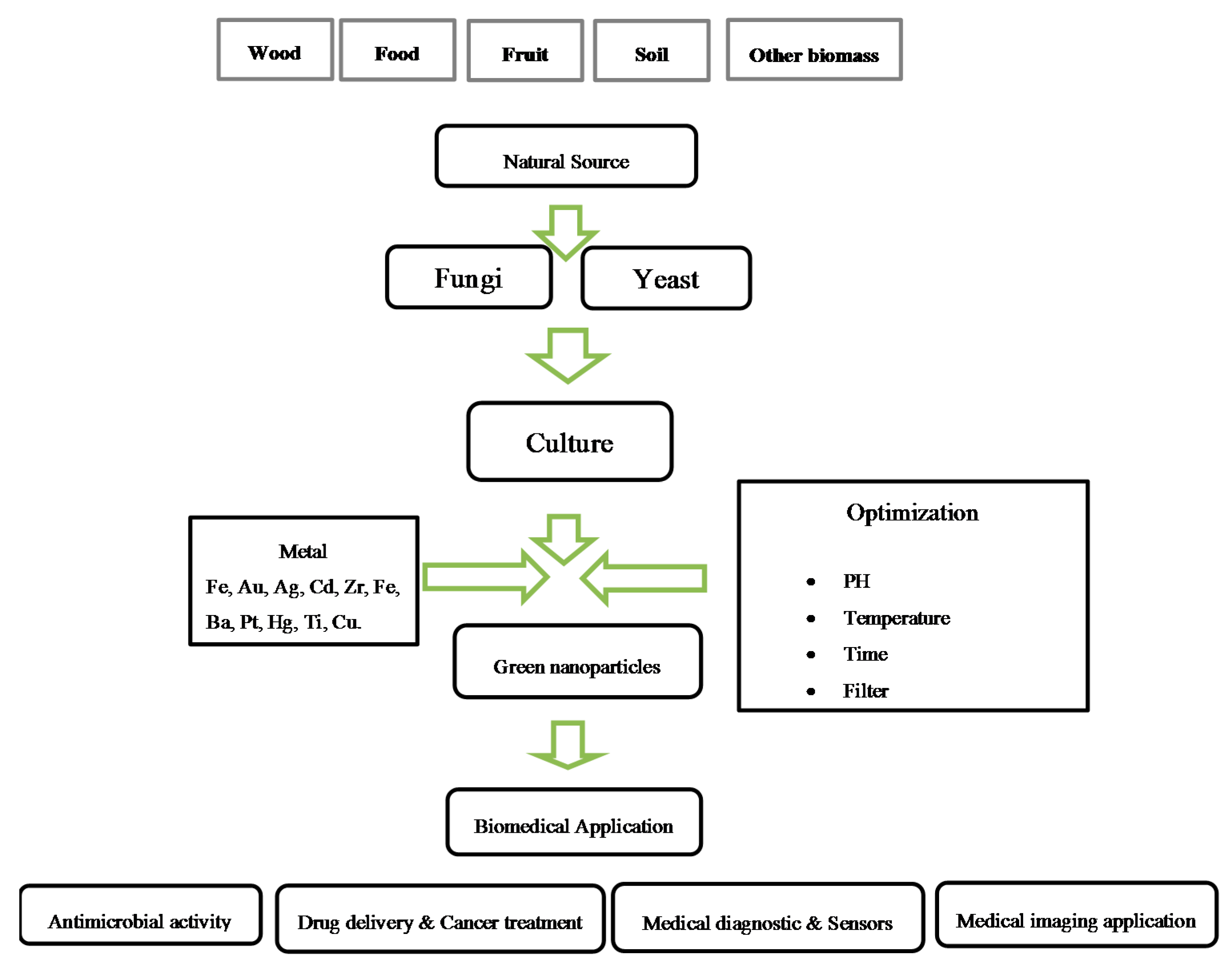
2. Biological Synthesis of Nanoparticles


| Biomass | Possible Mechanism of Nanoparticle Biosynthesis | Reference |
|---|---|---|
Plant
| Secondary metabolites (alkaloids, flavonoids, saponins, steroids, tannins and other nutritional compounds) acts as reducing and stabilizing agents | [17] |
Algae
| Polysaccharides have hydroxyl groups and other functionalities that can play important roles in both the reduction and the stabilization of nanoparticles | [18,19,20,21] |
| Fungi | Reducing enzyme intracellularly or extracellularly and the procedure of biomimetic mineralization | [22,23] |
| Yeast | Membrane bound (as well as cytosolic) oxido reductases and quinones | [24] |
| Bacteria | The microbial cell reduces metal ions by use of specific reducing enzymes like NADH-dependent reductase or nitrate dependent reductase | [25,26] |
3. Biosynthesis of Nanoparticles by Microorganisms
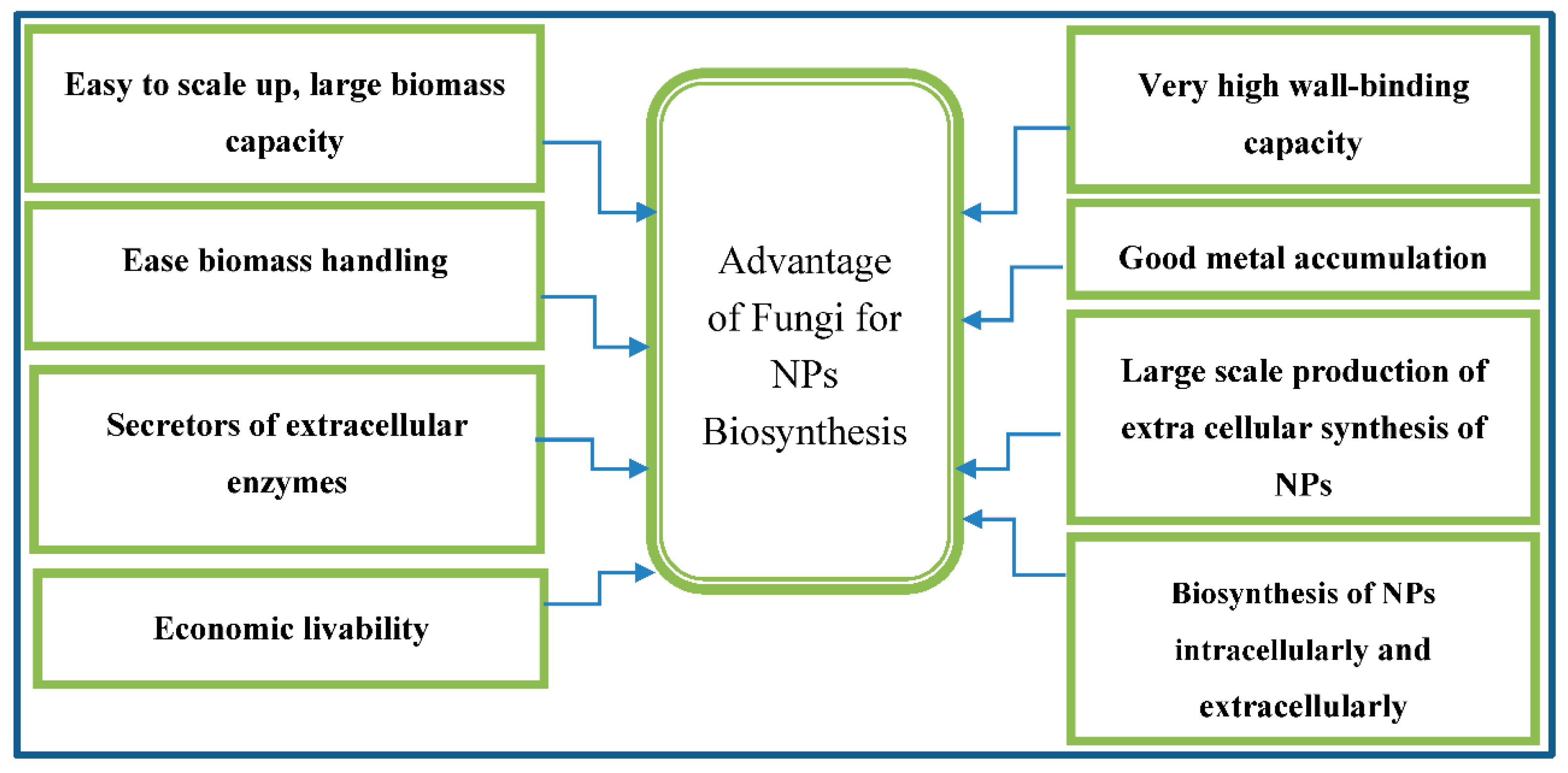
4. Biosynthesis of Nanoparticles by Fungi

| Fungus Species | NPs | Localization | Size (nm) | Shape | Application | Reference |
|---|---|---|---|---|---|---|
| Aspergillus fumigatus | ZnO | Extracellular | 1.2–6.8 | Spherical and hexagonal | Industrial, medical and agricultural sectors | [43] |
| Aspergillus oryzae | FeCl3 | - | 10–24.6 | Spherical | Agricultural, biomedical and engineering sectors | [44] |
| Aspergillus tubingensis | Ca3P2O8 | Extracellular | 28.2 | Spherical | Agricultural, biomedical and engineering sectors | [45] |
| Rhizopus oryzae | Au | Cell surface | 10 | Nanocrystalline | Pesticides | [46] |
| Rhizopus stolonifer | Au | - | 1–5 | Irregularly (uniform) | - | [47] |
| Aspergillus niger | Au | Extracellular | 10–20 | Polydispersed | - | [48] |
| Aspergillus niger | Au | Extracellular | 12.79 ± 5.61 | Spherical | - | [49] |
| Aureobasidium pullulans | Au | Intracellular | 29 ± 6 | Spherical | - | [50] |
| Colletotrichum sp. | Au | - | 20–40 | Decahedral and icosahedral | - | [51] |
| Fusarium semitectum | Au | - | 25 | Spherical | Optoelectronics | [52] |
| Fusarium oxysporum | Au | - | 2–50 | Spherical, monodispersity | - | [50] |
| Fusarium oxysporum | Au | Intracellular | 128 ± 70 a | Aggregates | - | [11] |
| Helminthosporum solani | Au | Extracellular | 2–70 | Polydispersed | Anti-cancer drug | [53] |
| Neurospora crassa | Au | - | 32 | Spherical | - | [36] |
| Penicillium brevicompactum | Au | - | 10−50 | Spherical | To target cancer cells | [54] |
| Verticillium sp. | Au | Cell wall | 20 ± 8 | Spherical | - | [55] |
| Verticillium sp. | Au | Cytoplsmicmembran | 20 ± 8 | Quasihexagonl | - | [55] |
| Verticillium luteoalbum | Au | Intracellular | <10 | Spheres and rods | - | [56] |
| Cylindrocladium floridanu | Au | Extracellular | 19.5 | Spherical | - | [57] |
| Phanerochaete chrysosporium | Au | Extracellular | 10–100 | Spherical | - | [58] |
| Volvariella volvacea | Au | - | 20–150 | Spherical | Therapeutic | [59] |
| Sclerotium rolfsii | Au | Extracellular | 25 | Triangles, decahedral, hexagonal and rods | - | [60] |
| Fusarium oxyporum | Au | Extracellular | 8–40 | Spherical and triangular | - | [61] |
| Fusarium oxyporum | Au | Extracellular | 46.21 | Spherical, triangular | - | [62] |
| Colletotrichum sp. | Au | Extracellular | 8–40 | Spherical | - | [51] |
| Rhizopus stolonifer | Au | - | 1–5 | Irregularly | - | [41] |
| Verticillium luteoalbum | Au | Intracellular | Various | Various | - | [63] |
| Coriolis versicolor | Au | Extra- and intracellular | 20–100, 100–300 | Spherical and ellipsoidal | - | [64] |
| Rhizopus oryzae | Au | - | Various | Triangular, hexagonal, pentagonal, spheroidal, sea urchin like, 2D nanowires, nanorods | - | [65] |
| Aspergillus niger | Au | - | Various | Plates, aggregates, spherical | - | [48] |
| Aspergillus niger | Au | - | Various | Nanowalls, spiral plates, spherical | - | [48] |
| Aspergillus niger | Au | - | 50–500 | Nanoplates | - | [48] |
| Candida albicans | Au | - | 20–40, 60–80 | Spherical & nonspherical | Detection of liver cancer | [66] |
| Verticillum sp. | Ag | Intracellular | 25 | Spherical | - | [38] |
| Fusarium oxyporum | Ag | Extracellular | 5–15 | Highly variable | - | [67] |
| Fusarium oxyporum | Ag | Extracellular | 20–50 | Spherical | Antibacterial | [23] |
| Fusarium oxyporum | Ag | - | 10–25 | Aggregates | - | [68] |
| Aspergillus fumigatus | Ag | - | 5–25 | Mostly spherical, some triangular | - | [69] |
| Aspergillus niger | Ag | Extracellular | 3–30 | Spherical | Antibacterial and antifungal activity | [70] |
| Aspergillus fumigatus | Ag | - | 15–45 | Mostly spherical | Antiviral against HIV-1 | [71] |
| Pleurotus sajor caju | Ag | Extracellular | 30.5 | Spherical | Antibacterial activity | [72] |
| Aspergillus flavus | Ag | On cell wall surface | 8.92 | Spherical | - | [73] |
| Aspergillus niger | Ag | - | 5–35 | Spherical | Antimicrobial | [32] |
| Trichoderma asperellum | Ag | - | 13–18 | Nanocrystalline | Agriculture | [74] |
| Volvariella volvaceae | Ag | - | 15 | Spherical | Medical applications | [15] |
| Penicillium fellutanum | Ag | Extracellular | 5–25 | Mostly spherical | - | [75] |
| Penicillium strain J3 | Ag | - | 10–100 | Mostly spherical | - | [76] |
| Cladosporium cladosporioides | Ag | - | 10–100 | Mostly spherical | - | [77] |
| Phoma glomerata | Ag | - | 60–80 | Spherical | Antibiotic | [78] |
| Coriolis versicolor | Ag | Extra- and intracellular | 25–75, 444–491 | Spherical | - | [79] |
| Trichoderma viride | Ag | - | 5–40 | Spherical, rod-like | Antibacterial activity | [80] |
| Trichoderma viride | Ag | - | 2–4, 10–40, 80–100 | Spherical | - | [81] |
| Trichoderma viride | Ag | - | 2–4 | Mostly spherical | Biosensor and bio imaging | [82] |
| Trichoderma viride | Ag | Extracellular | 5–40 | Spherical, rod-like | synergistic effect with antibiotics | [83] |
| Amylomyces rouxii KSU-09 | Ag | - | 5–27 | Spherical | Antimicrobial | [84] |
| Aspergillus clavitus | Ag | Extracellular | 550–650 | - | Antimicrobial | [85] |
| Aspergillus flavus NJP08 | Ag | - | 17 | Spherical | - | [86] |
| Rhizopus stolonifer | Ag | - | 25–30 | Quasi-spherical | - | [41] |
| Aspergillus terreus CZR-1 | Ag | Extracellular | 2.5 | Spherical | Agriculture, Biomedical and engineering sector | [87] |
| Volvariella volvaceae | Au-Ag | Extracellular | 20–150 | Triangular | Medical application | [15] |
| Fusarium oxyporum | Au-Ag | Extracellular | 8–14 | Quasi-spherical | - | [88] |
| Fusarium oxysporum | Fe3O4 | Extracellular | 20–50 | Irregular, quasi-spherical | - | [89] |
| Verticillium sp. | Fe3O4 | Extracellular | 100–400, 20–50 | Cubo-octahedral, quasi-spherical | - | [89] |
| Aspergillus flavus | TiO2 | - | 62–74 | Spherical | Antimicrobial | [90] |
| Aspergillus flavus TFR7 | TiO2 | 12–15 | Extracellular | Plant nutrient | [91] | |
| Fusarium oxyporum | BT | Extracellular | 4–5 | Quasi-spherical | - | [92] |
| Fusarium oxyporum | Cd | Extracellular | 9–15 | Spherical | - | [5] |
| Fusarium oxyporum | Pt | - | 70–180 | Rectangular, triangular, spherical and aggregates | - | [42] |
| Fusarium oxysporum f. sp. lycopersici | Pt | Extra-and intracellular | 10–100 | Hexagonal, pentagonal, circular, squares, rectangles | - | [40] |
| Fusarium spp. | Zn | Intracellular | 100–200 | Irregular, some spherical | - | [93] |
| Aspergillus versicolor mycelia | Hg | Surface of mycelia | 20.5 ± 1.82 | Alteration | - | [94] |
| Fungi isolated from the soil | Zn, Mg and Ti | extracellular | Various | - | - | [95] |
5. Production of Nanoparticles by Using Yeast
| Yeast | NPs | Localization | Size (nm) | Shape | Application | Reference |
|---|---|---|---|---|---|---|
| Candida glabrata | CdS | Extra- and intracellular | 20 Å, 29 Å | Hexamer | Physiological | [98] |
| Candida glabrata | CdS | Intracellular | - | - | - | [101] |
| Yeast strain MKY3 | Ag | Extracellular | 2–5 | Twinned or multitwinned, some hexagonal | - | [100] |
| Schizosaccharomyces pombe | Cds | Extra- and intracellular | 18 Å, 29 Å | - | - | [98] |
| Schizosaccharomyces pombe | Cds | Intracellular | 1–1.5 | Hexagonal | [99] | |
| Schizosaccharomyces pombe | Cds | Intracellular | - | - | [101] | |
| Pichia jadinii (Candida utilis) | Au | Intracellular | - | Various | - | [63] |
| Yarrowia lipolytica NCIM3589 | Au | Cell surface | Varying | Particles and plates | - | [102] |
| Yeast | Zr | - | - | Irregular mesoporous | Fuel cells | [103] |
| Yeast | Zn3(PO4)2 | Extracellular | 10–80, 80–200 | Rectangular | Antirust pigment and electronic luminophore | [104] |
6. Biomedical Applications of Green Synthesis Nanoparticles

6.1. Drug Delivery
6.2. Anticancer NPs
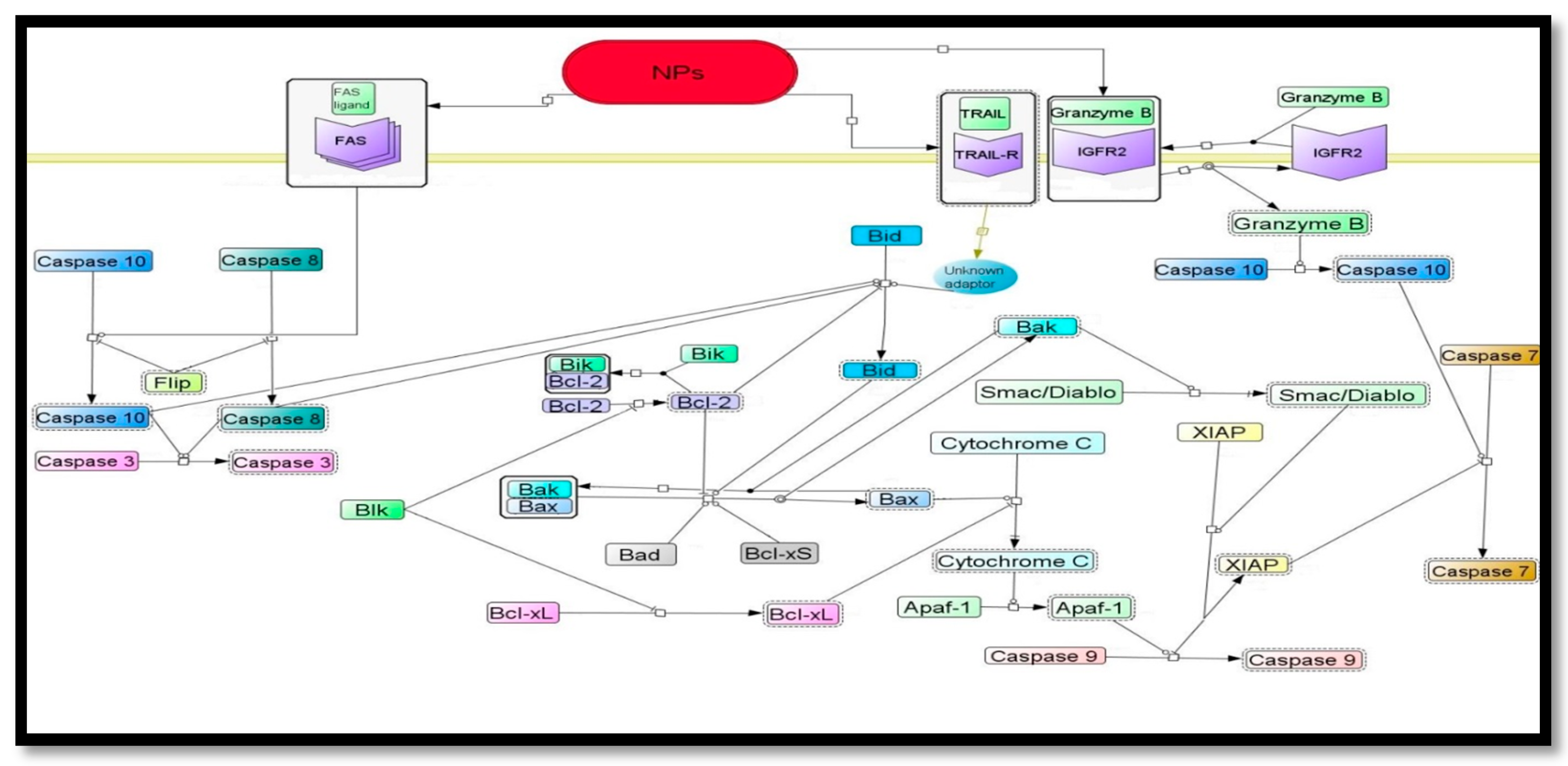
6.2.1.Proposed Signaling Pathways of Nanoparticles Induced Apoptosis
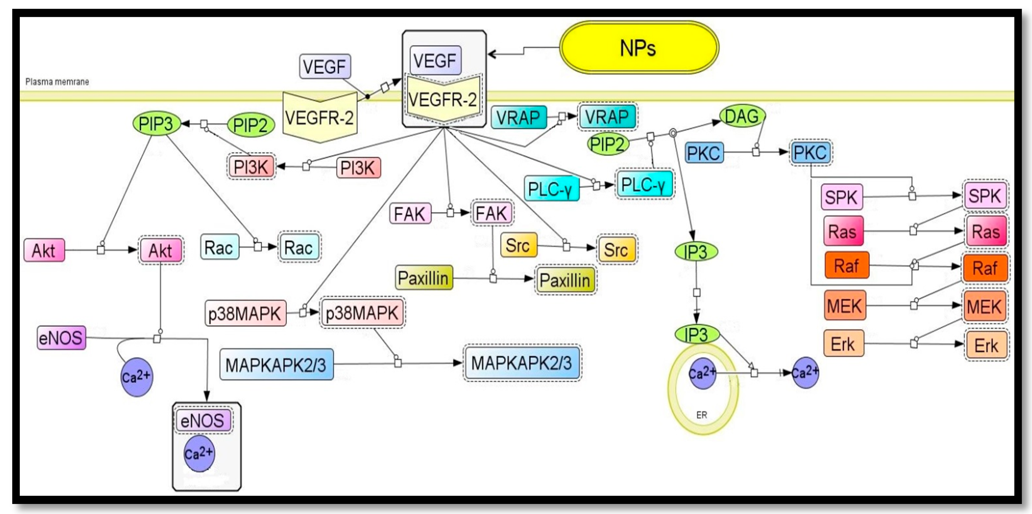
6.2.2. Anti-Angiogenesis Signaling Pathways Possibly Modulated by Nanoparticle


6.3. Antibacterial Agent
Antifungal Activity
6.4. Biosensor
6.5. Medical Imaging
7. Conclusions
Acknowledgments
Author Contributions
Conflicts of Interest
References
- Mohanpuria, P.; Rana, N.K.; Yadav, S.K. Biosynthesis of nanoparticles: Technological concepts and future applications. J. Nanopart. Res. 2008, 10, 507–517. [Google Scholar] [CrossRef]
- Liu, J.; Qiao, S.Z.; Hu, Q.H. Magnetic nanocomposites with mesoporous structures: Synthesis and applications. Small 2011, 7, 425–443. [Google Scholar] [CrossRef] [PubMed]
- Jain, K.K. Applications of nanobiotechnology in clinical diagnostics. Clin. Chem. 2007, 53, 2002–2009. [Google Scholar] [CrossRef] [PubMed]
- Kim, Y.C.; Park, N.C.; Shin, J.S.; Lee, S.R.; Lee, Y.J.; Moon, D.J. Partial oxidation of ethylene to ethylene oxide over nanosized Ag/α-Al2O3 catalysts. Catal. Today 2003, 87, 153–162. [Google Scholar] [CrossRef]
- Kumar, S.A.; Ansary, A.A.; Ahmad, A.; Khan, M.I. Extracellular biosynthesis of CdSe quantum dots by the fungus, Fusarium oxysporum. J. Biomed. Nanotechnol. 2007, 3, 190–194. [Google Scholar] [CrossRef]
- Anker, J.N.; Hall, W.P.; Lyandres, O.; Shah, N.C.; Zhao, J.; van Duyne, R.P. Biosensing with plasmonic nanosensors. Nat. Mater. 2008, 7, 442–453. [Google Scholar] [CrossRef] [PubMed]
- Pinna, N.; Niederberger, M. Oxide Synthesis as Cornerstone of Nanoscience. Eur. J. Inorg. Chem. 2008, 2008, 825. [Google Scholar] [CrossRef]
- Voldman, J.; Gray, M.L.; Schmidt, M.A. Microfabrication in biology and medicine. Annu. Rev. Biomed. Eng. 1999, 1, 401–425. [Google Scholar]
- Chen, Y.; Pépin, A. Nanofabrication: Conventional and nonconventional methods. Electrophoresis 2001, 22, 187–207. [Google Scholar] [CrossRef]
- Piner, R.D.; Zhu, J.; Xu, F.; Hong, S. “Dip-Pen” Nanolithography. Science 1999, 283, 661–664. [Google Scholar] [CrossRef] [PubMed]
- Mandal, D.; Bolander, M.E.; Mukhopadhyay, D.; Sarkar, G.; Mukherjee, P. The use of microorganisms for the formation of metal nanoparticles and their application. Appl. Microbiol. Biotechnol. 2006, 69, 485–492. [Google Scholar] [CrossRef] [PubMed]
- Sotiropoulou, S.; Sierra-Sastre, Y.; Mark, S.S.; Batt, C.A. Biotemplated Nanostructured Materials. Chem. Mater. 2008, 20, 821–834. [Google Scholar] [CrossRef]
- Klaus, T.; Joerger, R.; Olsson, E.; Granqvist, C.G. Silver-based crystalline nanoparticles, microbially fabricated. Proc. Natl. Acad. Sci. USA 1999, 96, 13611–13614. [Google Scholar] [CrossRef] [PubMed]
- Nagajyothi, P.C.; Lee, K.D. Synthesis of Plant-Mediated Silver Nanoparticles Using Dioscorea batatas Rhizome Extract and Evaluation of Their Antimicrobial Activities. J. Nanomater. 2011, 2011, 1–7. [Google Scholar] [CrossRef]
- Thakkar, K.N.; Mhatre, S.S.; Parikh, R.Y. Biological synthesis of metallic nanoparticles. Nanomedicine 2010, 6, 257–262. [Google Scholar] [CrossRef] [PubMed]
- Sriram, M.I.; Kalishwaralal, K.; Gurunathan, S. Biosynthesis of silver and gold nanoparticles using Bacillus licheniformis. Methods Mol. Biol. 2012, 906, 33–43. [Google Scholar] [PubMed]
- Kuppusamy, P.; Yousoff, M.M.; Manian, G.P.; Govindan, N. Biosynthesis of metallic nanoparticles using plant derivatives and their new avenues in pharmacological applications—An updated report. Saudi Pharm. J. 2014. [Google Scholar] [CrossRef]
- Ghodake, G.; Lee, D.S. Biological synthesis of gold nanoparticlesa using the aqueous extract of the brown algae Laminaria japonica. J. Nanoelectron. Optoelectron. 2011, 6, 268–271. [Google Scholar] [CrossRef]
- Azizi, S.; Ahmad, M.B.; Namvar, F.; Mohamad, R. Green biosynthesis and characterization of zinc oxide nanoparticles using brown marine macroalga Sargassum muticum aqueous extract. Mater. Lett. 2014, 116, 275–277. [Google Scholar] [CrossRef]
- Mahdavi, M.; Namvar, F.; Ahmad, M.B.; Mohammad, R. Green biosynthesis and characterization of magnetic iron oxide (Fe3O4) nanoaprticles using seaweed (Sargassum muticum) aqueous extract. Molecules 2013, 18, 5954–5964. [Google Scholar] [CrossRef] [PubMed]
- Azizi, S.; Namvar, F.; Mahdavi, M.; Ahmad, M.B.; Mohamad, R. Biosynthesis of silver nanoparticles using brown marine macroalga, Sargussum muticum aqueous extract. Materials 2013, 6, 5942–5950. [Google Scholar] [CrossRef]
- Ahmad, A.; Senapati, S.; Khan, M.I.; Kumar, R.; Ramani, R.; Srinivas, V.; Sastry, M. Intracellular synthesis of gold nanoparticles by a novel alkalotolerant actinomycete, Rhodococcus species. Nanotechnology 2003, 14, 824–828. [Google Scholar] [CrossRef]
- Durán, N.; Marcato, P.D.; Alves, O.L.; de Souza, G.I.H.; Esposito, E. Mechanistic aspects of biosynthesis of silver nanoparticles by several Fusarium oxysporum strains. J. Nanobiotechnol. 2005, 3. [Google Scholar] [CrossRef] [PubMed] [Green Version]
- Botham, K.M.; Mayes, P.A. Biologic Oxidation. In Harper’s Illustrared Biochemistry, 28th ed.; Lange-McGraw Hill: London, UK, 2006; p. 47. [Google Scholar]
- Durán, N.; Marcato, P.D.; Durán, M.; Yadav, A.; Gade, A.; Rai, M. Mechanistic aspects in the biogenic synthesis of extracellular metal nanoparticles by peptides, bacteria, fungi, and plants. Appl. Microbiol. Biotechnol. 2011, 90, 1609–1624. [Google Scholar] [CrossRef] [PubMed]
- Mishra, S.; Dixit, S.; Soni, S. Methods of nanoparticles biosynthesis for medical and commercial applications. Bio-Nanopart. Biosynth. Sustain. Biotechnol. Implic. 2015, 141–154. [Google Scholar] [CrossRef]
- Asmathunisha, N.; Kathiresan, K. A review on biosynthesis of nanoparticles by marine organisms. Colloids Surf. B Biointerfaces 2013, 103, 283–287. [Google Scholar] [CrossRef] [PubMed]
- Sharma, N.C.; Sahi, S.V.; Nath, S.; Parsons, J.G.; Gardea-Torresdey, J.L.; Pal, T. Synthesis of plant-mediated gold nanoparticles and catalytic role of biomatrix embedded nanomaterials. Environ. Sci. Technol. 2007, 41, 5137–5142. [Google Scholar] [CrossRef] [PubMed]
- Vigneshwaran, N.; Kathe, A.A.; Varadarajan, P.V.; Nachane, R.P.; Balasubramanya, R.H. Biomimetics of silver nanoparticles by white rot fungus, Phaenerochaete chrysosporium. Colloids Surf. B. Biointerfaces 2006, 53, 55–59. [Google Scholar] [CrossRef] [PubMed]
- Huang, X.; Neretina, S.; El-Sayed, M.A. Gold nanorods: From synthesis and properties to biological and biomedical applications. Adv. Mater. 2009, 21, 4880–4910. [Google Scholar] [CrossRef] [PubMed]
- MubarakAli, D.; Gopinath, V.; Rameshbabu, N.; Thajuddin, N. Synthesis and characterization of CdS nanoparticles using C-phycoerythrin from the marine cyanobacteria. Mater. Lett. 2012, 74, 8–11. [Google Scholar] [CrossRef]
- Kathiresan, K.; Alikunhi, N.M.; Pathmanaban, S.; Nabikhan, A.; Kandasamy, S. Analysis of antimicrobial silver nanoparticles synthesized by coastal strains of Escherichia coli and Aspergillus niger. Can. J. Microbiol. 2010, 56, 1050–1059. [Google Scholar] [CrossRef] [PubMed]
- Blackwell, M. The fungi: 1, 2, 3 ... 5.1 million species? Am. J. Bot. 2011, 98, 426–438. [Google Scholar] [CrossRef] [PubMed]
- Sastry, M.; Ahmad, A.; Islam Khan, M.; Kumar, R. Biosynthesis of metal nanoparticles using fungi and actinomycete. Curr. Sci. 2003, 85, 162–170. [Google Scholar]
- Castro-Longoria, E.; Moreno-Velásquez, S.D.; Vilchis-Nestor, A.R.; Arenas-Berumen, E.; Avalos-Borja, M. Production of Platinum Nanoparticles and Nanoaggregates Using Neurospora crassa. J. Microbiol. Biotechnol. 2012, 22, 1000–1004. [Google Scholar] [CrossRef] [PubMed]
- Castro-Longoria, E.; Vilchis-Nestor, A.R.; Avalos-Borja, M. Biosynthesis of silver, gold and bimetallic nanoparticles using the filamentous fungus Neurospora crassa. Colloids Surf. B Biointerfaces 2011, 83, 42–48. [Google Scholar] [CrossRef] [PubMed]
- Volesky, B.; Holan, Z.R. Biosorption of heavy metals. Biotechnol. Prog. 1995, 11, 235–250. [Google Scholar] [CrossRef] [PubMed]
- Mukherjee, P.; Ahmad, A.; Mandal, D.; Senapati, S.; Sainkar, S.R.; Khan, M.I.; Parishcha, R.; Ajaykumar, P.V.; Alam, M.; Kumar, R.; et al. Fungus-Mediated Synthesis of Silver Nanoparticles and Their Immobilization in the Mycelial Matrix: A Novel Biological Approach to Nanoparticle Synthesis. Nano Lett. 2001, 1, 515–519. [Google Scholar] [CrossRef]
- Ahmad, A.; Mukherjee, P.; Mandal, D.; Senapati, S.; Khan, M.I.; Kumar, R.; Sastry, M. Enzyme mediated extracellular synthesis of CdS nanoparticles by the fungus, Fusarium oxysporum. J. Am. Chem. Soc. 2002, 124, 12108–12109. [Google Scholar] [CrossRef] [PubMed]
- Riddin, T.L.; Gericke, M.; Whiteley, C.G. Analysis of the inter- and extracellular formation of platinum nanoparticles by Fusarium oxysporum f. sp. lycopersici using response surface methodology. Nanotechnology 2006, 17, 3482–3489. [Google Scholar] [PubMed]
- Binupriya, A.R.; Sathishkumar, M.; Yun, S.I. Biocrystallization of silver and gold ions by inactive cell filtrate of Rhizopus stolonifer. Colloids Surf. B. Biointerfaces 2010, 79, 531–534. [Google Scholar] [CrossRef] [PubMed]
- Govender, Y.; Riddin, T.; Gericke, M.; Whiteley, C.G. Bioreduction of platinum salts into nanoparticles: A mechanistic perspective. Biotechnol. Lett. 2009, 31, 95–100. [Google Scholar] [CrossRef] [PubMed]
- Raliya, R.; Tarafdar, J.C. ZnO nanoparticle biosynthesis and its effect on phosphorous-mobilizing enzyme secretion and gum contents in Clusterbean (Cyamopsis tetragonoloba L.). Agirc. Res. 2013, 2, 48–57. [Google Scholar] [CrossRef]
- Raliya, R. Rapid, low-cost, and ecofriendly approach her for iron nanoparticle synthesis using Aspergillus oryzae TFR9. J. Nanoparticles 2013, 2013. [Google Scholar] [CrossRef]
- Tarafdar, J.C.; Raliya, R.; Rathore, I. Microbial synthesis of phosphorous nanoparticle from tri-calcium phosphate using Aspergillus tubingensis TFR-5. J. Bionanosci. 2012, 6, 84–89. [Google Scholar] [CrossRef]
- Das, S.K.; Das, A.R.; Guha, A.K. Gold Nanoparticles: Microbial Synthesis and Application in Water Hygiene Management. Langmuir 2009, 25, 8192–8199. [Google Scholar] [CrossRef] [PubMed]
- Sarkar, J.; Ray, S.; Chattopadhyay, D.; Laskar, A.; Acharya, K. Mycogenesis of gold nanoparticles using a phytopathogen Alternaria alternata. Bioprocess Biosyst. Eng. 2012, 35, 637–643. [Google Scholar] [CrossRef] [PubMed]
- Xie, J.; Lee, J.Y.; Wang, D.I.C.; Ting, Y.P. High-yield synthesis of complex gold nanostructures in a fungal system. J. Phys. Chem. C 2007, 111, 16858–16865. [Google Scholar] [CrossRef]
- Bhambure, R.; Bule, M.; Shaligram, N.; Kamat, M.; Singhal, R. Extracellular biosynthesis of gold nanoparticles using Aspergillus niger—Its characterization and stability. Chem. Eng. Technol. 2009, 32, 1036–1041. [Google Scholar] [CrossRef]
- Zhang, X.; He, X.; Wang, K.; Yang, X. Different active biomolecules involved in biosynthesis of gold nanoparticles by three fungus species. J. Biomed. Nanotechnol. 2011, 7, 245–254. [Google Scholar] [CrossRef] [PubMed]
- Shankar, S.S.; Ahmad, A.; Pasricha, R.; Sastry, M. Bioreduction of chloroaurate ions by geranium leaves and its endophytic fungus yields gold nanoparticles of different shapes. J. Mater. Chem. 2003, 13, 1822–1826. [Google Scholar] [CrossRef]
- Sawle, B.D.; Salimath, B.; Deshpande, R.; Bedre, M.D.; Prabhakar, B.K.; Venkataraman, A. Biosynthesis and stabilization of Au and Au-Ag alloy nanoparticles by fungus, Fusarium semitectum. Sci. Technol. Adv. Mater. 2008, 9. [Google Scholar] [CrossRef]
- Kumar, S.A.; Peter, Y.A.; Nadeau, J.L. Facile biosynthesis, separation and conjugation of gold nanoparticles to doxorubicin. Nanotechnology 2008, 19. [Google Scholar] [CrossRef] [PubMed]
- Mishra, A.; Tripathy, S.; Wahab, R.; Jeong, S.H.; Hwang, I.; Yang, Y.B.; Kim, Y.S.; Shin, H.S.; Yun, S.I. Microbial synthesis of gold nanoparticles using the fungus Penicillium brevicompactum and their cytotoxic effects against mouse mayo blast cancer C2C12 cells. Appl. Microbiol. Biotechnol. 2011, 92, 617–630. [Google Scholar] [CrossRef] [PubMed]
- Mukherjee, P.; Ahmad, A.; Mandal, D.; Senapati, S.; Sainkar, S.R.; Khan, M.I.; Ramani, R.; Parischa, R.; Ajayakumar, P.V.; Alam, M. Bioreduction of AuCl4− ions by the fungus, Verticillium sp. and surface trapping of the gold nanoparticles formed. Angew. Chem. Int. Ed. 2001, 40, 3585–3588. [Google Scholar] [CrossRef]
- Gericke, M.; Pinches, A. Microbial Production of Gold Nanoparticles. Gold Bull. 2006, 39, 22–28. [Google Scholar] [CrossRef]
- Narayanan, K.; Sakthivel, N. Mycocrystallization of gold ions by the fungus Cylindrocladium floridanum. World J. Microbiol. Biotechnol. 2013, 29, 2207–2211. [Google Scholar] [CrossRef] [PubMed]
- Sanghi, R.; Verma, P.; Pouri, S. Enzymatic Formation of Gold Nanoparticles Using Phanerochaete chrysosporium. Sci. Res. 2011, 1, 154–162. [Google Scholar]
- Philip, D. Biosynthesis of Au, Ag and Au-Ag nanoparticles using edible mushroom extract. Spectrochim. Acta Part A Mol. Biomol. Spectrosc. 2009, 73, 374–381. [Google Scholar] [CrossRef] [PubMed]
- Narayanan, K.B.; Sakthivel, N. Facile green synthesis of gold nanostructures by NADPH-dependent enzyme from the extract of Sclerotium rolfsii. Colloids Surf. A Physicochem. Eng. Asp. 2011, 380, 156–161. [Google Scholar] [CrossRef]
- Mukherjee, P.; Senapati, S.; Mandal, D.; Ahmad, A.; Khan, M.I.; Kumar, R.; Sastry, M. Extracellular synthesis of gold nanoparticles by the fungus Fusarium oxysporum. ChemBioChem 2002, 3, 461–463. [Google Scholar] [CrossRef]
- Shankar, S.S.; Ahmad, A.; Pasricha, R.; Khan, M.I.; Kumar, R.; Sastry, M. Immobilization of biogenic gold nanoparticles in thermally evaporated fatty acid and amine thin films. J. Colloid Interface Sci. 2004, 274, 69–75. [Google Scholar] [CrossRef] [PubMed]
- Gericke, M.; Pinches, A. Biological synthesis of metal nanoparticles. Hydrometallurgy 2006, 83, 132–140. [Google Scholar] [CrossRef]
- Sanghi, R.; Verma, P. pH dependant fungal proteins in the “green” synthesis of gold nanoparticles. Adv. Mater. Lett. 2010, 1, 193–199. [Google Scholar] [CrossRef]
- Das, S.K.; Das, A.R.; Guha, A.K. Microbial synthesis of multishaped gold nanostructures. Small 2010, 6, 1012–1021. [Google Scholar] [CrossRef] [PubMed]
- Chuhan, A.; Zubair, S.; Tufail, S.; Sherwani, A.; Sajid, M.; Raman, S.C.; Azam, A.; Owais, M. Fungus-mediated biological synthesis of gold nanoparticles : Potential in detection of liver cancer. Int. J. Nanomed. 2011, 6, 2305–2319. [Google Scholar]
- Ahmad, A.; Mukherjee, P.; Senapati, S.; Mandal, D.; Khan, M.I.; Kumar, R.; Sastry, M. Extracellular biosynthesis of silver nanoparticles using the fungus Fusarium oxysporum. Colloids Surf. B Biointerfaces 2003, 28, 313–318. [Google Scholar] [CrossRef]
- Kumar, S.A.; Abyaneh, M.K.; Gosavi, S.W.; Kulkarni, S.K.; Pasricha, R.; Ahmad, A.; Khan, M.I. Nitrate reductase-mediated synthesis of silver nanoparticles from AgNO3. Biotechnol. Lett. 2007, 29, 439–445. [Google Scholar] [CrossRef] [PubMed]
- Bhainsa, K.C.; D’Souza, S.F. Extracellular biosynthesis of silver nanoparticles using the fungus Aspergillus fumigatus. Colloids Surf. B Biointerfaces 2006, 47, 160–164. [Google Scholar] [CrossRef] [PubMed]
- Jaidev, L.R.; Narasimha, G. Fungal mediated biosynthesis of silver nanoparticles, characterization and antimicrobial activity. Colloids Surf. B Biointerfaces 2010, 81, 430–433. [Google Scholar] [CrossRef] [PubMed]
- Alani, F.; Moo-Young, M.; Anderson, W. Biosynthesis of silver nanoparticles by a new strain of Streptomyces sp. compared with Aspergillus fumigatus. World J. Microbiol. Biotechnol. 2012, 28, 1081–1086. [Google Scholar] [CrossRef] [PubMed]
- Vigneshwaran, N.; Kathe, A. Silver-protein (core-shell) nanoparticle production using spent mushroom substrate. Langmuir 2007, 23, 7113–7117. [Google Scholar] [CrossRef] [PubMed]
- Vigneshwaran, N.; Ashtaputre, N.M.; Varadarajan, P.V.; Nachane, R.P.; Paralikar, K.M.; Balasubramanya, R.H. Biological synthesis of silver nanoparticles using the fungus Aspergillus flavus. Mater. Lett. 2007, 61, 1413–1418. [Google Scholar] [CrossRef]
- Mukherjee, P.; Roy, M.; Mandal, B.P.; Dey, G.K.; Mukherjee, P.K.; Ghatak, J.; Tyagi, A.K.; Kale, S.P. Green synthesis of highly stabilized nanocrystalline silver particles by a non-pathogenic and agriculturally important fungus T. asperellum. Nanotechnology. 2008, 19, 1–7. [Google Scholar] [CrossRef] [PubMed]
- Kathiresan, K.; Manivannan, S.; Nabeel, M.; Dhivya, B. Studies on silver nanoparticles synthesized by a marine fungus, Penicillium fellutanum isolated from coastal mangrove sediment. Colloids Surf. B Biointerfaces 2009, 71, 133–137. [Google Scholar] [CrossRef] [PubMed]
- Maliszewska, I.; Szewczyk, K.; Waszak, K. Biological synthesis of silver nanoparticles. J. Phys. Conf. Ser. 2009, 146. [Google Scholar] [CrossRef]
- Balaji, D.S.; Basavaraja, S.; Deshpande, R.; Mahesh, D.B.; Prabhakar, B.K.; Venkataraman, A. Extracellular biosynthesis of functionalized silver nanoparticles by strains of Cladosporium cladosporioides fungus. Colloids Surf. B Biointerfaces 2009, 68, 88–92. [Google Scholar] [CrossRef] [PubMed]
- Birla, S.S.; Tiwari, V.V.; Gade, A.K.; Ingle, A.P.; Yadav, A.P.; Rai, M.K. Fabrication of silver nanoparticles by Phoma glomerata and its combined effect against Escherichia coli, Pseudomonas aeruginosa and Staphylococcus aureus. Lett. Appl. Microbiol. 2009, 48, 173–179. [Google Scholar] [CrossRef] [PubMed]
- Sanghi, R.; Verma, P. Biomimetic synthesis and characterisation of protein capped silver nanoparticles. Bioresour. Technol. 2009, 100, 501–504. [Google Scholar] [CrossRef] [PubMed]
- Fayaz, A.M.; Balaji, K.; Girilal, M.; Kalaichelvan, P.T.; Venkatesan, R. Mycobased synthesis of silver nanoparticles and their incorporation into sodium alginate films for vegetable and fruit preservation. J. Agric. Food Chem. 2009, 57, 6246–6252. [Google Scholar] [CrossRef] [PubMed]
- Fayaz, A.M.; Balaji, K.; Kalaichelvan, P.T.; Venkatesan, R. Fungal based synthesis of silver nanoparticles—An effect of temperature on the size of particles. Colloids Surf. B Biointerfaces 2009, 74, 123–126. [Google Scholar] [CrossRef] [PubMed]
- Fayaz, M.; Tiwary, C.S.; Kalaichelvan, P.T.; Venkatesan, R. Blue orange light emission from biogenic synthesized silver nanoparticles using Trichoderma viride. Colloids Surf. B Biointerfaces 2010, 75, 175–178. [Google Scholar] [CrossRef] [PubMed]
- Fayaz, A.M.; Balaji, K.; Girilal, M.; Yadav, R.; Kalaichelvan, P.T.; Venketesan, R. Biogenic synthesis of silver nanoparticles and their synergistic effect with antibiotics: A study against gram-positive and gram-negative bacteria. Nanomed. Nanotechnol. Biol. Med. 2010, 6, 103–109. [Google Scholar] [CrossRef] [PubMed]
- Musarrat, J.; Dwivedi, S.; Singh, B.R.; Al-Khedhairy, A.A.; Azam, A.; Naqvi, A. Production of antimicrobial silver nanoparticles in water extracts of the fungus Amylomyces rouxii strain KSU-09. Bioresour. Technol. 2010, 101, 8772–8776. [Google Scholar] [CrossRef] [PubMed]
- Saravanan, M.; Nanda, A. Extracellular synthesis of silver bionanoparticles from Aspergillus clavatus and its antimicrobial activity against MRSA and MRSE. Colloids Surf. B Biointerfaces 2010, 77, 214–218. [Google Scholar] [CrossRef] [PubMed]
- Jain, N.; Bhargava, A.; Majumdar, S.; Tarafdar, J.C.; Panwar, J. Extracellular biosynthesis and characterization of silver nanoparticles using Aspergillus flavus NJP08: A mechanism perspective. Nanoscale 2011, 3, 635–641. [Google Scholar] [CrossRef] [PubMed]
- Raliya, R.; Tarafdar, J.C. Novel approach for silver nanoparticle synthesis using Aspergillus terreus CZR-1: mechanism perspective. J. Bionanosci. 2012, 6, 12–16. [Google Scholar] [CrossRef]
- Senapati, S.; Ahmad, A.; Khan, M.I.; Sastry, M.; Kumar, R. Extracellular biosynthesis of bimetallic Au-Ag alloy nanoparticles. Small 2005, 1, 517–520. [Google Scholar] [CrossRef] [PubMed]
- Bharde, A.; Rautaray, D.; Bansal, V.; Ahmad, A.; Sarkar, I.; Yusuf, S.M.; Sanyal, M.; Sastry, M. Extracellular biosynthesis of magnetite using fungi. Small 2006, 2, 135–141. [Google Scholar] [CrossRef] [PubMed]
- Rajakumar, G.; Rahuman, A.; Roopan, S.M.; Khanna, V.G.; Elango, G.; Kamaraj, C.; Zahir, A.A.; Velayutham, K. Fungus-mediated biosynthesis and characterization of TiO2 nanoparticles and their activity against pathogenic bacteria. Spectrochim. Acta A Mol. Biomol. Spectrosc. 2012, 91, 23–29. [Google Scholar] [CrossRef] [PubMed]
- Raliya, R.; Biswas, P.; Tarafdar, J.C. TiO2 nanoparticle biosynthesis and its physiological effect on mung bean (Vigna radiata L.). Biotechnol. Rep. 2015, 5, 22–26. [Google Scholar] [CrossRef]
- Bansal, V.; Poddar, P.; Ahmad, A.; Sastry, M. Room-Temperature Biosynthesis of Ferroelectric Barium Titanate Nanoparticles. J. Am. Chem. Soc. 2006, 128, 11958–11963. [Google Scholar] [CrossRef] [PubMed]
- Velmurugan, P.; Shim, J.; You, Y.; Choi, S.; Kamala-Kannan, S.; Lee, K.J.; Kim, H.J.; Oh, B.T. Removal of zinc by live, dead, and dried biomass of Fusarium spp. isolated from the abandoned-metal mine in South Korea and its perspective of producing nanocrystals. J. Hazard. Mater. 2010, 182, 317–324. [Google Scholar] [CrossRef] [PubMed]
- Das, S.; Das, A.; Guha, A. Adsorption behavior of mercury on functionalized Aspergillus versicolor mycelia: Atomic force microscopic study. Langmuir 2008, 25, 360–366. [Google Scholar] [CrossRef] [PubMed]
- Raliya, R.; Rathore, I.; Tarafdar, J.C. Developmental of microbial nanofactory for zinc, magnesium and titanium nanoparticles production using soil fungi. J. Bionanosci. 2013, 7, 59–96. [Google Scholar] [CrossRef]
- Sun, Y.; Xia, Y. Shape-controlled synthesis of gold and silver nanoparticles. Science 2002, 298, 2176–2179. [Google Scholar] [CrossRef] [PubMed]
- Kumar, D.; Karthik, L.; Kumar, G.; Roa, K.B. Biosynthesis of Silver anoparticles from Marine Yeast and Their Antimicrobial Activity Against Multidrug Resistant Pathogens. Pharmacologyonline 2011, 3, 1100–1111. [Google Scholar]
- Dameron, C.T.; Reese, R.N.; Mehra, R.K.; Kortan, A.R.; Carroll, P.J.; Steigerwald, M.L.; Brus, L.E.; Winge, D.R. Biosynthesis of cadmium sulphide quantum semiconductor crystallites. Nature 1989, 338, 596–597. [Google Scholar] [CrossRef]
- Kowshik, M.; Deshmukh, N.; Vogel, W.; Urban, J.; Kulkarni, S.K.; Paknikar, K.M. Microbial synthesis of semiconductor CdS nanoparticles, their characterization, and their use in the fabrication of an ideal diode. Biotechnol. Bioeng. 2002, 78, 583–588. [Google Scholar] [CrossRef] [PubMed]
- Kowshik, M.; Ashtaputre, S.; Kulkani, S.K.; Parknikar, K.M.M. Extracellular synthesis of silver nanoparticles by a silver-tolerant yeast strain MKY3. Nanotechnology 2003, 14, 95–100. [Google Scholar] [CrossRef]
- Krumov, N.; Oder, S.; Perner-Nochta, I.; Angelov, A.; Posten, C. Accumulation of CdS nanoparticles by yeasts in a fed-batch bioprocess. J. Biotechnol. 2007, 132, 481–486. [Google Scholar] [CrossRef] [PubMed]
- Pimprikar, P.S.; Joshi, S.S.; Kumar, A.R.; Zinjarde, S.S.; Kulkarni, S.K. Influence of biomass and gold salt concentration on nanoparticle synthesis by the tropical marine yeast Yarrowia lipolytica NCIM 3589. Colloids Surf. B Biointerfaces 2009, 74, 309–316. [Google Scholar] [CrossRef] [PubMed]
- Tian, X.; He, W.; Cui, J.; Zhang, X.; Zhou, W.; Yan, S.; Sun, X.; Han, X.; Han, S.; Yue, Y. Mesoporous zirconium phosphate from yeast biotemplate. J. Colloid Interface Sci. 2010, 343, 344–349. [Google Scholar] [CrossRef] [PubMed]
- Yan, S.; He, W.; Sun, C.; Zhang, X.; Zhao, H.; Li, Z.; Zhou, W.; Tian, X.; Sun, X.; Han, X. The biomimetic synthesis of zinc phosphate nanoparticles. Dye Pigment 2009, 80, 254–258. [Google Scholar] [CrossRef]
- Fadeel, B.; Garcia-Bennett, A.E. Better safe than sorry: Understanding the toxicological properties of inorganic nanoparticles manufactured for biomedical applications. Adv. Drug Deliv. Rev. 2010, 62, 362–374. [Google Scholar] [CrossRef] [PubMed]
- Chan, W.C.W.; Nie, S. Quantum dot bioconjugates for ultrasensitive nonisotopic detection. Science 1998, 281, 2016–2018. [Google Scholar] [CrossRef] [PubMed]
- Tian, F.; Tian, F.; Prina-Mello, A.; Estrada, G.; Beyerle, A.; Möller, W.; Schulz, H.; Kreyling, W.; Stoeger, T. A novel assay for the quantification of internalized nanoparticles in macrophages. Nanotoxicology 2008, 2, 232–242. [Google Scholar] [CrossRef]
- Cui, D.; Tian, F.; Coyer, S.R.; Wang, J.; Pan, B.; Gao, F.; He, R.; Zhang, Y. Effects of Antisense-Myc-Conjugated Single-Walled Carbon Nanotubes on HL-60Cells. J. Nanosci. Nanotechnol. 2007, 7, 1639–1646. [Google Scholar] [CrossRef] [PubMed]
- Pantarotto, D.; Partidos, C.D.; Hoebeke, J.; Brown, F.; Kramer, E.D.; Briand, J.P.; Muller, S.; Prato, M.; Bianco, A. Immunization with peptide-functionalized carbon nanotubes enhances virus-specific neutralizing antibody responses. Chem. Biol. 2003, 10, 961–966. [Google Scholar] [CrossRef] [PubMed]
- De La Isla, A.; Brostow, W.; Bujard, B.; Estevez, M.; Rodriguez, J.R.; Vargas, S.; Castano, V.M. Nanohybrid scratch resistant coatings for teeth and bone viscoelasticity manifested in tribology. Mater. Res. Innov. 2003, 7, 110–114. [Google Scholar]
- Ma, J.; Wong, H.; Kong, L.B.; Peng, K.W. Biomimetic processing of nanocrystallite bioactive apatite coating on titanium. Nanotechnology 2003, 14, 619. [Google Scholar] [CrossRef]
- Shinkai, M.; Yanase, M.; Suzuki, M.; Honda, H.; Wakabayashi, T.; Yoshida, J.; Kobayashi, T. Intracellular hyperthermia for cancer using magnetite cationic liposomes. J. Magn. Magn. Mater. 1999, 194, 176–184. [Google Scholar] [CrossRef]
- Weissleder, R.; Elizondo, G.; Wittenberg, J.; Rabito, C.A.; Bengele, H.H.; Josephson, L. Ultrasmall superparamagnetic iron oxide: Characterization of a new class of contrast agents for MR imaging. Radiology 1990, 175, 489–493. [Google Scholar] [CrossRef] [PubMed]
- Parak, W.J.; Boudreau, R.; le Gros, M.; Gerion, D.; Zanchet, D.; Micheel, C.M.; Williams, S.C.; Alivisatos, A.P.; Larabell, C. Cell motility and metastatic potential studies based on quantum dot imaging of phagokinetic tracks. Adv. Mater. 2002, 14, 882–885. [Google Scholar] [CrossRef]
- Chaloupka, K.; Malam, Y.; Seifalian, A.M. Nanosilver as a new generation of nanoproduct in biomedical applications. Trends Biotechnol. 2010, 28, 580–588. [Google Scholar] [CrossRef] [PubMed]
- Emerich, D.F.; Thanos, C.G. The pinpoint promise of nanoparticle-based drug delivery and molecular diagnosis. Biomol. Eng. 2006, 23, 171–184. [Google Scholar] [CrossRef] [PubMed]
- Häfeli, U.O.; Riffle, J.S.; Harris-Shekhawat, L.; Carmichael-Baranauskas, A.; Mark, F.; Dailey, J.P.; Bardenstein, D. Cell uptake and in vitro toxicity of magnetic nanoparticles suitable for drug delivery. Mol. Pharm. 2009, 6, 1417–1428. [Google Scholar] [CrossRef] [PubMed]
- Vaidyanathan, R.; Kalishwaralal, K.; Gopalram, S.; Gurunathan, S. RETRACTED: Nanosilver—The burgeoning therapeutic molecule and its green synthesis. Biotechnol. Adv. 2009, 27, 924–937. [Google Scholar] [CrossRef] [PubMed]
- Xiang, L.; Wei, J.; Jianbo, S.; Guili, W.; Feng, G.; Ying, L. Purified and sterilized magnetosomes from Magnetospirillum gryphiswaldense MSR-1 were not toxic to mouse fibroblasts in vitro. Lett. Appl. Microbiol. 2007, 45, 75–81. [Google Scholar] [CrossRef] [PubMed]
- Giljohann, D.A.; Seferos, D.S.; Daniel, W.L.; Massich, M.D.; Patel, P.C.; Mirkin, C.A. Gold nanoparticles for biology and medicine. Angew. Chemie Int. Ed. 2010, 49, 3280–3294. [Google Scholar] [CrossRef] [PubMed]
- Patra, C.R.; Bhattacharya, R.; Mukhopadhyay, D.; Mukherjee, P. Fabrication of gold nanoparticles for targeted therapy in pancreatic cancer. Adv. Drug Deliv. Rev. 2010, 62, 346–361. [Google Scholar] [CrossRef] [PubMed]
- Kalishwaralal, K.; Banumathi, E.; Pandian, S.R.K.; Deepak, V.; Muniyandi, J.; Eom, S.H.; Gurunathan, S. Silver nanoparticles inhibit VEGF induced cell proliferation and migration in bovine retinal endothelial cells. Colloids Surf. B: Biointerfaces 2009, 73, 51–57. [Google Scholar] [CrossRef] [PubMed]
- Chertok, B.; Moffat, B.A.; David, A.E.; Yu, F.; Bergemann, C.; Ross, B.D.; Yang, V.C. Iron oxide nanoparticles as a drug delivery vehicle for MRI monitored magnetic targeting of brain tumors. Biomaterials 2008, 29, 487–496. [Google Scholar] [CrossRef] [PubMed]
- Amarnath, K.; Mathew, N.L.; Nellore, J.; Siddarth, C.R.V.; Kumar, J. Facile synthesis of biocompatible gold nanoparticles from Vites vinefera and its cellular internalization against HBL-100 cells. Cancer Nanotechnol. 2011, 2, 121–132. [Google Scholar] [CrossRef] [PubMed]
- Jeyaraj, M.; Sathishkumar, G.; Sivanandhan, G.; MubarakAli, D.; Rajesh, M.; Arun, R.; Kapildev, G.; Manickavasagam, M.; Thajuddin, N.; Premkumar, K. Biogenic silver nanoparticles for cancer treatment: an experimental report. Colloids Surf. B Biointerfaces 2013, 106, 86–92. [Google Scholar] [CrossRef] [PubMed]
- Hsin, Y.H.; Chen, C.F.; Huang, S.; Shih, T.S.; Lai, P.S.; Chueh, P.J. The apoptotic effect of nanosilver is mediated by a ROS- and JNK-dependent mechanism involving the mitochondrial pathway in NIH3T3 cells. Toxicol. Lett. 2008, 179, 130–139. [Google Scholar] [CrossRef] [PubMed]
- Vamanu, C.I.; Cimpan, M.R.; Høl, P.J.; Sørnes, S.; Lie, S.A.; Gjerdet, N.R. Induction of cell death by TiO2 nanoparticles: Studies on a human monoblastoid cell line. Toxicol. In Vitro 2008, 22, 1689–1696. [Google Scholar] [CrossRef] [PubMed]
- Park, E.J.; Yi, J.; Chung, K.H.; Ryu, D.Y.; Choi, J.; Park, K. Oxidative stress and apoptosis induced by titanium dioxide nanoparticles in cultured BEAS-2B cells. Toxicol. Lett. 2008, 180, 222–229. [Google Scholar] [CrossRef] [PubMed]
- Zhao, J.; Bowman, L.; Zhang, X.; Vallyathan, V.; Young, S.H.; Castranova, V.; Ding, M. Titanium dioxide (TiO2) nanoparticles induce JB6 cell apoptosis through activation of the caspase-8/Bid and mitochondrial pathways. J. Toxicol. Environ. Heal. Part A 2009, 72, 1141–1149. [Google Scholar] [CrossRef] [PubMed]
- Huang, D.M.; Chung, T.H.; Hung, Y.; Lu, F.; Wu, S.H.; Mou, C.Y.; Yao, M.; Chen, Y.C. Internalization of mesoporous silica nanoparticles induces transient but not sufficient osteogenic signals in human mesenchymal stem cells. Toxicol. Appl. Pharmacol. 2008, 231, 208–215. [Google Scholar] [CrossRef] [PubMed]
- Zhu, Y.; Eaton, J.W.; Li, C. Titanium Dioxide (TiO2) Nanoparticles Preferentially Induce Cell Death in Transformed Cells in a Bak/Bax-Independent Fashion. PLoS ONE 2012, 7, 1–11. [Google Scholar] [CrossRef] [PubMed]
- Nishida, N.; Yano, H.; Nishida, T.; Kamura, T.; Kojiro, M. Angiogenesis in cancer. Vasc. Health Risk Manag. 2006, 2, 213–219. [Google Scholar] [CrossRef] [PubMed]
- Ghosh, P.; Han, G.; De, M.; Kim, C.K.; Rotello, V.M. Gold nanoparticles in delivery applications. Adv. Drug Deliv. Rev. 2008, 60, 1307–1315. [Google Scholar] [CrossRef] [PubMed]
- Will, S.E.A.; Favaron, P.O.; Pavez, M.A.; Florentino, L.C.; Soares, D.; Oliveira, F.C.; Rici, R.E.G.; Miglino, M.A.; Alcântara, D.; Mamizuka, E.M. Bactericidal silver nanoparticles present an antiangiogenic effect in the Chorioallantoic Membrane Model (CAM). Sci. Against Microb. Pathog. Commun. Curr. Res. Technol. Adv. 2011, 1, 219–227. [Google Scholar]
- Gurunathan, S.; Lee, K.-J.; Kalishwaralal, K.; Sheikpranbabu, S.; Vaidyanathan, R.; Eom, S.H. Antiangiogenic properties of silver nanoparticles. Biomaterials 2009, 30, 6341–6350. [Google Scholar] [CrossRef] [PubMed]
- Sheikpranbabu, S.; Kalishwaralal, K.; Venkataraman, D.; Eom, S.H.; Park, J.; Gurunathan, S. Silver nanoparticles inhibit VEGF-and IL-1-beta-induced vascular permeability via Src dependent pathway in porcine retinal endothelial cells. J. Nanobiotechnol. 2009, 7. [Google Scholar] [CrossRef] [PubMed]
- Mohammadzadeh, R. Hypothesis: Silver nanoparticles as an adjuvant for cancertherapy. Adv. Pharm. Bull. 2012, 2, 2. [Google Scholar] [CrossRef]
- Durán, N.; Marcato, P.D.; de Souza, G.I.H.; Alves, O.L.; Esposito, E. Antibacterial effect of silver nanoparticles produced by fungal process on textile fabrics and their effluent treatment. J. Biomed. Nanotechnol. 2007, 3, 203–208. [Google Scholar] [CrossRef]
- Gajbhiye, M.; Kesharwani, J.; Ingle, A.; Gade, A.; Rai, M. Fungus-mediated synthesis of silver nanoparticles and their activity against pathogenic fungi in combination with fluconazole. Nanomed. Nanotechnol. Biol. Med. 2009, 5, 382–386. [Google Scholar] [CrossRef] [PubMed]
- Bankar, A.; Joshi, B.; Kumar, A.R.; Zinjarde, S. Banana peel extract mediated synthesis of gold nanoparticles. Colloids Surf. B Biointerfaces 2010, 80, 45–50. [Google Scholar] [CrossRef] [PubMed]
- Zheng, D.; Hu, C.; Gan, T.; Dang, X.; Hu, S. Preparation and application of a novel vanillin sensor based on biosynthesis of Au-Ag alloy nanoparticles. Sens. Actuators B Chem. 2010, 148, 247–252. [Google Scholar] [CrossRef]
- Zheng, B.; Qian, L.; Yuan, H.; Xiao, D.; Yang, X.; Paau, M.C.; Choi, M.M.F. Preparation of gold nanoparticles on eggshell membrane and their biosensing application. Talanta 2010, 82, 177–183. [Google Scholar] [CrossRef] [PubMed]
- Iskandar, F. Nanoparticle processing for optical applications–A review. Adv. Powder Technol. 2009, 20, 283–292. [Google Scholar] [CrossRef]
- Zhu, T.; Cloutier, S.G.; Ivanov, I.; Knappenberger, K.L.; Robel, I.; Zhang, F. Nanocrystals for electronic and optoelectronic applications. J. Nanomater. 2012, 2012. [Google Scholar] [CrossRef]
- Sarkar, R.; Kumbhakar, P.; Mitra, A.K. Green synthesis of silver nanoparticles and its optical properties. Dig. J. Nanomater. Biostruct. 2010, 5, 491–496. [Google Scholar]
- Podgaetsky, V.M.; Tereshchenko, S.A.; Reznichenko, A.V.; Selishchev, S.V. Laser-limiting materials for medical use. In Optical Technologies for Industrial, Environmental, and Biological Sensing; International Society for Optics and Photonics: Bellingham, WA, USA, 2004; pp. 183–191. [Google Scholar]
- Bao, H.; Hao, N.; Yang, Y.; Zhao, D. Biosynthesis of biocompatible cadmium telluride quantum dots using yeast cells. Nano Res. 2010, 3, 481–489. [Google Scholar] [CrossRef]
- Bao, H.; Lu, Z.; Cui, X.; Qiao, Y.; Guo, J.; Anderson, J.M.; Li, C.M. Extracellular microbial synthesis of biocompatible CdTe quantum dots. Acta Biomater. 2010, 6, 3534–3541. [Google Scholar] [CrossRef] [PubMed]
© 2015 by the authors. Licensee MDPI, Basel, Switzerland. This article is an open access article distributed under the terms and conditions of the Creative Commons Attribution license ( http://creativecommons.org/licenses/by/4.0/).
Share and Cite
Boroumand Moghaddam, A.; Namvar, F.; Moniri, M.; Md. Tahir, P.; Azizi, S.; Mohamad, R. Nanoparticles Biosynthesized by Fungi and Yeast: A Review of Their Preparation, Properties, and Medical Applications. Molecules 2015, 20, 16540-16565. https://doi.org/10.3390/molecules200916540
Boroumand Moghaddam A, Namvar F, Moniri M, Md. Tahir P, Azizi S, Mohamad R. Nanoparticles Biosynthesized by Fungi and Yeast: A Review of Their Preparation, Properties, and Medical Applications. Molecules. 2015; 20(9):16540-16565. https://doi.org/10.3390/molecules200916540
Chicago/Turabian StyleBoroumand Moghaddam, Amin, Farideh Namvar, Mona Moniri, Paridah Md. Tahir, Susan Azizi, and Rosfarizan Mohamad. 2015. "Nanoparticles Biosynthesized by Fungi and Yeast: A Review of Their Preparation, Properties, and Medical Applications" Molecules 20, no. 9: 16540-16565. https://doi.org/10.3390/molecules200916540