
Green Synthesized Chitosan/Chitosan Nanoforms/Nanocomposites for Drug Delivery Applications
by
 , ,
, ,
Iyyakkannu Sivanesan
1 ,
,
Judy Gopal
2,
Manikandan Muthu
2,
Juhyun Shin
3,
Selvaraj Mari
4 and
Jaewook Oh
3,* 1
Department of Bioresources and Food Science, Konkuk University, Hwayang-dong, Gwangjin-gu, Seoul 05029, Korea
2
Laboratory of Neo Natural Farming, Chunnampet, Tamil Nadu 603 401, India
3
Department of Stem Cell and Regenerative Biotechnology, Konkuk University, Seoul 143-701, Korea
4
Department of Chemistry, Guru Nanak College, Chennai 600 042, India
*
Author to whom correspondence should be addressed.
Polymers 2021, 13(14), 2256; https://doi.org/10.3390/polym13142256
Submission received: 22 June 2021
/
Revised: 5 July 2021
/
Accepted: 6 July 2021
/
Published: 9 July 2021
(This article belongs to the Special Issue Marine Biomolecules from Food By-Products: Chitosan and Gelatine)
Abstract
:Chitosan has become a highlighted polymer, gaining paramount importance and research attention. The fact that this valuable polymer can be extracted from food industry-generated shell waste gives it immense value. Chitosan, owing to its biological and physicochemical properties, has become an attractive option for biomedical applications. This review briefly runs through the various methods involved in the preparation of chitosan and chitosan nanoforms. For the first time, we consolidate the available scattered reports on the various attempts towards greens synthesis of chitosan, chitosan nanomaterials, and chitosan nanocomposites. The drug delivery applications of chitosan and its nanoforms have been reviewed. This review points to the lack of systematic research in the area of green synthesis of chitosan. Researchers have been concentrating more on recovering chitosan from marine shell waste through chemical and synthetic processes that generate toxic wastes, rather than working on eco-friendly green processes—this is projected in this review. This review draws the attention of researchers to turn to novel and innovative green processes. More so, there are scarce reports on the application of green synthesized chitosan nanoforms and nanocomposites towards drug delivery applications. This is another area that deserves research focus. These have been speculated and highlighted as future perspectives in this review.
1. Introduction
Chitosan, a natural polysaccharide is second after cellulose in terms of abundance, usage, and distribution [1]. Chemically, chitosan consists of glucosamine and N-acetyl glucosamine residues [2]. Chitosan [(1,4)-2-amino-2-deoxy-D-glucan] is a linear polyaminosaccharide obtained subsequent to N-deacetylation of chitin. Chitin is the structural component of the exoskeleton of shrimps, lobsters, and crabs; it is also present in the cell walls of fungi and yeast, squid pens [3], green algae, and cuticles of insects and arachnids. Chitosan has gained great impetus owing to its biological properties and applications in the medical, food, and agricultural sectors [4,5,6]. The recovery of chitosan from marine shell waste and from marine food waste generated by food-processing industries makes this polymer one of the most important renewable assets.
Chitosan’s therapeutic properties, such as inhibition of microorganisms, pain alleviation [7,8], promotion of hemostasis, and epidermal cell growth [9] are rather unique. Much interest is targetted towards their potential applications in medical and pharmaceutical roles. The increased interest in chitosan is attributed to its favorable properties, such as biocompatibility, ability to bind some organic compounds, susceptibility to enzymatic hydrolysis, and intrinsic physiological activity combined with nontoxicity [10,11,12]. These properties come handy for biomedical applications such as drug delivery and targeting, wound healing and tissue engineering, as well as in nanobiotechnology. Chitosan has, because of its biological and physicochemical properties, become an attractive option as a drug delivery element and for delivery of other macromolecules [13,14,15]. Chitosan-based delivery systems range from microparticles to nanoparticles (NPs), nanoparticle (NP) composites, nanofibres, and films. Chitosan combines with nanometals such as iron (Fe), copper (Cu), silver (Ag), silicon (Si), zinc (Zn), zinc oxide (ZnO), and titanium dioxide (TiO2) to form nanocomposites, leading to enhanced anti-microbial properties [16,17,18,19,20,21].
The advent of nanotechnology and introduction of NPs has resulted in their diverse applications [22]: biomedical, drug delivery, antitumour activity, tissue engineering, sensor development, pathogen detection, protein detection, gene delivery, environmental remediation and water purification, and various similar applications [23,24,25,26,27,28,29]. NPs have become very attractive options for diagnosis and therapeutics, owing to their unique properties. Small sizes with large surface area to volume ratio, as well as their stability and enhanced translocation into the cells, are handy assets equipping these NPs for diverse applications. NPs exhibit exceptional optical properties, rendering them suitable for imaging applications (based on their ability to produce quantum effects) [27,28,29]. The most commonly studied metallic NPs include gold (Au), Ag, aluminum (Al), Zn, Fe, and TiO2 NPs [30]. Researchers in an attempt to enhance the properties of these metallic NPs have conjugated/modified/functionalized these with various biomolecules and ligands [30,31,32].
Chitosan NPs (Ch NPs) have been recently gaining a lot of popularity. Its bio nature, abundance, degradability, and special properties are its strengths. Chitosan can be easily processed in diverse forms, such as films, threads, tablets, membranes, and microparticles/nanoparticles. This allows to design a variety of medical and pharmacological devices adaptable to end purposes. Ch NPs are reported to exhibit antitumor properties by improving the body’s immune function [33,34,35,36,37,38]. Chitosan is useful in bandages to reduce bleeding and as an antibacterial agent and for drug delivery.
NPs synthesis requires the use of reactive and toxic reducing agents, which may result in death and adversely affect the surrounding environment and organisms [39]. Green synthesis is an alternative approach. The green synthesis method has an edge over chemical and physical methods because it is cost-effective, is environment friendly, and there is no requirement for high pressure, energy, temperature, and toxic chemicals [40]. Furthermore, the synthesis of NPs using biological resources is even more attractive, since it is rapid, eco-friendly, and non-toxic. Biological material includes microorganisms such as bacteria [41] and fungus [42], algae [43,44], while plants have also become an alternative option. Nowadays, plant-mediated synthesis is preferable compared to microbe-mediated synthesis due to its simplicity, rapidity, and avoidance of cumbersome culture maintenance processes [45,46]. Plant materials like leaves [45,47], flowers [48,49], seeds [50,51], stems [52,53], fruits [54,55,56], peels [57,58], and weeds [59] have been used for the synthesis of NPs.
In the following paper, we review the various methods for recovery of chitosan from chitin. The methods used to prepare chitosan nanoparticles and nanoforms are also presented briefly. The current status on the available techniques for green synthesis of chitosan/nanochitosan has been reviewed for the first time. The drug delivery applications of chitosan and nanochitosan have been discussed and the need for introducing green synthesized chitosan nanomaterial for drug delivery options has been proposed.
2. Chitosan/Chitosan NPs Synthesis
2.1. Preparation of Chitosan
Chitosan is not generally available in nature; it is recovered from chitin, a naturally available polymer. Mucor, Absidia, and Rhizopus species, that are representative members of Mucorales are the only exceptions, where the polymer chitosan naturally exists [60]. Chitin, in turn, is extracted from discarded remnants of shrimp, squid, prawn, lobster, crab, and domestic marine shell wastes. Crustacean shells are composed of about 15–40% chitin and 20–40% protein, 20–50% calcium and magnesium carbonate and astaxanthin, lipids, and other minerals in trace quantities [61]. Isolation techniques are diverse because the sources are diverse and their composition is variable [62]. A chemical process is mostly initiated for extracting the protein and removing inorganic matter. A bleaching step is optional, using a solvent or through oxidation of pigments [63]; this is followed by demineralization of shells using dilute HCl/HNO3/H2SO4/CH3COOH [64]. Deacetylation of chitin by hydrolysis of the acetamide groups with concentrated NaOH or KOH at temperatures above 100 °C yields chitosan. The degree of acetylation (DA) of chitosan depends on the deacetylation conditions [65]. Lertwattanaseri et al. report a microwave technique to obtain chitin whiskers [66].
Alternative treatments use ethylene diamine tetra acetic acid (EDTA) [67] or ionic liquid extraction [68,69]. Lactic acid fermentation has been reported for the extraction of chitin from prawn shells [70]. Enzymatic extracts or isolated enzymes and microbiological fermentation have been tested [71], but is time-consuming and results in 1–7% of residual protein [72]. Other biotechnological processes that use enzymatic deacetylation of chitin have been demonstrated as alternatives for chemical processing. Chitin deacetylases have been used to hydrolyze N-acetamide bonds, resulting in chitosan [73]. These enzymes are obtained from some select fungi and insects. The enzymatic deacetylation process is reported to enhance the degree of acetylation and average molecular mass of chitosan. However, this alternative is still in the lab [62,74].
2.2. Preparation of Ch NPs
Ch NPs have been reported to be prepared by emulsion droplet coalescence [75], a reverse micellar method, ionic gelation [76,77], precipitation [78], sieving, and spray drying [79]. All of the above-described techniques follow a bottom-up approach. Bottom–up techniques arrange smaller components into complex assemblies and top-down approaches begin with large sized materials and break them into smaller ones. Routine conventional NPs synthesis usually follow bottom–up techniques.
Chitosan micro- and nanoparticles have been prepared using varied techniques. The particle size, stability of the active constituent and the final product, residual toxicity present in the final product, and their drug release kinetics are what go into the selection of an appropriate preparation method [78]. It is confirmed that the size of the prepared particles depends on the molecular weight and chemical structure and degree of deacetylation (DDA) of chitosan, including the method used. The higher the molecular weight of chitosan, the larger the particle size [80,81]. The most common methods for obtaining Ch NPs are: ionotropic gelation, microemulsion, emulsification solvent diffusion, and emulsion-based solvent evaporation. Each of these methods influence the particle size and surface charge of nanochitosan and impact the molecular weight and degree of acetylation. The coacervation method involves the separation of spherical particles by mixing electrostatically driven liquids [82,83]. In the polyelectrolyte complex (PEC) method, an anionic solution is added to the cationic polymer, under mechanical stirring, to obtain nanoparticles [84,85]. The coprecipitation method involves the addition of a chitosan in low pH to a high pH solution, resulting in coprecipitation of highly monodisperses chitosan nanoparticles [86]. In the microemulsion method, chitosan in acetic acid solution and glutaraldehyde are added to a surfactant in an organic solvent such as hexane. NPs form overnight as the cross-linking process is completed, resulting in the formation of small-sized nanoparticles [87]. The Emulsification Solvent Diffusion Method is where an o/w emulsion is prepared with mechanical stirring and high pressure homogenization [88,89] to achieve 300–500 nm sized Ch NPs. Emulsion Based Solvent Evaporation Method is a slight modification of the above method but avoids high shear forces. In reverse micellar method, the surfactant is dissolved in an organic solvent, to which chitosan, and drug and crosslinking agents are added under constant overnight vortex mixing, leading to the formation of Ch NPs of fine sizes [90].
3. Green Synthesis of Chitin/Chitosan/Chitin and Chitosan NPs
Chemical extraction of chitosan has its own drawbacks: (i) the physico-chemical properties of chitin are affected and MW and DA decrease, negatively affecting intrinsic properties; (ii) wastewater effluents contain some chemicals, and (iii) increased cost of purification processes. This is why biological/green extraction techniques are gaining popularity. Biological synthesis uses enzymes and microorganisms for chitin extraction and chitosan recovery. Usually, as in the case of all nanomaterial synthesis, two distinct categories are nominated—chemical synthesis or green synthesis. Here, in the case of chitin/chitosan-based materials, chemical synthesis was the pioneering technique; biological methods of chitin and chitosan preparation then gained popularity. With the existence of a middle term, already established by numerous publications, green synthesis as such in the case of chitin research has become hard to clearly demarcate. As supporters of the fact that biological methods are indeed green synthesis methods, we categorize biological methods of preparation within the topic of green synthesis.
3.1. Biological Method
Khanafari et al. [91] compared chemical versus biological extraction of chitin from shrimp shells. The biological method (using microorganisms) was demonstrated to be better than the chemical method, since the structural integrity of chitin was preserved. Bustos and Healy [92] confirmed that chitin obtained by the deproteinization of shrimp shells with various proteolytic microorganisms has higher molecular weight. The biological extraction of chitin has the following advantages: high reproducibility in shorter time, simple manipulation, less solvent consumption, and lower energy input. However, the biological method is still limited to laboratory scale studies. Recently, two reports have elaborately reviewed the most common biological methods used for chitin extraction [71,93]. The lactic acid bacterial fermentation process has been studied more extensively by Guerrero Legarreta et al. [94] and Cira et al. [95]. Enzymatic deproteinization of chitin requires the use of proteases. Proteolytic enzymes such as alcalase, pepsin, papain, pancreatine, devolvase, and trypsin are mainly obtained from plant, microbe, and animal sources; these are involved in deproteinization of crustacean shells. Alcalase 2.4 L (Novo Nordisk A/S) is a serine endopeptidase obtained from Bacillus licheniformis; this has been used for the isolation of chitin containing about 4% protein impurities [72]. Such purity is sufficient for many non-medical applications of chitin [96]. Manni et al. [97] reported the isolation of chitin from shrimp waste using Bacillus cereus SV1 crude alkaline proteases. In another study, enzymatic deproteinization was optimized by Younes et al. [98]. In this study six alkaline crude microbial proteases from Bacillus mojavensis, Bacillus subtilis, B. licheniformis, B. licheniformis, Vibrio metschnikovii, and Aspergillus clavatus were used. Mukhin and Novikov [99] used crude proteases isolated from the hepatopancreas of crab. Younes et al. [62] used alkaline proteases from the red scorpionfish Scorpaena scrofa [100]. Here, the excessive cost of using enzymes can be decreased by performing deproteinization using a fermentation process, using an endogeneous microorganisms (auto-fermentation) as fermenter or by adding selected strains of microorganisms [71]. Fermentation methods could be via lactic acid fermentation or non-lactic acid fermentation. (a) Lactic acid fermentation of crustacean shells utilizes Lactobacillus sp. that produce lactic acid and proteases. (b) In non-lactic acid fermentation, both bacteria and fungi were used: Bacillus sp. [101,102,103], Pseudomonas sp. [70,104,105], and Aspergillus sp. [106].
Sini et al. [102] studied the fermentation of shrimp shells in jaggery broth using B. subtilis [104,107,108]. Ghorbel-Bellaaj et al. [109] elaborately studied the fermentation efficiency of P. aeruginosa. Teng et al. [110] evaluated the production of chitin from shrimp shells and fungi in a one-pot fermentation process, where fungal proteases hydrolyze proteins into amino acids. Recently, Younes and Rinaudo extensively reviewed the preparation methods involved in the recovery of chitin and chitosan from marine sources [98]. Their review has dealt with the state-of-the-art methodologies involved in the recovery of chitin/chitosan and their future perspectives.
3.2. Green Synthesis for Chitin/Chitosan/Chitin NF/Ch NPs and Ch NF
Actually there are quite a number of studies that describe the use of chitin/chitosan in the green synthesis of inorganic metal NPs. They have also been used as stabilizers during NPs synthesis. However, the term ‘green synthesis’ of chitin/chitosan/Ch NPs’ did not show many results. Green synthesis of chitin and its associates, as we reviewed, was observed to be only supported by scattered scanty reports. We use the term ‘scattered’ because these reports are single publications on this topic, which have not been further researched or developed on. Here, we present the scattered information available on this subject.
Recently, Madalloni et al., 2020, reviewed the green synthesis of chitin using deep Eutectic Solvents (DESs) and Natural Deep Eutectic Solvents (NADESs) [111,112,113]. In this extraction procedure, shrimp shells are treated with 10% citric acid (this is an edible, weak acid that can be extracted from natural sources, used instead of HCl), for demineralization [114]. Deproteinization includes using microwave irradiation, on the above-mentioned pretreated samples suspended in different DES solutions. Finally, simple centrifugation allows for the separation of chitin from DES. Another group [115] developed a zero-waste method to convert shrimp shell waste into chitin in NADESs rather than in DESs. Liu et al. [116] reported an efficient and green chemical process using glycerol to reduce the NaOH content that is usually utilized. Glycerol is a recyclable, stable, green solvent that can be obtained as a by-product of biodiesel. The chitin to chitosan reaction involves the treatment of chitin with 30% NaOH and glycerol, keeping a 1:40 chitin/glycerol ratio, with water being the only other additive. The superiority of this method is that both glycerol and NaOH can be recovered and reused again.
Green synthesis of Ch NPs is also represented by just a few reports. Ch NPs are generated by ionic cross-linking between chitosan and sodium tri-polyphosphate (TPP); there is a report where antimicrobial Ch NPs were synthesized by chemical cross-linking with cinnamaldehyde, another eco-friendly bactericidal agent [117]. Bacterial leaf blight caused by Xanthomonas oryzae devastates rice crops. The antibacterial activity of biosynthesized Ch NPs against this rice pathogen has been reported [118].
A pair of grinding stones can effectively disintegrate chitin organization in crab shells. Mechanical grinding is another simple, yet powerful method allowing the recovery of chitin NFs from waste crab shell in large amounts [119]. The chitin consisted of highly uniform NFs with a width of approximately 10 nm. The same Ifuku group also reported the recovery of chitin NFs from shrimp shells, mushrooms, and squid pens [120,121,122]. Grinding does not involve any complicated chemicals and hence is an eco-friendly process [123]. Few reports of chitin nanofiber synthesis have also been noted with diameters of 3 nm being fabricated in hexafluoroisopropanol (HFIP) through a self-assembly strategy [124,125]. Although chitin NFs have been prepared in HFIP using self-assembly, HFIP is toxic. “Green” solvents such as ionic liquids and urea–NaOH mixtures are preferred in place of HFIP. Qin et al. successfully reported the use of environmentally friendly ionic liquids to obtain high molecular weight purified chitin and chitin films and nanofibers [126,127,128]. Chitosan nanofibers (Ch NFs) have also been synthesized via electrospinning [129]. Polyethylene oxide and poly (vinyl alcohol) (PVA) are often used to blend with chitosan solutions [130,131]. The authors of [132] report a simple and green method for the preparation of chitosan and chitosan-based nanofibers by freeze-drying dilute aqueous solutions, without the use of organic solvents, high concentration of acid solutions, or the need to pre-treat chitosan. Ch NFs with diameters ranging from 100 to 700 nm were obtained from aqueous chitosan solutions. Chitosan/PVA blend NFs with different mass ratios were produced by freeze-drying [130,131].
Our research group [133] recently reported an eco-friendly, sustainable phytomediated one pot recovery of chitosan from commercial chitin and from crab and shrimp shells and squid pen solid wastes. Graviola extracts were employed for the recovery of Ch NFs. Graviola contains acetogenins that actively interact with chitin in insects. With that as the core idea, the graviola extracts were chosen for orchestrating chitin recovery and a possible chitin to chitosan transformation mediated under magnetic stirring on a hot plate.
Silver nanoparticles (AgNPs), particularly those entrapped in polymeric nanosystems, have arisen as options for managing plant bacterial diseases. Chitosan, owing to its low cost, good biocompatibility, antimicrobial properties, and biodegradability, is the polymer under high consideration. Authors have reported green-synthesized Ag NPs using different concentrations of aqueous extract of tomato leaves, followed by entrapment of AgNPs with chitosan (Ch-AgNPs). They used green synthesized systems for controlling tomato bacterial wilt caused by Ralstonia solanacearum [134]. Green synthesis procedures have also been demonstrated for the synthesis of chitosan bionanocomposites. Silver-based chitosan bionanocomposites have been synthesized using the stem extract of Saccharum officinarum [135]. The antibacterial activity of these silver-based chitosan bionanocomposites was evaluated against Bacillus subtilis, Klebsiella planticola, Streptococcus faecalis, Pseudomonas aeruginosa, and Escherichia coli. Kim et al. [136] used a green route to produce Au NPs in a chitosan matrix, whose functional groups favor the interaction with caffeic acid. Shameli et al. [137] demonstrated the green synthesis of Ag/montmorillonite (MMT)/chitosan bionanocomposites using the UV irradiation method and demonstrated their antibacterial activity against Gram-positive and Gram-negative bacteria. Ch NFs with silica phase (Ch NFs/silica) were synthesized by an electrospinning technique to obtain highly porous 3D NF scaffolds. Silver nanoparticles in the form of a well-dispersed metallic phase were synthesized in an external preparation step and embedded in the Ch NFs /silica to form Ag/Ch/silica nanocomposites [138]. Green synthesis of Ag-Ch nanocomposites using chitosan as a reducing agent as well as a stabilizing agent and NaOH as accelerator is reported [139]. The Ag/Ch nanocomposite gel is transformed into colloid by dissolving into chitosan solution and its antibacterial activity against E. coli and S. aureus bacteria was demonstrated. In another work [140], colloidal Au NPs were stabilized into a chitosan matrix and prepared using a green route. The use of chitosan, with a large number of amino and hydroxyl functional groups, enables the simultaneous synthesis and surface modification of AuNPs in one pot. These hybrid nanocomposite films were used as sensors for the determination of caffeic acid, an antioxidant that has recently attracted much attention because of its benefits to human health. A novel Ch-Fe2O3 nanocomposite was synthesized by a facile one pot green route [141]. The nanocomposite showed excellent recyclable efficiency up to five cycles. The nanocomposite has also been proved to be an excellent potential sorbent for recovery of toxic elements from industrial and medical wastewater. Using cinnamaldehyde (CA), a natural preservative, researchers have prepared CA/SA/Ch NPs nanocomposites. In vitro release experiments showed that CA/SA/Ch NPs had the function of sustained release, indicating that SA/Ch NPs can be used as a promising carrier for CA [142]. Antimicrobial carboxymethyl chitosan-nanosilver (CMC-Ag) hybrids with controlled silver release was fabricated by Huang et al. [143]. Under principles of green chemistry, the synthesis was conducted in an aqueous medium exposed to microwave irradiation for 10 min with non-toxic chemicals. Their antibacterial activity was tested against Staphylococcus aureus and Escherichia coli.
Ultrasound-induced synthesis of Ch-modified nano-scale graphene oxide (Ch-NGO) hybrid nanosheets, which has great potential pharmaceutical applications, in supercritical CO2 without catalyst is reported [144]. Ciprofloxacin hydrochloride (CIP) was incorporated into a green based nano-composite (HS/Ch-NC) to control the antibiotic release and increase its bioavailability [145]. An in vitro drug release study of the nano-composite HS/Ch-NC showed high activity against gram negative bacterial strains due to the successful release of CIP from the chitosan composite into the tested bacterial strains. Chitosan nanoparticles (NPs) are widely studied as vehicles for drug, protein, and gene delivery. However, lack of sufficient stability, particularly under physiological conditions, render chitosan NPs limited pharmaceutical utility. Stable Ch NPs suitable for drug delivery applications [146] were prepared by grafting to phthalic or phenylsuccinic acids. Subsequently, polyphosphoric acid (PPA), hexametaphosphate (HMP), or tripolyphosphate (TPP) were used to achieve tandem ionotropic/covalently crosslinked chitosan NPs in the presence of 1-ethyl-3-(3-dimethylaminopropyl)-carbodiimide (EDC). Chitosan-zinc oxide nanoparticle composite [147] and CuO/Chitosan composites [148] are also reported. Table 1 presents a consolidated list of chitin family of green synthesized materials.
4. Drug Delivery Applications of Chitosan/Nanochitosan
4.1. Basics of Polymers in Drug Delivery
Nanomaterials or nanoparticles offer new opportunities in material science and biomedicine. The small size of nanoparticles allows them to enter cells and organelles for targeted drug delivery [149]. NPs can be conjugated with ligands or antibodies to enable recognition and binding to specific receptors on cell targets [150,151]. Different methods are available to prepare chitosan micro-/nanoparticles in which the drug is mostly bound to chitosan by hydrogen bonding, electrostatic interaction, or hydrophobic linkage. There are several mechanisms which govern drug release from chitosan nanoparticles, such as swelling of the polymer [152], diffusion of the adsorbed drug, drug diffusion through the polymeric matrix, polymer erosion or degradation, and a combination of both erosion and degradation [147], as represented in Figure 1. The swelling of the polymer is characterized by the imbibition of water into the polymer until the polymer dissolves. This drug release mechanism is characterized by the solubility of the polymer in water, or the surrounding biological medium [153]. Erosion and degradation of polymers are interrelated features. Sometimes, degradation of the polymer may cause subsequent physical erosion as bonds break. Erosion of polymers is a complex phenomenon as it involves swelling, diffusion, and dissolution.
4.2. Chitosan Microparticles-Based Drug Delivery
Among the novel drug delivery systems investigated, chitosan micro-/NPs offer great promise in oral, parenteral, topical, and nasal applications. In these systems, the drug is either confined and surrounded by a polymeric membrane or is uniformly dispersed in the polymer matrix. Drug release at a specific site and for an extended period of time could also be achieved by mucoadhesion, where chitosan adheres to specific mucosal surfaces in the body, such as buccal, nasal, and vaginal cavities [154,155,156]. Chitosan microparticles have shown varied applications in the delivery of a range of compounds owing to particle size reduction by micronization and their mucoadhesive properties. Dastan and Turan [157] developed chitosan–DNA microparticles and reported a sustained-release profile of DNA, with a potential transfer of the DNA into human embryonic kidney, Swiss 3T3, and HeLa cell lines. Another research group prepared chitosan–DNA microparticles for mucosal vaccination in simulated intestinal fluid and simulated gastric fluid [158]. Luteinizing hormone-releasing chitosan-based microparticles as a vaccine delivery vehicle are also reported. Successful delivery of hormones by these particles extends their application for induction of immunity against some tumor antigens and microorganisms such as bacteria and viruses [159].
Insulin delivery via the nasal route using chitosan microparticles was demonstrated by Varshosaz et al. [160]. They demonstrated that insulin-loaded microspheres exhibited a 67% lowering in blood glucose level, compared to insulin administered intravenously. Many research groups have thus described the applicability of chitosan microparticles in drug delivery. The encapsulation of diclofenac sodium [161], 5-flurouracil [162], cisplatin [163], felodipine [164], and hydroquinone [165] into these carriers has been reported, and the designed microparticles generally exhibit a controlled-release effect. Chitosan magnetic microparticles (CMM) are a special class of chitosan microparticles that have been developed and used for the delivery of anticancer drugs or radionuclide atoms to a targeted tissue [166] by binding the drug or the radioactive atom to a magnetic compound, which is then injected into the blood and stopped at the targeted tissue by an externally applied magnetic field [167,168]. Attapulgite, a nanosized silicate clay naturally occurring polymer used in drug delivery [169], has been introduced into cross-linked diclofenac sodium chitosan microspheres in which the prepared chitosan/attapulgite hybrid microspheres exhibited narrow size distribution and minimum drug release in the simulated gastric fluid [170].
4.3. Ch NPs-Based Drug Delivery
The preparation of curcumin-loaded Ch NPs has been reported to enhance the drug solubility and stability in the gastro intestinal tract [171]. Facilitation of the transmucosal delivery of two hydrophobic drugs, triclosan and furosemide, has been achieved by developing drug-loaded Ch NPs [172]. Low-molecular weight heparin (LMWH) has been loaded into Ch NPs and showed improved oral absorption and relative bioavailability, compared to a solution of LMWH [173]. Recently, chitosan nanotherapeutics have received great attention in the field of oncology because of their enhanced tumor targeting, ability to load different hydrophobic anticancer drugs, and the ability to control anticancer drug release rates [174,175]. Chitosan-loaded paclitaxel NPs exhibited excellent tumor-homing ability in tumor-bearing mice [176,177,178,179,180]. Protein/siRNA-loaded Ch NPs have been prepared and have shown 98% entrapment efficiency with adequate stability [164]. BSA has been encapsulated into Ch NPs [181]. Genetic immunization using Ch NPs-loaded plasmid DNA was investigated, and the results showed measurable and quantifiable levels of gene expression and considerable antigen titer [182]. Other workers investigated the potential of these nanoparticles as carriers for antigens by using recombinant hepatitis B surface antigen [183]. Ch NPs for oral delivery of insulin have been successfully formulated and demonstrated for enhanced in vitro as well as in vivo absorption and improved insulin bioavailability. [184] Magnetic nanosized Ch NPs were also developed as a special type of Ch NPs and showed sustained drug release with minimal toxicity. Camptothecin magnetic Ch NPs in which polyethylene glycol was coupled with magnetic nanoparticles to increase their biocompatibility has been reported [185].
Ch NPs have been successfully used for drug delivery. Mohammed et al. elaborately reviewed the extensive milestones achieved through Ch NPs-based drug delivery [186]. The effective delivery of catechin and epigallocatechin across interstinal membranes was achieved by encapsulation within Ch NPs [187]. Permeation of tamoxifen across intestinal epithelium was enabled via formulating tamoxifen into lecithin-chitosan NPs [188]. Feng et al. [189] reported successful oral delivery of anticancer drugs, using nanoparticles of doxorubicin hydroxide (DOX)/chitosan/ carboxymethyl chitosan formulations, which could enable enhanced absorption of DOX throughout the intestine. Alendronate sodium is used in the treatment of osteoporosis, and alendronate sodium was encapsulated in Ch NPs. This overcame the low oral bioavailability and gastrointestinal side-effects that this drug confronted during drug delivery [190]. Sustained delivery of sunitinib, a tyrosine kinase inhibitor, up to 72 h, was achieved though encapsulation of Ch NPs [191]. Insulin--loaded Ch NPs crosslinked with TPP increased their uptake by the intestinal epithelium [192]. Bay 41-4109, an active inhibitor of hepatitis B virus, was formulated as Ch NPs to improve drug solubility and oral bioavailability [193]. Chitosan and carboxymethyl Ch NPs were found to be excellent carriers for oral vaccine delivery of extracellular products of V. anguillarum (pathogenic bacteria). This chitosan-based formulation increased its stability, resulted in sustained release, and protected the antigenic protein from entering the spleen and the kidney, which is critical for immune response [194].
Chitosan is biodegradable, biocompatible, exhibits low toxicity, adheres to mucus, and opens the tight junctions of the nasal membrane. Owing to these properties, chitosan has applications in nasal delivery [195]. Carboxymethyl Ch NPs of carbamazepine (treats epilepsy) enhanced the bioavailability and brain targeting via the nasal route. The bioavailability of leuprolide (used to treat prostate cancer and hormone-dependent diseases), increased when formulated as thiolated-Ch NPs [196,197]. Islam and Ferro [198] reviewed the various modes of Ch NPs-based nanoparticle-aided drug delivery to the lungs. The authors claimed that the positive charge on the surface of chitosan provides mucoadhesive properties, increasing the potential for drug absorption. In addition, the positively charged chitosan are able to open the intercellular tight junctions of the lung epithelium, increasing uptake. More so, chitosan binds to phosphoryl groups and lipopolysaccharides on bacterial cell membranes, and helps in fighting pulmonary bacterial infections. A dry powder inhalation (DPI) of rifampicin, an antitubercular drug, was formulated with chitosan, bringing about sustained drug release until 24 h and no toxicity to cells nor organs [199]. The antifungal drug Itraconazole, which is used to treat pulmonary infections, suffers from low solubility. Its aerosolization properties, which would enable its pulmonary deposition, was improved by formulating it with spray dried chitosan NPs with lactose, mannitol, and leucine [200].
Giovino and co-workers have investigated chitosan buccal films of insulin-loaded poly (ethylene glycol) methyl ether-block-polylactide (PEG-b-PLA) NP [201]. Polysaccharide-based NP chitosan [202] and curcumin prepared as polycaprolactone nanoparticles coated with chitosan and NP encapsulation of enriched flavonoid fraction (EFF-Cg) obtained from Cecropia glaziovii were successfully demonstrated for buccal delivery [84]. Chitosan-vancomycin NPs for colon delivery were prepared by two different methods: ion gelation and spray drying [203]. Coco et al. proved the ability of NPs made with chitosan for inflamed colon drug delivery [204]. Several batches of NPs were prepared by entrapping ovalbumin (OVA) into Eudragit S, trimethylchitosan, PLGA, PEG-PLGA and PEG-PCL, separately. Of all the NPs made, NPs with trimethyl chitosan have shown the highest permeability of OVA. However, high permeability was also seen with PEG-PLGA NPs as they were coated with mannose for active targeting of the area of inflammation. As another example, chitosan-carboxymethyl starch nanoparticles of 5-aminosalicylic acid, another drug for inflammatory bowel disease, was prepared, which achieved high entrapment efficiency as well as controlled drug release [205]. Systemic absorption of insulin was demonstrated by the formulation in Ch NPs and administration by the nasal route. Insulin loading up to 55% was achieved and nasal absorption of insulin was greater from Ch NP [206]. Rosmarinic acid-loaded chitosan NPs was prepared by an ion gelation method for ocular delivery. Imiquimod was formulated as chitosan-coated PCL nanocapsules embedded in chitosan hydrogel for vaginal delivery to treat human papillomavirus infection [207]. The pharmacokinetics of chitosan-based formulations has been studied by a few authors [89,208] and a couple of clinical vaccine trials of chitosan formulations have been reported [209,210]. Quinones et al. exhaustively reviewed the chitosan-based self-assembled NPs in drug delivery [211].
Metronidazole (MZ) is an antibiotic with common side-effects of nausea, vomiting, epigastric pain, and mouth dryness, most likely caused by high concentrations of residual MZ in the saliva [212]. To protect MZ from dissolution in saliva, the drug was loaded into Ch NPs of 200–300 nm in size, showing controlled release [213]. Ch NPs loaded with insulin were also developed to improve the systemic delivery of insulin through the nasal passage [214]. The NPs were shown to reduce blood glucose levels by 52.9% in rats and 72.6% in sheep. Ch was chemically modified with the hydrophobic n-hexanoic anhydride to form an amphiphilic Ch derivative that showed better blood compatibility [215]. Cefadroxil drug-loaded nanofibers CFX-CPNFs were successfully fabricated by the freeze drying method. The antimicrobial activity indicated that the CFXCPNFs had excellent bacterial activity against 16 strains of Staphylococcus bacteria [216].
Electrospun Ch NFs have been reported for their role in drug delivery [217]. Ch/PVA NFs has been successfully used for enzyme lipase immobilization [218], Ch/ PVA NFs have been used as novel biomedicated nanofibers for preventing wound infections and local chemotherapy [219], Chitosan Carboxymethyl-β-CycloDextrin/PVA has been demonstrated for slow release of CD drug [220], Ch/Polylactic-Co Glycolic Acid (PLGA) showed better fibroblast attachment and proliferation compared with PLGA alone, and Chitosan/phospholipid NFs were attributed to be used as platforms for transdermal drug delivery. The release rate of the model drugs used in this study depended on their solubility [221,222]. Figure 2 gives an overview of the overall drug delivery options successfully demonstrated using Ch NPs.
5. Challenges and Future Perspectives
Ch-based nanomaterials are among the most promising polymeric biomaterials being synthesized, because of their distinctive characteristics, biodegradability, non-toxicity, and antimicrobial properties. Ch NPs are extensively studied for biological, biomedical, and pharmaceutical applications, including drug delivery and gene delivery, as well as a therapeutic delivery system and nanosystem for cancer, for wound healing, and as bactericidal agents. Current research is focused on improving the stability, biocompatibility, and synthesis of novel Ch NPs to enhance their effectiveness in biomedical applications [223]. Commonly, Ch was found to be relatively safe due to its biodegradable and biocompatible properties. However, several studies showed the cytotoxicity of Ch NPs in vitro and in vivo. Thus, the present knowledge on Ch-based nanomaterials is not adequate. More research is required to comprehensively investigate the toxicity of Ch NPs for human beings and other living organisms. Moreover, green and environmentally benign synthesis methods for Ch derivatives should be developed to protect the environment [224]. Recently, the concepts of “eco-sustainability”, “reuse”, and “recycling” have become the basis of holistic research approaches.
There are several drawbacks in the use of chitosan for drug delivery systems. The main drawback is its poor solubility at physiological pH owing to the partial protonation of amino groups. To overcome these inherent drawbacks, various derivatives of chitosan, such as carboxylated, different conjugates, thiolated, and acylated chitosan have been devised [225,226]. Researchers reported on the goals of using chitosan as an excipient for drug delivery systems [227,228] and in the development of chitosan drug control releasing systems, including chitosan sponges, chitosan film, chitosan beads, chitosan microbeads (microspheres), and chitosan nanoparticles.
To date, chitosan has shown little or no toxicity in animal models and there have been no reports of major adverse effects in healthy human volunteers; however, clinical data are lacking. Even though chitosan is approved in dietary use, wound dressing applications and cartilage formulations, chitosan-based drug formulation are yet to be approved for mass marketing [228]. Of all the various biomedical applications, we observed that drug delivery applications of Ch NPs are that which are being highly researched and investigated. Ch NPs targeted cancer theranostics, dermatological applications, and targeted parenteral drug delivery systems need to be seriously looked into [229,230,231,232,233,234,235,236]. With the advent of new strategies in overcoming the limitations of chitosan, we expect to see more chitosan research work in near future, especially in nasal and pulmonary drug delivery.
As this review exposes, there are only a few scattered reports on the green synthesis of Ch NPs; this being the latest trend in nanomaterial synthesis, it is rather absurd that not much attention is paid in this direction. This review expects a hike in research publication in the direction of green synthesized Ch NPs, and Ch nanocomposites. In addition, not much work is available on the use of green synthesized Ch NPs for biomedical applications; this is an interesting area worth working on. If a material recovered from marine wastes can be extracted using green methods and put to use for valuable biomedical causes, what are we waiting for?
6. Conclusions
The general scenario of chitosan recovery was briefly reviewed in this paper. The scattered reports on green synthesis of chitosan and Ch NPs have been consolidated and presented. The drug delivery milestones by chitosan and its nanoforms has been presented and the lacunae in the application of green synthesized towards drug delivery applications has been highlighted.
Author Contributions
Conceptualization, writing—original draft preparation, writing—review and editing, I.S., M.M., and J.G.; supervision, and review, J.S. and S.M. and funding acquisition, supervision, and review, J.O. All authors have read and agreed to the published version of the manuscript.
Funding
This research received no external funding.
Institutional Review Board Statement
Not applicable.
Informed Consent Statement
Not applicable.
Acknowledgments
This work was supported by the KU Research Professor Program of Konkuk University.
Conflicts of Interest
The authors declare no conflict of interest.
References
- Mincea, M.; Negrulescu, A.; Ostafe, V. Preparation, modification, and applications of chitin nanowhiskers: A review. Rev. Adv. Mater. Sci. 2012, 30, 225–242. [Google Scholar]
- Sashiwa, H.; Aiba, S.-I. Chemically modified chitin and chitosan as biomaterials. Prog. Polym. Sci. 2004, 29, 887–908. [Google Scholar] [CrossRef]
- Illum, L.; Jabbal-Gill, I.; Hinchcliffe, M.; Fisher, A.N.; Davis, S.S. Chitosan as a novel nasal delivery system for vaccines. Adv. Drug Deliv. Rev. 2001, 51, 81–96. [Google Scholar] [CrossRef]
- Allan, G.G.; Altman, L.C.; Bensinger, R.E.; Ghosh, D.K.; Hirabayashi, Y.; Neogi, A.N.; Neogi, S. Biomedical applications of chitin and chitosan. In Chitin, Chitosan, and Related Enzymes; Zikakis, J.P., Ed.; Academic Press: New York, NY, USA, 1984; pp. 119–133. [Google Scholar]
- Nishimura, K.; Nishimura, S.; Nishi, N.; Saiki, I.; Tokura, S.; Azuma, I. Immunological activity of chitin and its derivatives. Vaccine 1984, 2, 93–99. [Google Scholar] [CrossRef]
- Li, Q.; Dunn, E.T.; Grandmaison, E.W. Applications of chitin and chitosan. In Applications and Properties of Chitosan; Goosen, M.F.A., Ed.; Technomic Publishing Co.: Lancaster, PA, USA, 1997; pp. 3–29. [Google Scholar]
- Balakrishnan, B.; Mohanty, M.; Umashankar, P.R.; Jayakrishnan, A. Evaluation of an in situ forming hydrogel wound dressing based on oxidized alginate and gelatin. Biomaterials 2005, 26, 6335–6342. [Google Scholar] [CrossRef] [PubMed]
- Badawy, M.E.; Rabea, E.I.; Rogge, T.M.; Stevens, C.V.; Smagghe, G.; Steurbaut, W.; Hofte, M. Synthesis and fungicidal activity of new N,O-acyl chitosan derivatives. Biomacromolecules 2004, 5, 589–595. [Google Scholar] [CrossRef]
- Howling, G.I.; Dettmar, P.W.; Goddard, P.A.; Hampson, F.C.; Dornish, M.; Wood, E.J. The effect of chitin and chitosan on the proliferation of human skin fibroblasts and keratinocytes in vitro. Biomaterials 2001, 22, 2959–2966. [Google Scholar] [CrossRef]
- Wang, X.H.; Cui, F.Z.; Feng, Q.L.; Li, J.C.; Zhang, Y.H. Preparation and characterization of collagen/chitosan matrices as potential biomaterials. J. Bioact. Compat. Pol. 2003, 18, 453–467. [Google Scholar] [CrossRef]
- Kumar, M.N.V.R. A review of chitin and chitosan applications. React. Funct. Polym. 2000, 46, 1–27. [Google Scholar] [CrossRef]
- Li, Q.; Grandmaison, E.W.; Goosen, M.F.A.; Dunn, E.T. Applications and properties of chitosan. J. Bioact. Compat. Pol. 1992, 7, 370–397. [Google Scholar] [CrossRef]
- Bernkop-Schnurch, A.; Walker, G. Multifunctional matrices for oral peptide delivery. Crit. Rev. Ther. Drug 2001, 18, 459–501. [Google Scholar] [CrossRef]
- George, M.; Abraham, T.E. Polyionic hydrocolloids for the intestinal delivery of protein drugs: Alginate and chitosan—A review. J. Control. Release 2006, 114, 1–14. [Google Scholar] [CrossRef]
- Patel, M.; Shah, T.; Amin, A. Therapeutic opportunities in colon-specific drug-delivery systems. Crit. Rev. Ther. Drug 2007, 24, 147–202. [Google Scholar] [CrossRef] [PubMed]
- Wen, Y.Z.; Ma, J.Q.; Chen, J.; Shen, C.S.; Li, H.; Liu, W.P. Carbonaceous sulfur-containing chitosan-Fe(III): A novel adsorbent for efficient removal of copper (II) from water. Chem. Eng. J. 2015, 259, 372–380. [Google Scholar] [CrossRef]
- Wang, M.; Sun, R.R.; Li, C.; Wang, Q.L.; Zhang, B.H. MicroRNA expression profiles during cotton (Gossypium hirsutum L.) fiber early development. Sci. Rep. 2017, 7. [Google Scholar] [CrossRef] [Green Version]
- Ou, C.Y.; Chen, S.; Liu, Y.H.; Shao, J.J.; Li, S.D.; Fu, T.M.; Fan, W.L.; Zheng, H.; Lu, Q.; Bi, X.L. Study on the thermal degradation kinetics and pyrolysis characteristics of chitosan-Zn complex. J. Anal. Appl. Pyrol. 2016, 122, 268–276. [Google Scholar] [CrossRef]
- Elwakeel, K.Z.; Guibal, E. Arsenic(V) sorption using chitosan/Cu(OH)(2) and chitosan/CuO composite sorbents. Carbohydr. Polym. 2015, 134, 190–204. [Google Scholar] [CrossRef] [PubMed]
- Chen, C.; Lee, S.H.; Cho, M.; Kim, J.; Lee, Y. Cross-linked chitosan as an efficient binder for Si anode of Li-ion batteries. ACS Appl. Mater. Interfaces 2016, 8, 2658–2665. [Google Scholar] [CrossRef]
- Biao, L.; Tan, S.; Wang, Y.; Guo, X.; Fu, Y.; Xu, F.; Zu, Y.; Liu, Z. Synthesis, characterization and antibacterial study on the chitosan-functionalized Ag nanoparticles. Mater. Sci. Eng. C Mater. Biol. Appl. 2017, 76, 73–80. [Google Scholar] [CrossRef]
- Ghadi, A.; Mahjoub, S.; Tabandeh, F.; Talebnia, F. Synthesis and optimization of chitosan nanoparticles: Potential applications in nanomedicine and biomedical engineering. Casp. J. Intern. Med. 2014, 5, 156–161. [Google Scholar]
- Savage, N.; Diallo, M.S. Nanomaterials and water purification: Opportunities and challenges. J. Nanopart. Res. 2005, 7, 331–342. [Google Scholar] [CrossRef]
- Tratnyek, P.G.; Johnson, R.L. Nanotechnologies for environmental cleanup. Nano Today 2006, 1, 44–48. [Google Scholar] [CrossRef]
- Gupta, A.K.; Gupta, M. Synthesis and surface engineering of iron oxide nanoparticles for biomedical applications. Biomaterials 2005, 26, 3995–4021. [Google Scholar] [CrossRef] [PubMed]
- Wang, J.J.; Zeng, Z.W.; Xiao, R.Z.; Xie, T.A.; Zhou, G.L.; Zhan, X.R.; Wang, S.L. Recent advances of chitosan nanoparticles as drug carriers. Int. J. Nanomed. 2011, 6, 765–774. [Google Scholar] [CrossRef] [Green Version]
- Salata, O. Applications of nanoparticles in biology and medicine. J. Nanobiotechnol. 2004, 2, 3. [Google Scholar] [CrossRef] [PubMed] [Green Version]
- Saini, R.; Saini, S.; Sharma, S. Nanotechnology: The future medicine. J. Cutan Aesthet. Surg. 2010, 3, 32–33. [Google Scholar] [CrossRef] [PubMed]
- Li, X.; Gao, J.; Yang, Y.; Fang, H.; Han, Y.; Wang, X.; Ge, W. Nanomaterials in the application of tumor vaccines: Advantages and disadvantages. OncoTargets Ther. 2013, 6, 629–634. [Google Scholar] [CrossRef] [Green Version]
- Katz, E.; Willner, I. Integrated nanoparticle-biomolecule hybrid systems: Synthesis, properties, and applications. Angew. Chem. Int. Ed. Engl. 2004, 43, 6042–6108. [Google Scholar] [CrossRef]
- Ito, A.; Shinkai, M.; Honda, H.; Kobayashi, T. Medical application of functionalized magnetic nanoparticles. J. Biosci. Bioeng. 2005, 100, 1–11. [Google Scholar] [CrossRef] [Green Version]
- Tiwari, P.M.; Vig, K.; Dennis, V.A.; Singh, S.R. Functionalized gold nanoparticles and their biomedical applications. Nanomaterials 2011, 1, 31–63. [Google Scholar] [CrossRef]
- Fang, G.J.; Hong, Y.; Jiang, Y.Y. Comparison of antitumor effects of chitosan nanoparticles from different sources in vitro. J. Clin. Rehab. Tissue Eng. Res. 2007, 11, 9696–9699. [Google Scholar]
- Maeda, Y.; Kimura, Y. Antitumor effects of various low-molecular-weight chitosans are due to increased natural killer activity of intestinal intraepithelial lymphocytes in sarcoma 180-bearing mice. J. Nutr. 2004, 134, 945–950. [Google Scholar] [CrossRef] [PubMed]
- Torzsas, T.L.; Kendall, C.W.; Sugano, M.; Iwamoto, Y.; Rao, A.V. The influence of high and low molecular weight chitosan on colonic cell proliferation and aberrant crypt foci development in CF1 mice. Food Chem. Toxicol. 1996, 34, 73–77. [Google Scholar] [CrossRef]
- Zhou, S.H.; Hong, Y.; Fang, G.J. Preparation, characterization and anticancer effect of chitosan nanoparticles. J. Clin. Rehab. Tissue Eng. Res. 2007, 11, 9688–9691. [Google Scholar]
- Cao, J.; Zhou, N.J. Progress in antitumor studies of chitosan. Chin. J. Biochem. Pharm. 2005, 26, 127. [Google Scholar]
- Aruna, U.; Rajalakshmi, R.; Muzib, Y.I.; Vinesha, V.; Sushma, M.; Vandana, K.R.; Kumar, N.V. Role of chitosan nanoparticles in cancer therapy. Int. J. Innov. Pharm. Res. 2013, 4, 318–324. [Google Scholar]
- Gurunathan, S.; Kalishwaralal, K.; Vaidyanathan, R.; Deepak, V.; Pandian, S.R.K.; Muniyandi, J.; Hariharan, N.; Eom, S.H. Biosynthesis, purification and characterization of silver nanoparticles using Escherichia coli. Colloid Surf. B 2009, 74, 328–335. [Google Scholar] [CrossRef]
- Ahmad, A.; Senapati, S.; Khan, M.I.; Kumar, R.; Sastry, M. Extracellular biosynthesis of monodisperse gold nanoparticles by a novel extremophilic actinomycete, Thermomonospora sp. Langmuir 2003, 19, 3550–3553. [Google Scholar] [CrossRef]
- Gou, Y.; Zhou, R.; Ye, X.; Gao, S.; Li, X. Highly efficient in vitro biosynthesis of silver nanoparticles using Lysinibacillus sphaericus MR-1 and their characterization. Sci. Technol. Adv. Mater. 2015, 16, 015004. [Google Scholar] [CrossRef]
- Govindappa, M.; Farheen, H.; Chandrappa, C.P.; Channabasava; Rai, R.V.; Raghavendra, V.B. Mycosynthesis of silver nanoparticles using extract of endophytic fungi, Penicillium species of Glycosmis mauritiana, and its antioxidant, antimicrobial, anti-inflammatory and tyrokinase inhibitory activity. Adv. Nat. Sci. Nanosci. Nanotechnol. 2016, 7. [Google Scholar] [CrossRef]
- Selvi, B.C.G.; Amutha, J.M.; Santhanam, A. Cytotoxic effect of silver nanoparticles synthesized from Padina tetrastromatica on breast cancer cell line. Adv. Nat. Sci. Nanosci. Nanotechnol. 2016, 7. [Google Scholar] [CrossRef]
- Ebrahiminezhad, A.; Bagheri, M.; Taghizadeh, S.M.; Berenjian, A.; Ghasemi, Y. Biomimetic synthesis of silver nanoparticles using microalgal secretory carbohydrates as a novel anticancer and antimicrobial. Adv. Nat. Sci. Nanosci. Nanotechnol. 2016, 7. [Google Scholar] [CrossRef]
- Mittal, A.K.; Chisti, Y.; Banerjee, U.C. Synthesis of metallic nanoparticles using plant extracts. Biotechnol. Adv. 2013, 31, 346–356. [Google Scholar] [CrossRef]
- Saxena, A.; Tripathi, R.M.; Zafar, F.; Singh, P. Green synthesis of silver nanoparticles using aqueous solution of Ficus benghalensis leaf extract and characterization of their antibacterial activity. Mater. Lett. 2012, 67, 91–94. [Google Scholar] [CrossRef]
- Ravichandran, V.; Vasanthi, S.; Shalini, S.; Shah, S.A.A.; Harish, R. Green synthesis of silver nanoparticles using Atrocarpus altilis leaf extract and the study of their antimicrobial and antioxidant activity. Mater. Lett. 2016, 180, 264–267. [Google Scholar] [CrossRef]
- Padalia, H.; Moteriya, P.; Chanda, S. Green synthesis of silver nanoparticles from marigold flower and its synergistic antimicrobial potential. Arab. J. Chem. 2015, 8, 732–741. [Google Scholar] [CrossRef] [Green Version]
- Vidhu, V.K.; Philip, D. Spectroscopic, microscopic and catalytic properties of silver nanoparticles synthesized using Saraca indica flower. Spectrochim. Acta A Mol. Biomol. Spectrosc. 2014, 117, 102–108. [Google Scholar] [CrossRef] [PubMed]
- Bar, H.; Bhui, D.K.; Sahoo, G.P.; Sarkar, P.; Pyne, S.; Misra, A. Green synthesis of silver nanoparticles using seed extract of Jatropha curcas. Colloid Surf. A 2009, 348, 212–216. [Google Scholar] [CrossRef]
- Dhand, V.; Soumya, L.; Bharadwaj, S.; Chakra, S.; Bhatt, D.; Sreedhar, B. Green synthesis of silver nanoparticles using Coffea arabica seed extract and its antibacterial activity. Mater. Sci. Eng. C Mater. Biol. Appl. 2016, 58, 36–43. [Google Scholar] [CrossRef]
- Paulkumar, K.; Gnanajobitha, G.; Vanaja, M.; Rajeshkumar, S.; Malarkodi, C.; Pandian, K.; Annadurai, G. Piper nigrum leaf and stem assisted green synthesis of silver nanoparticles and evaluation of its antibacterial activity against agricultural plant pathogens. Sci. World J. 2014, 2014. [Google Scholar] [CrossRef] [Green Version]
- Ankanna, S.; Prasad, T.N.V.K.V.; Elumalai, E.K.; Savithramma, N. Production of biogenic silver nanoparticles using Boswellia ovalifoliolata stem bark. Dig. J. Nanomater. Bios. 2010, 5, 369–372. [Google Scholar]
- Gavade, S.J.M.; Nikam, G.H.; Dhabbe, R.S.; Sabale, S.R.; Tamhankar, B.V.; Mulik, G.N. Green synthesis of silver nanoparticles by using carambola fruit extract and their antibacterial activity. Adv. Nat. Sci. Nanosci. Nanotechnol. 2015, 6. [Google Scholar] [CrossRef]
- Gnanajobitha, G.; Paulkumar, K.; Vanaja, M.; Rajeshkumar, S.; Malarkodi, C.; Annadurai, G.; Kannan, C. Fruit-mediated synthesis of silver nanoparticles using Vitis vinifera and evaluation of their antimicrobial efficacy. J. Nanostruct. Chem. 2013, 3, 1–6. [Google Scholar] [CrossRef] [Green Version]
- Ankamwar, B.; Damle, C.; Ahmad, A.; Sastry, M. Biosynthesis of gold and silver nanoparticles using Emblica officinalis fruit extract, their phase transfer and transmetallation in an organic solution. J. Nanosci. Nanotechnol. 2005, 5, 1665–1671. [Google Scholar] [CrossRef] [PubMed]
- Kahrilas, G.A.; Wally, L.M.; Fredrick, S.J.; Hiskey, M.; Prieto, A.L.; Owens, J.E. Microwave-assisted green synthesis of silver nanoparticles using orange peel extract. ACS Sustain. Chem. Eng. 2014, 2, 367–376. [Google Scholar] [CrossRef]
- Kaviya, S.; Santhanalakshmi, J.; Viswanathan, B.; Muthumary, J.; Srinivasan, K. Biosynthesis of silver nanoparticles using citrus sinensis peel extract and its antibacterial activity. Spectrochim. Acta A Mol. Biomol. Spectrosc. 2011, 79, 594–598. [Google Scholar] [CrossRef] [PubMed]
- Kumar, V.A.; Uchida, T.; Mizuki, T.; Nakajima, Y.; Katsube, Y.; Hanajiri, T.; Maekawa, T. Synthesis of nanoparticles composed of silver and silver chloride for a plasmonic photocatalyst using an extract from a weed Solidago altissima (goldenrod). Adv. Nat. Sci. Nanosci. Nanotechnol. 2016, 7. [Google Scholar] [CrossRef]
- Steinbüchel, A.; de Baets, S.; Vandamme, E.J. Polysaccharides II: Polysaccharides from Eukaryotes. In Biopolymers; Wiley-Blackwell: Weinheim, Germany, 2002; Volume 6. [Google Scholar]
- Khoushab, F.; Yamabhai, M. Chitin research revisited. Mar. Drugs 2010, 8, 1988–2012. [Google Scholar] [CrossRef] [Green Version]
- Younes, I.; Nasri, R.; Bkhairia, I.; Jellouli, K.; Nasri, M. New proteases extracted from red scorpionfish (Scorpaena scrofa) viscera: Characterization and application as a detergent additive and for shrimp waste deproteinization. Food Bioprod. Process. 2015, 94, 453–462. [Google Scholar] [CrossRef]
- Chen, X.; Yang, H.; Yan, N. Shell biorefinery: Dream or reality? Chemistry 2016, 22, 13402–13421. [Google Scholar] [CrossRef]
- Roberts, G.A.F. Chitin Chemistry; Macmillan: London, UK, 1992. [Google Scholar]
- Tsaih, M.L.; Chen, R.H. The effect of reaction time and temperature during heterogenous alkali deacetylation on degree of deacetylation and molecular weight of resulting chitosan. J. Appl. Polym. Sci. 2003, 88, 2917–2923. [Google Scholar] [CrossRef]
- Lertwattanaseri, T.; Ichikawa, N.; Mizoguchi, T.; Tanaka, Y.; Chirachanchai, S. Microwave technique for efficient deacetylation of chitin nanowhiskers to a chitosan nanoscaffold. Carbohydr. Res. 2009, 344, 331–335. [Google Scholar] [CrossRef]
- Brugnerotto, J.; Lizardi, J.; Goycoolea, F.M.; Arguelles-Monal, W.; Desbrieres, J.; Rinaudo, M. An infrared investigation in relation with chitin and chitosan characterization. Polymer 2001, 42, 3569–3580. [Google Scholar] [CrossRef]
- Setoguchi, T.; Kato, T.; Yamamoto, K.; Kadokawa, J. Facile production of chitin from crab shells using ionic liquid and citric acid. Int. J. Biol. Macromol. 2012, 50, 861–864. [Google Scholar] [CrossRef]
- Muzzarelli, R.A.A. Biomedical Exploitation of Chitin and Chitosan via Mechano-Chemical Disassembly, Electrospinning, Dissolution in Imidazolium Ionic Liquids, and Supercritical Drying. Mar. Drugs 2011, 9, 1510–1533. [Google Scholar] [CrossRef] [PubMed] [Green Version]
- Beaney, P.; Lizardi-Mendoza, J.; Healy, M. Comparison of chitins produced by chemical and bioprocessing methods. J. Chem. Technol. Biotechnol. 2005, 80, 145–150. [Google Scholar] [CrossRef]
- Arbia, W.; Arbia, L.; Adour, L.; Amrane, A. Chitin Extraction from crustacean shells using biological methods—A review. Food Technol. Biotechnol. 2013, 51, 12–25. [Google Scholar]
- Synowiecki, J.; Al-Khateeb, N.A.A.Q. The recovery of protein hydrolysate during enzymatic isolation of chitin from shrimp Crangon crangon processing discards. Food Chem. 2000, 68, 147–152. [Google Scholar] [CrossRef]
- Hamer, S.N.; Cord-Landwehr, S.; Biarnes, X.; Planas, A.; Waegeman, H.; Moerschbacher, B.M.; Kolkenbrock, S. Enzymatic production of defined chitosan oligomers with a specific pattern of acetylation using a combination of chitin oligosaccharide deacetylases. Sci. Rep. 2015, 5, 8716. [Google Scholar] [CrossRef] [Green Version]
- Antonino, R.S.C.M.D.; Fook, B.R.P.L.; Lima, V.A.D.; Rached, R.I.D.; Lima, E.P.N.; Lima, R.J.D.; Covas, C.A.P.; Fook, M.V.L. Preparation and characterization of chitosan obtained from shells of shrimp (Litopenaeus vannamei Boone). Mar. Drugs 2017, 15, 141. [Google Scholar] [CrossRef] [Green Version]
- Mitra, S.; Gaur, U.; Ghosh, P.C.; Maitra, A.N. Tumour targeted delivery of encapsulated dextran-doxorubicin conjugate using chitosan nanoparticles as carrier. J. Control. Release 2001, 74, 317–323. [Google Scholar] [CrossRef]
- Calvo, P.; Remunan-Lopez, C.; Vila-Jato, J.L.; Alonso, M.J. Chitosan and chitosan/ethylene oxide-propylene oxide block copolymer nanoparticles as novel carriers for proteins and vaccines. Pharm. Res. 1997, 14, 1431–1436. [Google Scholar] [CrossRef]
- Calvo, P.; Remunan-Lopez, C.; Vila-Jato, J.L.; Alonso, M. Novel hydrophilic chitosan-polyethylene oxide nanoparticles as protein carriers. J. Appl. Polym. Sci. 1998, 63, 125–132. [Google Scholar] [CrossRef]
- Agnihotri, S.A.; Mallikarjuna, N.N.; Aminabhavi, T.M. Recent advances on chitosan-based micro- and nanoparticles in drug delivery. J. Control. Release 2004, 100, 5–28. [Google Scholar] [CrossRef] [PubMed]
- He, P.; Davis, S.S.; Illum, L. Chitosan microspheres prepared by spray drying. Int. J. Pharm. 1999, 187, 53–95. [Google Scholar] [CrossRef]
- Luangtana-anan, M.; Opanasopit, P.; Ngawhirunpat, T.; Nunthanid, J.; Sriamornsak, P.; Limmatvapirat, S.; Lim, L.Y. Effect of chitosan salts and molecular weight on a nanoparticulate carrier for therapeutic protein. Pharm. Dev. Technol. 2005, 10, 189–196. [Google Scholar] [CrossRef] [PubMed]
- Chan, H.K.; Kwok, P.C.L. Production methods for nanodrug particles using the bottom-up approach. Adv. Drug Deliv. Rev. 2011, 63, 406–416. [Google Scholar] [CrossRef] [PubMed]
- Zhuo, Y.; Han, J.; Tang, L.; Liao, N.; Gui, G.F.; Chai, Y.Q.; Yuan, R. Quenching of the emission of peroxydisulfate system by ferrocene functionalized chitosan nanoparticles: A sensitive “signal off” electrochemiluminescence immunosensor. Sens. Actuators B Chem. 2014, 192, 791–795. [Google Scholar] [CrossRef]
- Zhao, K.; Shi, X.; Zhao, Y.; Wei, H.; Sun, Q.; Huang, T.; Zhang, X.; Wang, Y. Preparation and immunological effectiveness of a swine influenza DNA vaccine encapsulated in chitosan nanoparticles. Vaccine 2011, 29, 8549–8556. [Google Scholar] [CrossRef]
- Hembram, K.C.; Prabha, S.; Chandra, R.; Ahmed, B.; Nimesh, S. Advances in preparation and characterization of chitosan nanoparticles for therapeutics. Artif. Cells Nanomed. Biotechnol. 2016, 44, 305–314. [Google Scholar] [CrossRef]
- Nagpal, K.; Singh, S.K.; Mishra, D.N. Chitosan nanoparticles: A promising system in novel drug delivery. Chem. Pharm. Bull. 2010, 58, 1423–1430. [Google Scholar] [CrossRef] [PubMed] [Green Version]
- Tiyaboonchai, W. Chitosan nanoparticles: A promising system for drug delivery. Naresuan Univ. J. 2003, 11, 51–66. [Google Scholar]
- Wang, Y.; Wang, X.; Luo, G.; Dai, Y. Adsorption of bovin serum albumin (BSA) onto the magnetic chitosan nanoparticles prepared by a microemulsion system. Bioresour. Technol. 2008, 99, 3881–3884. [Google Scholar] [CrossRef]
- Niwa, T.; Takeuchi, H.; Hino, T.; Kunou, N.; Kawashima, Y. Preparations of biodegradable nanospheres of water-soluble and insoluble drugs with D,L-lactide/glycolide copolymer by a novel spontaneous emulsification solvent diffusion method, and the drug release behavior. J. Control. Release 1993, 25, 89–98. [Google Scholar] [CrossRef]
- El-Shabouri, M.H. Positively charged nanoparticles for improving the oral bioavailability of cyclosporin-A. Int. J. Pharm. 2002, 249, 101–108. [Google Scholar] [CrossRef]
- Banerjee, T.; Mitra, S.; Kumar Singh, A.; Sharma, R.K.; Maitra, A. Preparation, characterization and biodistribution of ultrafine chitosan nanoparticles. Int. J. Pharm. 2002, 243, 93–105. [Google Scholar] [CrossRef]
- Khanafari, A.; Marandi, R.; Sanatei, S. Recovery of chitin and chitosan from shrimp waste by chemical and microbial methods. Iran. J. Environ. Health 2008, 5, 19–24. [Google Scholar]
- Bustos, R.O.; Healy, M.G. Microbial deproteinization of waste prawn shell. In Proceedings of the Second International Symposium on Environmental Biotechnology, Brighton, UK, 4–6 July 1994; pp. 15–25. [Google Scholar]
- Gortari, M.C.; Hours, R.A. Biotechnological processes for chitin recovery out of crustacean waste: A mini-review. Electron. J. Biotechnol. 2013, 16. [Google Scholar] [CrossRef]
- Legarrenta, G.I.; Zakaria, Z.; Hall, G.M. Lactic fermentation of prawn waste: Comparison of commercial and isolated starter culture. In Advances in Chitin Science; Domard, A., Jeuniaux, C., Muzzarelli, R., Roberts, G., Eds.; Jacques Andre: Lyon, France, 1996; Volume 1, pp. 399–406. [Google Scholar]
- Cira, L.A.; Huerta, S.; Guerrero, I.; Rosas, R.; Shirai, K. Scaling up of lactic acid fermentation of prawn wastes in packed-bed column reactor for chitin recovery. In Advances in Chitin Science; Peter, M.G., Domard, A., Muzzarelli, R.A.A., Eds.; Potsdam University: Postdam, Germany, 2000; Volume 4, pp. 2–27. [Google Scholar]
- Gildberg, A.; Stenberg, E. A new process for advanced utilisation of shrimp waste. Process. Biochem. 2001, 36, 809–812. [Google Scholar] [CrossRef]
- Manni, L.; Ghorbel-Bellaaj, O.; Jellouli, K.; Younes, I.; Nasri, M. Extraction and characterization of chitin, chitosan, and protein hydrolysates prepared from shrimp waste by treatment with crude protease from Bacillus cereus SV1. Appl. Biochem. Biotechnol. 2010, 162, 345–357. [Google Scholar] [CrossRef] [PubMed]
- Younes, I.; Ghorbel-Bellaaj, O.; Nasri, R.; Chaabouni, M.; Rinaudo, M.; Nasri, M. Chitin and chitosan preparation from shrimp shells using optimized enzymatic deproteinization. Process. Biochem. 2012, 47, 2032–2039. [Google Scholar] [CrossRef]
- Mukhin, V.A.; Novikov, V.Y. Enzymatic hydrolysis of proteins from crustaceans of the Barents Sea. Appl. Biochem. Microbiol. 2001, 37, 538–542. [Google Scholar] [CrossRef]
- Kaur, S.; Dhillon, G.S. Recent trends in biological extraction of chitin from marine shell wastes: A review. Crit. Rev. Biotechnol. 2015, 35, 44–61. [Google Scholar] [CrossRef]
- Yang, J.K.; Shih, I.L.; Tzeng, Y.M.; Wang, S.L. Production and purification of protease from a Bacillus subtilis that can deproteinize crustacean wastes. Enzyme Microb. Technol. 2000, 26, 406–413. [Google Scholar] [CrossRef]
- Sini, T.K.; Santhosh, S.; Mathew, P.T. Study on the production of chitin and chitosan from shrimp shell by using Bacillus subtilis fermentation. Carbohydr. Res. 2007, 342, 2423–2429. [Google Scholar] [CrossRef]
- Ghorbel-Bellaaj, O.; Younes, I.; Maalej, H.; Hajji, S.; Nasri, M. Chitin extraction from shrimp shell waste using Bacillus bacteria. Int. J. Biol. Macromol. 2012, 51, 1196–1201. [Google Scholar] [CrossRef]
- Oh, K.T.; Kim, Y.J.; Nguyen, V.N.; Jung, W.J.; Park, R.D. Demineralization of crab shell waste by Pseudomonas aeruginosa F722. Process. Biochem. 2007, 42, 1069–1074. [Google Scholar] [CrossRef]
- Wang, S.L.; Chio, S.H. Deproteinization of shrimp and crab shell with the protease of Pseudomonas aeruginosa K-187. Enzyme Microb. Technol. 1998, 22, 629–633. [Google Scholar] [CrossRef]
- Mahmoud, N.S.; Ghaly, A.E.; Arab, F. Unconventional approach for demineralization of deproteinized crustacean shells for chitin production. Am. J. Biochem. Biotechnol. 2007, 3, 1–9. [Google Scholar] [CrossRef]
- Choorit, W.; Patthanamanee, W.; Manurakchinakorn, S. Use of response surface method for the determination of demineralization efficiency in fermented shrimp shells. Bioresour. Technol. 2008, 99, 6168–6173. [Google Scholar] [CrossRef]
- Jung, W.J.; Kuk, J.H.; Kim, K.Y.; Park, R.D. Demineralization of red crab shell waste by lactic acid fermentation. Appl. Microbiol. Biotechnol. 2005, 67, 851–854. [Google Scholar] [CrossRef]
- Ghorbel-Bellaaj, O.; Jellouli, K.; Younes, I.; Manni, L.; Ouled Salem, M.; Nasri, M. A solvent-stable metalloprotease produced by Pseudomonas aeruginosa A2 grown on shrimp shell waste and its application in chitin extraction. Appl. Biochem. Biotechnol. 2011, 164, 410–425. [Google Scholar] [CrossRef]
- Teng, W.L.; Khor, E.; Tan, T.K.; Lim, L.Y.; Tan, S.C. Concurrent production of chitin from shrimp shells and fungi. Carbohydr. Res. 2001, 332, 305–316. [Google Scholar] [CrossRef]
- Maddaloni, M.; Vassalini, I.; Alessandri, I. Green routes for the development of chitin/chitosan sustainable hydrogels. Sustain. Chem. 2020, 1, 22. [Google Scholar] [CrossRef]
- Zhang, Q.; de Oliveira Vigier, K.; Royer, S.; Jérôme, F. Deep eutectic solvents: Syntheses, properties and applications. Chem. Soc. Rev. 2012, 41, 7108–7146. [Google Scholar] [CrossRef]
- Dai, Y.; van Spronsen, J.; Witkamp, G.J.; Verpoorte, R.; Choi, Y.H. Ionic liquids and deep eutectic solvents in natural products research: Mixtures of solids as extraction solvents. J. Nat. Prod. 2013, 76, 2162–2173. [Google Scholar] [CrossRef]
- Zhao, D.; Huang, W.C.; Guo, N.; Zhang, S.; Xue, C.; Mao, X. Two-step separation of chitin from shrimp shells using citric acid and deep eutectic solvents with the assistance of microwave. Polymers 2019, 11, 409. [Google Scholar] [CrossRef] [Green Version]
- Bradić, B.; Novak, U.; Likozar, B. Crustacean shell bio-refining to chitin by natural deep eutectic solvents. Green Process. Synth. 2020, 9, 13–25. [Google Scholar] [CrossRef]
- Liu, C.; Wang, G.; Sui, W.; An, L.; Si, C. Preparation and characterization of chitosan by a novel deacetylation approach using glycerol as green reaction solvent. ACS Sustain. Chem. Eng. 2017, 5, 4690–4698. [Google Scholar] [CrossRef]
- Gadkari, R.R.; Suwalka, S.; Yogi, M.R.; Ali, W.; Das, A.; Alagirusamy, R. Green synthesis of chitosan-cinnamaldehyde cross-linked nanoparticles: Characterization and antibacterial activity. Carbohydr. Polym. 2019, 226, 115298. [Google Scholar] [CrossRef]
- Abdallah, Y.; Liu, M.J.; Ogunyemi, S.O.; Ahmed, T.; Fouad, H.; Abdelazez, A.; Yan, C.Q.; Yang, Y.; Chen, J.P.; Li, B. Bioinspired green synthesis of chitosan and zinc oxide nanoparticles with strong antibacterial activity against rice pathogen Xanthomonas oryzae pv. oryzae. Molecules 2020, 25, 4795. [Google Scholar] [CrossRef]
- Ifuku, S.; Nogi, M.; Abe, K.; Yoshioka, M.; Morimoto, M.; Saimoto, H.; Yano, H. Simple preparation method of chitin nanofibers with a uniform width of 10–20 nm from prawn shell under neutral conditions. Carbohydr. Polym. 2011, 84, 762–764. [Google Scholar] [CrossRef]
- Ifuku, S.; Nomura, R.; Morimoto, M.; Saimoto, H. Preparation of chitin nanofibers from mushrooms. Materials 2011, 4, 1417–1425. [Google Scholar] [CrossRef] [Green Version]
- Fan, Y.; Saito, T.; Isogai, A. Preparation of chitin nanofibers from squid pen β-chitin by simple mechanical treatment under acid conditions. Biomacromolecules 2008, 9, 1919–1923. [Google Scholar] [CrossRef]
- Dutta, A.K.; Izawa, H.; Morimoto, M.; Saimoto, H.; Ifuku, S. Simple preparation of chitin nanofibers from dry squid pen β-chitin powder by the star burst system. J. Chitin Chitosan Sci. 2013, 1, 186–191. [Google Scholar] [CrossRef]
- Ifuku, S.; Nogi, M.; Abe, K.; Yoshioka, M.; Morimoto, M.; Saimoto, H.; Yano, H. Preparation of chitin nanofibers with a uniform width as a-chitin from crab shells. Biomacromolecules 2009, 10, 1584–1588. [Google Scholar] [CrossRef]
- Cooper, A.; Zhong, C.; Kinoshita, Y.; Morrison, R.S.; Rolandi, M.; Zhang, M. Self-assembled chitin nanofiber templates for artificial neural networks. J. Mater. Chem. 2012, 22, 3105–3109. [Google Scholar] [CrossRef]
- Jin, J.; Hassanzadeh, P.; Perotto, G.; Sun, W.; Brenckle, M.A.; Kaplan, D.; Omenetto, F.G.; Rolandi, M. A biomimetic composite from solution self-assembly of chitin nanofibers in a silk fibroin matrix. Adv. Mater. 2013, 25, 4482–4487. [Google Scholar] [CrossRef]
- Ding, F.; Shi, X.; Li, X.; Cai, J.; Duan, B.; Du, Y. Homogeneous synthesis and characterization of quaternized chitin in NaOH/urea aqueous solution. Carbohydr. Polym. 2012, 87, 422–426. [Google Scholar] [CrossRef]
- Qin, Y.; Lu, X.M.; Sun, N.; Rogers, R.D. Dissolution or extraction of crustacean shells using ionic liquids to obtain high molecular weight purified chitin and direct production of chitin films and fibers. Green Chem. 2010, 12, 968–971. [Google Scholar] [CrossRef]
- Ding, F.; Shi, X.; Jiang, Z.; Liu, L.; Cai, J.; Li, Z.; Chen, S.; Du, Y. Electrochemically stimulated drug release from dual stimuli responsive chitin hydrogel. J. Mater. Chem. B 2013, 1, 1729–1737. [Google Scholar] [CrossRef]
- Zhou, Y.S.; Yang, D.Z.; Chen, X.M.; Xu, Q.; Lu, F.M.; Nie, J. Electrospun water-soluble carboxyethyl chitosan/poly(vinyl alcohol) nanofibrous membrane as potential wound dressing for skin regeneration. Biomacromolecules 2008, 9, 349–354. [Google Scholar] [CrossRef] [PubMed]
- Homayoni, H.; Ravandi, S.A.H.; Valizadeh, M. Electrospinning of chitosan nanofibers: Processing optimization. Carbohydr. Polym. 2009, 77, 656–661. [Google Scholar] [CrossRef]
- Geng, X.Y.; Kwon, O.H.; Jang, J.H. Electrospinning of chitosan dissolved in concentrated acetic acid solution. Biomaterials 2005, 26, 5427–5432. [Google Scholar] [CrossRef]
- Qian, L.; Zhang, H.F. Green synthesis of chitosan-based nanofibers and their applications. Green Chem. 2010, 12, 1207–1214. [Google Scholar] [CrossRef]
- Gopal, J.; Muthu, M.; Dhakshanamurthy, T.; Kim, K.J.; Hasan, N.; Kwon, S.J.; Chun, S. Sustainable ecofriendly phytoextract mediated one pot green recovery of chitosan. Sci. Rep. 2019, 9. [Google Scholar] [CrossRef] [Green Version]
- Santiago, T.R.; Bonatto, C.C.; Rossato, M.; Lopes, C.A.P.; Lopes, C.A.; Mizubuti, E.S.G.; Silva, L.P. Green synthesis of silver nanoparticles using tomato leaf extract and their entrapment in chitosan nanoparticles to control bacterial wilt. J. Sci. Food Agric. 2019, 99, 4248–4259. [Google Scholar] [CrossRef] [PubMed]
- Paulkumar, K.; Gnanajobitha, G.; Vanaja, M.; Pavunraj, M.; Annadurai, G. Green synthesis of silver nanoparticle and silver based chitosan bionanocomposite using stem extract of Saccharum officinarum and assessment of its antibacterial activity. Adv. Nat. Sci. Nanosci. Nanotechnol. 2017, 8, 035019. [Google Scholar] [CrossRef]
- Kim, J.H.; Yu, D.; Eom, S.H.; Kim, S.H.; Oh, J.; Jung, W.K.; Kim, Y.M. Synergistic antibacterial effects of chitosan-caffeic acid conjugate against antibiotic-resistant acne-related bacteria. Mar. Drugs 2017, 15, 167. [Google Scholar] [CrossRef]
- Shameli, K.; Ahmad, M.B.; Yunus, W.M.Z.W.; Rustaiyan, A.; Ibrahim, N.A.; Zargar, M.; Abdollahi, Y. Green synthesis of silver/montmorillonite/chitosan bionanocomposites using the UV irradiation method and evaluation of antibacterial activity. Int. J. Nanomed. 2010, 5, 875–887. [Google Scholar] [CrossRef] [Green Version]
- Zienkiewicz-Strzalka, M.; Derylo-Marczewska, A.; Skorik, Y.A.; Petrova, V.A.; Choma, A.; Komaniecka, I. Silver nanoparticles on chitosan/silica nanofibers: Characterization and antibacterial activity. Int. J. Mol. Sci. 2019, 21, 166. [Google Scholar] [CrossRef] [Green Version]
- Susilowati, E.; Ashadi, M. Green synthesis of silver-chitosan nanocomposite and their application as antibacterial material. J. Phys. Conf. Ser. 2019, 1153, 012135. [Google Scholar] [CrossRef]
- Di Carlo, G.; Curulli, A.; Toro, R.G.; Bianchini, C.; de Caro, T.; Padeletti, G.; Zane, D.; Ingo, G.M. Green synthesis of gold-chitosan nanocomposites for caffeic acid sensing. Langmuir 2012, 28, 5471–5479. [Google Scholar] [CrossRef]
- Ahmad, R.; Mirza, A. Facile one pot green synthesis of Chitosan-Iron oxide (CS-Fe2O3) nanocomposite: Removal of Pb(II) and Cd(II) from synthetic and industrial wastewater. J. Clean. Prod. 2018, 186, 342–352. [Google Scholar] [CrossRef]
- Ji, M.Y.; Sun, X.Y.; Guo, X.B.; Zhu, W.J.; Wu, J.L.; Chen, L.; Wang, J.H.; Chen, M.M.; Cheng, C.; Zhang, Q.Q. Green synthesis, characterization and in vitro release of cinnamaldehyde/sodium alginate/chitosan nanoparticles. Food Hydrocoll. 2019, 90, 515–522. [Google Scholar] [CrossRef]
- Huang, S.Q.; Yu, Z.M.; Zhang, Y.; Qi, C.S.; Zhang, S.F. In situ green synthesis of antimicrobial carboxymethyl chitosan-nanosilver hybrids with controlled silver release. Int. J. Nanomed. 2017, 12, 3181–3191. [Google Scholar] [CrossRef] [Green Version]
- Jia, J.; Gai, Y.; Wang, W.; Zhao, Y. Green synthesis of biocompatiable chitosan-graphene oxide hybrid nanosheet by ultrasonication method. Ultrason. Sonochem. 2016, 32, 300–306. [Google Scholar] [CrossRef]
- Shehabeldine, A.; Hasanin, M. Green synthesis of hydrolyzed starch-chitosan nano-composite as drug delivery system to gram negative bacteria. Environ. Nanotechnol. Monit. Manag. 2019, 12, 100252. [Google Scholar] [CrossRef]
- Saeed, R.M.; Dmour, I.; Taha, M.O. Stable chitosan-based nanoparticles using polyphosphoric acid or hexametaphosphate for tandem ionotropic/covalent crosslinking and subsequent investigation as novel vehicles for drug delivery. Front. Bioeng. Biotechnol. 2020, 8. [Google Scholar] [CrossRef] [Green Version]
- Al-Naamani, L.; Dobretsov, S.; Dutta, J. Chitosan-zinc oxide nanoparticle composite coating for active food packaging applications. Innov. Food Sci. Emerg. Technol. 2016, 38, 231–237. [Google Scholar] [CrossRef]
- Syame, S.M.; Mohamed, W.S.; Mahmoud, R.K.; Omara, S.T. Synthesis of copper-chitosan nanocomposites and its application in treatment of local pathogenic isolates bacteria. Orient. J. Chem. 2017, 33, 2959–2969. [Google Scholar] [CrossRef]
- Singh, R.; Lillard, J.W. Nanoparticle-based targeted drug delivery. Exp. Mol. Pathol. 2009, 86, 215–223. [Google Scholar] [CrossRef] [PubMed] [Green Version]
- Ahmad, Z.; Pandey, R.; Sharma, S.; Khuller, G.K. Pharmacokinetic and pharmacodynamic behaviour of antitubercular drugs encapsulated in alginate nanoparticles at two doses. Int. J. Antimicrob. Agents 2006, 27, 409–416. [Google Scholar] [CrossRef] [PubMed]
- He, C.; Hu, Y.; Yin, L.; Tang, C.; Yin, C. Effects of particle size and surface charge on cellular uptake and biodistribution of polymeric nanoparticles. Biomaterials 2010, 31, 3657–3666. [Google Scholar] [CrossRef] [PubMed]
- Liu, S.S.; Yang, S.L.; Ho, P.C. Intranasal administration of carbamazepine-loaded carboxymethyl chitosan nanoparticles for drug delivery to the brain. Asian J. Pharm. Sci. 2018, 13, 72–81. [Google Scholar] [CrossRef]
- Fonseca-Santos, B.; Chorilli, M. An overview of carboxymethyl derivatives of chitosan: Their use as biomaterials and drug delivery systems. Mater. Sci. Eng. C Mater. Biol. Appl. 2017, 77, 1349–1362. [Google Scholar] [CrossRef] [Green Version]
- Chowdary, K.P.; Rao, Y.S. Design and in vitro and in vivo evaluation of mucoadhesive microcapsules of glipizide for oral controlled release: A technical note. AAPS PharmSciTech 2003, 4, e39. [Google Scholar] [CrossRef] [Green Version]
- Lehr, C.M.; Bouwstra, J.A.; Schacht, E.H.; Junginger, H.E. In vitro evaluation of mucoadhesive properties of chitosan and some other natural polymers. Int. J. Pharm. 1992, 78, 43–48. [Google Scholar] [CrossRef]
- Rao, S.B.; Sharma, C.P. Use of chitosan as a biomaterial: Studies on its safety and hemostatic potential. J. Biomed. Mater. Res. 1997, 34, 21–28. [Google Scholar] [CrossRef]
- Dastan, T.; Turan, K. In vitro characterization and delivery of chitosan-DNA microparticles into mammalian cells. J. Pharm. Pharm. Sci. 2004, 7, 205–214. [Google Scholar]
- Li, Y.H.; Fan, M.W.; Bian, Z.; Chen, Z.; Zhang, Q.; Yang, H.R. Chitosan-DNA microparticles as mucosal delivery system: Synthesis, characterization and release in vitro. Chin. Med. J. 2005, 118, 936–941. [Google Scholar] [PubMed]
- Chua, B.Y.; Al Kobaisi, M.; Zeng, W.G.; Mainwaring, D.; Jackson, D.C. Chitosan microparticles and nanoparticles as biocompatible delivery vehicles for peptide and protein-based immunocontraceptive vaccines. Mol. Pharm. 2012, 9, 81–90. [Google Scholar] [CrossRef]
- Varshosaz, J.; Sadrai, H.; Alinagari, R. Nasal delivery of insulin using chitosan microspheres. J. Microencapsul. 2004, 21, 761–774. [Google Scholar] [CrossRef]
- Acikgoz, M.; Kas, H.S.; Orman, M.; Hincal, A.A. Chitosan microspheres of diclofenac sodium.1. Application of factorial design and evaluation of release kinetics. J. Microencapsul. 1996, 13, 141–159. [Google Scholar] [CrossRef]
- Akbuga, J.; Bergisadi, N. 5-fluorouracil-loaded chitosan microspheres: Preparation and release characteristics. J. Microencapsul. 1996, 13, 161–168. [Google Scholar] [CrossRef] [PubMed]
- Wang, Y.M.; Sato, H.; Adachi, I.; Horikoshi, I. Optimization of the formulation design of chitosan microspheres containing cisplatin. J. Pharm. Sci. 1996, 85, 1204–1210. [Google Scholar] [CrossRef]
- Ko, J.A.; Park, H.J.; Hwang, S.J.; Park, J.B.; Lee, J.S. Preparation and characterization of chitosan microparticles intended for controlled drug delivery. Int. J. Pharm. 2002, 249, 165–174. [Google Scholar] [CrossRef]
- Dini, E.; Alexandridou, S.; Kiparissides, C. Synthesis and characterization of cross-linked chitosan microspheres for drug delivery applications. J. Microencapsul. 2003, 20, 375–385. [Google Scholar] [CrossRef]
- Jansen, J.; Maibach, H.I. Encapsulation to deliver topical actives. In Handbook of Cosmetic Science and Technology; Marcel Dekker: New York, NY, USA, 2001. [Google Scholar]
- Kofuji, K.; Qian, C.J.; Murata, Y.; Kawashima, S. Preparation of chitosan microparticles by water-in-vegetable oil emulsion coalescence technique. React. Funct. Polym. 2005, 62, 77–83. [Google Scholar] [CrossRef]
- Wang, Q.; Xie, X.; Zhang, X.; Zhang, J.; Wang, A. Preparation and swelling properties of pH-sensitive composite hydrogel beads based on chitosan-g-poly (acrylic acid)/vermiculite and sodium alginate for diclofenac controlled release. Int. J. Biol. Macromol. 2010, 46, 356–362. [Google Scholar] [CrossRef]
- Krekeler, M.P.S.; Guggenheim, S. Defects in microstructure in palygorskite-sepiolite minerals: A transmission electron microscopy (TEM) study. Appl. Clay Sci. 2008, 39, 98–105. [Google Scholar] [CrossRef]
- Wang, Q.; Wu, J.; Wang, W.; Wang, A. Preparation, characterization and drug-release behaviors of crosslinked chitosan/attapulgite hybrid microspheres by a facile spray-drying technique. J. Biomater. Nanobiotechnol. 2011, 2, 250–257. [Google Scholar] [CrossRef] [Green Version]
- Guzman-Villanueva, D.; El-Sherbiny, I.M.; Herrera-Ruiz, D.; Smyth, H.D.C. Design and in vitro evaluation of a new nano-microparticulate system for enhanced aqueous-phase solubility of curcumin. Biomed Res. Int. 2013, 2013. [Google Scholar] [CrossRef] [PubMed] [Green Version]
- Maestrelli, F.; Garcia-Fuentes, M.; Mura, P.; Alonso, M.J. A new drug nanocarrier consisting of chitosan and hydoxypropylcyclodextrin. Eur. J. Pharm. Biopharm. 2006, 63, 79–86. [Google Scholar] [CrossRef]
- Wang, J.; Tan, H.N.; Yu, A.H.; Ling, P.X.; Lou, H.X.; Zhai, G.X.; Wu, J. Preparation of chitosan-based nanoparticles for delivery of low molecular weight heparin. J. Biomed. Nanotechnol. 2011, 7, 696–703. [Google Scholar] [CrossRef]
- Prabaharan, M. Chitosan-based nanoparticles for tumor-targeted drug delivery. Int. J. Biol. Macromol. 2015, 72, 1313–1322. [Google Scholar] [CrossRef]
- Lee, S.J.; Park, K.; Oh, Y.K.; Kwon, S.H.; Her, S.; Kim, I.S.; Choi, K.; Lee, S.J.; Kim, H.; Lee, S.G.; et al. Tumor specificity and therapeutic efficacy of photosensitizer-encapsulated glycol chitosan-based nanoparticles in tumor-bearing mice. Biomaterials 2009, 30, 2929–2939. [Google Scholar] [CrossRef]
- Kwon, I.C. Chitosan-based nanoparticles for cancer therapy; tumor specificity and enhanced therapeutic efficacy in tumor-bearing mice. J. Control. Release 2008, 132, E69–E70. [Google Scholar] [CrossRef]
- Pan, Y.; Li, Y.J.; Zhao, H.Y.; Zheng, J.M.; Xu, H.; Wei, G.; Hao, J.S.; Cui, F.D. Bioadhesive polysaccharide in protein delivery system: Chitosan nanoparticles improve the intestinal absorption of insulin in vivo. Int. J. Pharm. 2002, 249, 139–147. [Google Scholar] [CrossRef]
- Vila, A.; Sanchez, A.; Tobio, M.; Calvo, P.; Alonso, M.J. Design of biodegradable particles for protein delivery. J. Control. Release 2002, 78, 15–24. [Google Scholar] [CrossRef]
- Garcia-Fuentes, M.; Prego, C.; Torres, D.; Alonso, M.J. A comparative study of the potential of solid triglyceride nanostructures coated with chitosan or poly(ethylene glycol) as carriers for oral calcitonin delivery. Eur. J. Pharm. Sci. 2005, 25, 133–143. [Google Scholar] [CrossRef]
- Katas, H.; Raja, M.A.; Lam, K.L. Development of chitosan nanoparticles as a stable drug delivery system for protein/siRNA. Int. J. Biomater. 2013, 2013, 146320. [Google Scholar] [CrossRef] [Green Version]
- Gan, Q.; Wang, T. Chitosan nanoparticle as protein delivery carrier—Systematic examination of fabrication conditions for efficient loading and release. Colloid Surf. B 2007, 59, 24–34. [Google Scholar] [CrossRef]
- Cui, Z.R.; Mumper, R.J. Chitosan-based nanoparticles for topical genetic immunization. J. Control. Release 2001, 75, 409–419. [Google Scholar] [CrossRef]
- Prego, C.; Paolicelli, P.; Diaz, B.; Vicente, S.; Sanchez, A.; Gonzalez-Fernandez, A.; Alonso, M.J. Chitosan-based nanoparticles for improving immunization against hepatitis B infection. Vaccine 2010, 28, 2607–2614. [Google Scholar] [CrossRef]
- Ma, Z.S.; Lim, T.M.; Lim, L.Y. Pharmacological activity of peroral chitosan-insulin nanoparticles in diabetic rats. Int. J. Pharm. 2005, 293, 271–280. [Google Scholar] [CrossRef]
- Qu, J.B.; Shao, H.H.; Jing, G.L.; Huang, F. PEG-chitosan-coated iron oxide nanoparticles with high saturated magnetization as carriers of 10-hydroxycamptothecin: Preparation, characterization and cytotoxicity studies. Colloid Surf. B 2013, 102, 37–44. [Google Scholar] [CrossRef]
- Mohammed, M.A.; Syeda, J.T.M.; Wasan, K.M.; Wasan, E.K. An overview of chitosan nanoparticles and its application in non-parenteral drug delivery. Pharmaceutics 2017, 9, 53. [Google Scholar] [CrossRef] [Green Version]
- Dube, A.; Nicolazzo, J.A.; Larson, I. Chitosan nanoparticles enhance the intestinal absorption of the green tea catechins (+)-catechin and (−)-epigallocatechin gallate. Eur. J. Pharm. Sci. 2010, 41, 219–225. [Google Scholar] [CrossRef]
- Barbieri, S.; Buttini, F.; Rossi, A.; Bettini, R.; Colombo, P.; Ponchel, G.; Sonvico, F.; Colombo, G. Ex vivo permeation of tamoxifen and its 4-OH metabolite through rat intestine from lecithin/chitosan nanoparticles. Int. J. Pharm. 2015, 491, 99–104. [Google Scholar] [CrossRef]
- Feng, C.; Wang, Z.; Jiang, C.; Kong, M.; Zhou, X.; Li, Y.; Cheng, X.; Chen, X. Chitosan/o-carboxymethyl chitosan nanoparticles for efficient and safe oral anticancer drug delivery: In vitro and in vivo evaluation. Int. J. Pharm. 2013, 457, 158–167. [Google Scholar] [CrossRef]
- Miladi, K.; Sfar, S.; Fessi, H.; Elaissari, A. Enhancement of alendronate encapsulation in chitosan nanoparticles. J. Drug Deliv. Sci. Technol. 2015, 30, 391–396. [Google Scholar] [CrossRef]
- Joseph, J.J.; Sangeetha, D.; Gomathi, T. Sunitinib loaded chitosan nanoparticles formulation and its evaluation. Int. J. Biol. Macromol. 2016, 82, 952–958. [Google Scholar] [CrossRef]
- Diop, M.; Auberval, N.; Viciglio, A.; Langlois, A.; Bietiger, W.; Mura, C.; Peronet, C.; Bekel, A.; David, D.J.; Zhao, M.; et al. Design, characterisation, and bioefficiency of insulin-chitosan nanoparticles after stabilisation by freeze-drying or cross-linking. Int. J. Pharm. 2015, 491, 402–408. [Google Scholar] [CrossRef]
- Xue, M.Y.; Hu, S.; Lu, Y.F.; Zhang, Y.; Jiang, X.T.; An, S.; Guo, Y.B.; Zhou, X.; Hou, H.M.; Jiang, C. Development of chitosan nanoparticles as drug delivery system for a prototype capsid inhibitor. Int. J. Pharm. 2015, 495, 771–782. [Google Scholar] [CrossRef]
- Gao, P.; Xia, G.X.; Bao, Z.X.; Feng, C.; Cheng, X.J.; Kong, M.; Liu, Y.; Chen, X.G. Chitosan based nanoparticles as protein carriers for efficient oral antigen delivery. Int. J. Biol. Macromol. 2016, 91, 716–723. [Google Scholar] [CrossRef] [Green Version]
- Casettari, L.; Illum, L. Chitosan in nasal delivery systems for therapeutic drugs. J. Control. Release 2014, 190, 189–200. [Google Scholar] [CrossRef]
- Shahnaz, G.; Vetter, A.; Barthelmes, J.; Rahmat, D.; Laffleur, F.; Iqbal, J.; Perera, G.; Schlocker, W.; Dunnhaput, S.; Augustijns, P.; et al. Thiolated chitosan nanoparticles for the nasal administration of leuprolide: Bioavailability and pharmacokinetic characterization. Int. J. Pharm. 2012, 428, 164–170. [Google Scholar] [CrossRef]
- Illum, L. Nasal drug delivery—Possibilities, problems and solutions. J. Control. Release 2003, 87, 187–198. [Google Scholar] [CrossRef]
- Islam, N.; Ferro, V. Recent advances in chitosan-based nanoparticulate pulmonary drug delivery. Nanoscale 2016, 8, 14341–14358. [Google Scholar] [CrossRef] [PubMed] [Green Version]
- Rawal, T.; Parmar, R.; Tyagi, R.K.; Butani, S. Rifampicin loaded chitosan nanoparticle dry powder presents an improved therapeutic approach for alveolar tuberculosis. Colloid Surf. B 2017, 154, 321–330. [Google Scholar] [CrossRef]
- Jafarinejad, S.; Gilani, K.; Moazeni, E.; Ghazi-Khansari, M.; Najafabadi, A.R.; Mohajel, N. Development of chitosan-based nanoparticles for pulmonary delivery of itraconazole as dry powder formulation. Powder Technol. 2012, 222, 65–70. [Google Scholar] [CrossRef]
- Giovino, C.; Ayensu, I.; Tetteh, J.; Boateng, J.S. An integrated buccal delivery system combining chitosan films impregnated with peptide loaded PEG-b-PLA nanoparticles. Colloid Surf. B 2013, 112, 9–15. [Google Scholar] [CrossRef]
- Giovino, C.; Ayensu, I.; Tetteh, J.; Boateng, J.S. Development and characterisation of chitosan films impregnated with insulin loaded PEG-b-PLA nanoparticles (NPs): A potential approach for buccal delivery of macromolecules. Int. J. Pharm. 2012, 428, 143–151. [Google Scholar] [CrossRef]
- Cerchiara, T.; Abruzzo, A.; di Cagno, M.; Bigucci, F.; Bauer-Brandl, A.; Parolin, C.; Vitali, B.; Gallucci, M.C.; Luppi, B. Chitosan based micro- and nanoparticles for colon-targeted delivery of vancomycin prepared by alternative processing methods. Eur. J. Pharm. Biopharm. 2015, 92, 112–119. [Google Scholar] [CrossRef] [PubMed]
- Coco, R.; Plapied, L.; Pourcelle, V.; Jerome, C.; Brayden, D.J.; Schneider, Y.J.; Preat, V. Drug delivery to inflamed colon by nanoparticles: Comparison of different strategies. Int. J. Pharm. 2013, 440, 3–12. [Google Scholar] [CrossRef]
- Fernandez-Urrusuno, R.; Calvo, P.; Remunan-Lopez, C.; Vila-Jato, J.L.; Alonso, M.J. Enhancement of nasal absorption of insulin using chitosan nanoparticles. Pharm. Res. 1999, 16, 1576–1581. [Google Scholar] [CrossRef] [PubMed]
- Frank, L.A.; Chaves, P.S.; D’Amore, C.M.; Contri, R.V.; Frank, A.G.; Beck, R.C.R.; Pohlmann, A.R.; Button, A.; Guterres, S.S. The use of chitosan as cationic coating or gel vehicle for polymeric nanocapsules: Increasing penetration and adhesion of imiquimod in vaginal tissue. Eur. J. Pharm. Biopharm. 2017, 114, 202–212. [Google Scholar] [CrossRef]
- Bagre, A.P.; Jain, K.; Jain, N.K. Alginate coated chitosan core shell nanoparticles for oral delivery of enoxaparin: In vitro and in vivo assessment. Int. J. Pharm. 2013, 456, 31–40. [Google Scholar] [CrossRef] [PubMed]
- Ramirez, K.; Wahid, R.; Richardson, C.; Bargatze, R.F.; El-Kamary, S.S.; Sztein, M.B.; Pasetti, M.F. Intranasal vaccination with an adjuvanted Norwalk virus-like particle vaccine elicits antigen-specific B memory responses in human adult volunteers. Clin. Immunol. 2012, 144, 98–108. [Google Scholar] [CrossRef] [PubMed]
- El-Kamary, S.S.; Pasetti, M.F.; Mendelman, P.M.; Frey, S.E.; Bernstein, D.I.; Treanor, J.J.; Ferreira, J.; Chen, W.H.; Sublett, R.; Richardson, C.; et al. Adjuvanted intranasal norwalk virus-like particle vaccine elicits antibodies and antibody-secreting cells that express homing receptors for mucosal and peripheral lymphoid tissues. J. Infect. Dis. 2010, 202, 1649–1658. [Google Scholar] [CrossRef]
- Mills, K.H.; Cosgrove, C.; McNeela, E.A.; Sexton, A.; Giemza, R.; Jabbal-Gill, I.; Church, A.; Lin, W.; Illum, L.; Podda, A.; et al. Protective levels of diphtheria-neutralizing antibody induced in healthy volunteers by unilateral priming-boosting intranasal immunization associated with restricted ipsilateral mucosal secretory immunoglobulin a. Infect. Immun. 2003, 71, 726–732. [Google Scholar] [CrossRef] [PubMed] [Green Version]
- Quiñones, J.P.; Peniche, H.; Peniche, C. Chitosan based self-assembled nanoparticles in drug delivery. Polymers 2018, 10, 235. [Google Scholar] [CrossRef] [Green Version]
- Loh, J.W.; Yeoh, G.; Saunders, M.; Lim, L.Y. Uptake and cytotoxicity of chitosan nanoparticles in human liver cells. Toxicol. Appl. Pharm. 2010, 249, 148–157. [Google Scholar] [CrossRef]
- Eizatahry, A.A.; Eldin, M.S.M. Preparation and characterization of metronidazole loaded chitosan nanoparticles for drug delivery application. Polym. Adv. Technol. 2008, 19, 1787–1791. [Google Scholar] [CrossRef]
- Abbad, S.; Zhang, Z.H.; Waddad, A.Y.; Munyendo, W.L.L.; Lv, H.X.; Zhou, J.P. Chitosan-modified cationic amino acid nanoparticles as a novel oral delivery system for insulin. J. Biomed. Nanotechnol. 2015, 11, 486–499. [Google Scholar] [CrossRef] [PubMed]
- Aranaz, I.; Harris, R.; Heras, A. Chitosan amphiphilic derivatives. chemistry and applications. Curr. Org. Chem. 2010, 14, 308–330. [Google Scholar] [CrossRef]
- Iqbal, H.; Mahar, F.K.; Razzaq, A.; Kamal, R.; Khan, N.U.; Ullah, K.; Iqbal, S. Green synthesis of Cefadroxil loaded chitosan/PVA nanofibers by freeze drying. Mater. Res. Express 2019, 6. [Google Scholar] [CrossRef]
- Nawzat, D.; Al-Jboura, M.; Bega, D.; Gimbuna, J.; Alama, A.K.M.M. An overview of chitosan nanofibers and their applications in the drug delivery process. Curr. Drug Deliv. 2019, 16, 272–294. [Google Scholar]
- Li, L.; Hsieh, Y.-L. Chitosan bicomponent nanofibers and nanoporous fibers. Carbohydr. Res. 2006, 341, 374–381. [Google Scholar] [CrossRef]
- Sedghi, R.; Shaabani, A.; Mohammadi, Z.; Samadi, F.Y.; Isaei, E. Biocompatible electrospinning chitosan nanofibers: A novel delivery system with superior local cancer therapy. Carbohydr. Polym. 2017, 159, 1–10. [Google Scholar] [CrossRef] [PubMed]
- Bazhban, M.; Nouri, M.; Mokhtari, J. Electrospinning of cyclodextrin functionalized chitosan/PVA nanofibers as a drug delivery system. Chin. J. Polym. Sci. 2013, 31, 1343–1351. [Google Scholar] [CrossRef]
- Ajalloueian, F.; Tavanai, H.; Hilborn, J.; Donzel-Gargand, O.; Leifer, K.; Wickham, A.; Arpanaei, A. Emulsion electrospinning as an approach to fabricate PLGA/chitosan nanofibers for biomedical applications. Biomed Res. Int. 2014, 2014, 475280. [Google Scholar] [CrossRef] [PubMed]
- Mendes, A.C.; Gorzelanny, C.; Halter, N.; Schneider, S.W.; Chronakis, I.S. Hybrid electrospun chitosan-phospholipids nanofibers for transdermal drug delivery. Int. J. Pharm. 2016, 510, 48–56. [Google Scholar] [CrossRef]
- Rizeq, B.R.; Younes, N.N.; Rasool, K.; Nasrallah, G.K. Synthesis, bioapplications, and toxicity evaluation of chitosan-based nanoparticles. Int. J. Mol. Sci. 2019, 20, 5776. [Google Scholar] [CrossRef] [PubMed] [Green Version]
- Elgadir, M.A.; Uddin, M.S.; Ferdosh, S.; Adam, A.; Chowdhury, A.J.K.; Sarker, M.Z.I. Impact of chitosan composites and chitosan nanoparticle composites on various drug delivery systems: A review. J. Food Drug Anal. 2015, 23, 619–629. [Google Scholar] [CrossRef] [Green Version]
- Riva, R.; Ragelle, H.; des Rieux, A.; Duhem, N.; Jerome, C.; Preat, V. Chitosan and chitosan derivatives in drug delivery and tissue engineering. Adv. Polym. Sci. 2011, 244, 19–44. [Google Scholar] [CrossRef]
- Chopra, S.; Mahdi, S.; Kaur, J.; Iqbal, Z.; Talegaonkar, S.; Ahmad, F.J. Advances and potential applications of chitosan derivatives as mucoadhesive biomaterials in modern drug delivery. J. Pharm. Pharmacol. 2006, 58, 1021–1032. [Google Scholar] [CrossRef]
- Termsarasab, U.; Cho, H.J.; Kim, D.H.; Chong, S.; Chung, S.J.; Shim, C.K.; Moon, H.T.; Kim, D.D. Chitosan oligosaccharide-arachidic acid-based nanoparticles for anti-cancer drug delivery. Int. J. Pharm. 2013, 441, 373–380. [Google Scholar] [CrossRef]
- Pawar, H.; Douroumis, D.; Boateng, J.S. Preparation and optimization of PMAA-chitosan-PEG nanoparticles for oral drug delivery. Colloid Surf. B 2012, 90, 102–108. [Google Scholar] [CrossRef]
- Ferrari, P.C.; Souza, F.M.; Giorgetti, L.; Oliveira, G.F.; Chaud, M.V.; Ferraz, H.G.; Evangelista, R.C. In vitro drug permeation from chitosan pellets. Carbohydr. Polym. 2012, 87, 2526–2531. [Google Scholar] [CrossRef]
- Chen, M.M.; Liu, Y.; Yang, W.Z.; Li, X.M.; Liu, L.R.; Zhou, Z.M.; Wang, Y.S.; Li, R.F.; Zhang, Q.Q. Preparation and characterization of self-assembled nanoparticles of 6-O-cholesterol-modified chitosan for drug delivery. Carbohydr. Polym. 2011, 84, 1244–1251. [Google Scholar] [CrossRef]
- Anitha, A.; Maya, S.; Deepa, N.; Chennazhi, K.P.; Nair, S.V.; Tamura, H.; Jayakumar, R. Efficient water soluble O-carboxymethyl chitosan nanocarrier for the delivery of curcumin to cancer cells. Carbohydr. Polym. 2011, 83, 452–461. [Google Scholar] [CrossRef]
- Bellich, B.; D’Agostino, I.; Semeraro, S.; Gamini, A.; Cesaro, A. “The Good, the Bad and the Ugly” of Chitosans. Mar. Drugs 2016, 14, 99. [Google Scholar] [CrossRef] [Green Version]
- Swierczewska, M.; Han, H.S.; Kim, K.; Park, J.H.; Lee, S. Polysaccharide-based nanoparticles for theranostic nanomedicine. Adv. Drug Deliv. Rev. 2016, 99, 70–84. [Google Scholar] [CrossRef] [PubMed] [Green Version]
- Ahmed, T.A.; Aljaeid, B.M. Preparation, characterization, and potential application of chitosan, chitosan derivatives, and chitosan metal nanoparticles in pharmaceutical drug delivery. Drug Des. Dev. Ther. 2016, 10, 483–507. [Google Scholar] [CrossRef] [Green Version]
- Zhang, X.; Yang, X.; Ji, J.; Liu, A.; Zhai, G. Tumor targeting strategies for chitosan-based nanoparticles. Colloids Surf. B Biointerfaces 2016, 148, 460–473. [Google Scholar] [CrossRef] [PubMed]
- Key, J.; Park, K. Multicomponent, tumor-homing chitosan nanoparticles for cancer imaging and therapy. Int. J. Mol. Sci. 2017, 18, 594. [Google Scholar] [CrossRef]
Figure 1.
The predominant three processes involved in chitosan polymer-based drug delivery.

Figure 2.
Overview of various drug delivery applications demonstrated by chitosan and its nanoforms.
Figure 2.
Overview of various drug delivery applications demonstrated by chitosan and its nanoforms.


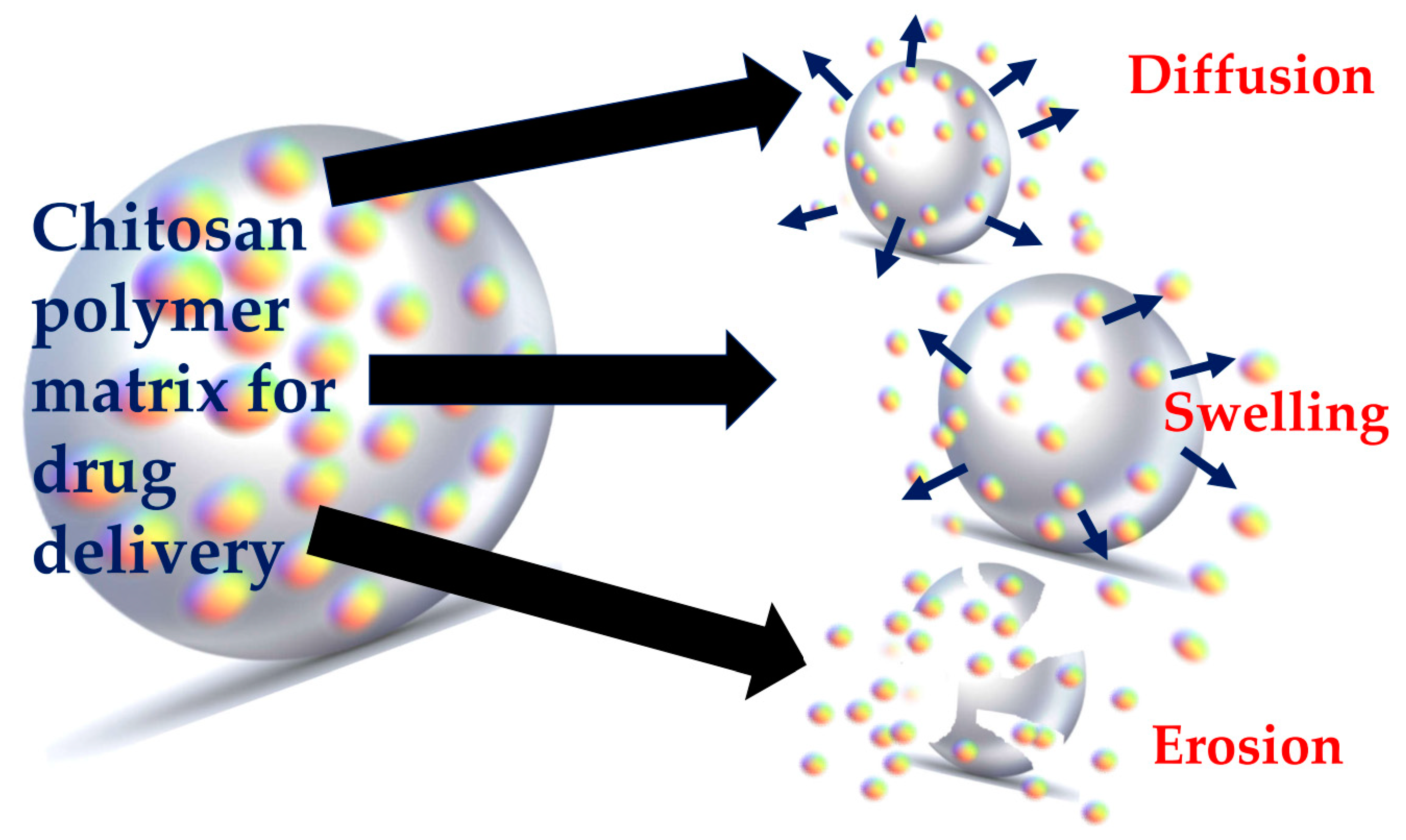
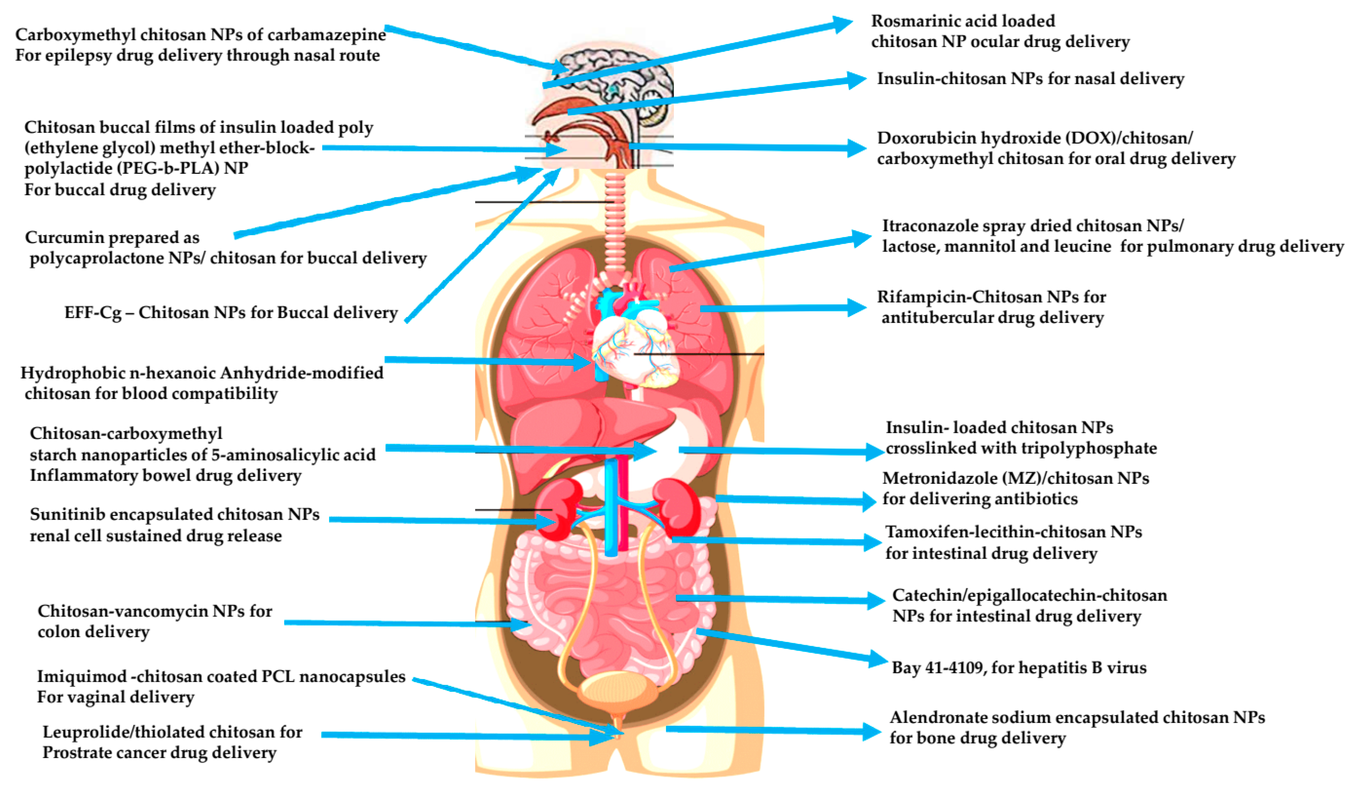
Table 1.
List of green synthesized chitin-based materials.
| Green Synthesized Chitin Based Material | Chitin/Chitosan/Composites/Chitin NF/Ch Nps | Green Synthesis Method | References |
|---|---|---|---|
| Chitin | Chitin recovered from shrimp shells | NADES, DES and citric acid-based microwave assisted method | [111] |
| Chitin | Chitin recovered from shrimp shells | NADES-based zero waste method | [115] |
| Chitosan | Chitin to chitosan | Glycerol-based | [116] |
| Ch NPs | Ch NPs/ cinnamaldehyde | Crosslinking with ecofriendly cinnamaldehyde | [117] |
| Chitin NFs | NFs from crab/shrimp/Squid pens | Mechanical grinding | [119,120,121,122] |
| Chitin NFs | NFs | Using ionic liquids instead of HFIP | [124,125,126,127,128] |
| Chitosan NFs | Ch NFs | Electrospinning | [130] |
| Chitosan NFs | Ch NFs | Freeze drying process | [132] |
| Chitosan NFs | NFs Extracted from crab/shrimp/squid pens | Graviola plant extract mediated recovery | [133] |
| Ag NPs/Chitosan | Green synthesized Ag NPs entrapped in chitosan | Aqueous extract of tomato plant | [134] |
| Chitosan based nanocomposite | Silver based chitosan bionanocomposites | synthesized using the stem extract of Saccharum officinarum | [135] |
| AuNPs/Ch composite | Au NPs in a chitosan matrix | Green route | [136] |
| Chitosan nanocomposite | Ag/montmorillonite (MMT)/chitosan bionanocomposites | UV irradiation | [137] |
| Chitosan nanocomposite | Ag/Ch/silica nanocomposites | Electrospinning | [138] |
| Chitosan nanocomposite | Ag-Ch nanocomposites | Using chitosan | [139] |
| Chitosan nanocomposite | Au NPs were stabilized into a chitosan matrix | Using chitosan | [140] |
| Chitosan nanocomposite | Ch-Fe2O3 nanocomposite | Facile one pot green route | [141] |
| Chitosan nanocomposite | CA/SA/Ch NPs nanocomposites | Using cinnamaldehyde | [142] |
| Chitosan nanocomposite | carboxymethyl chitosan-nanosilver | Microwave irradiation | [143] |
| Chitosan nanocomposite | Ch-modified nano-scale graphene oxide (Ch-NGO) hybrid nanosheets | Ultrasound | [144] |
| Chitosan nanocomposite | HS/Ch-Nanocomposite | Green route | [145] |
Publisher’s Note: MDPI stays neutral with regard to jurisdictional claims in published maps and institutional affiliations. |
© 2021 by the authors. Licensee MDPI, Basel, Switzerland. This article is an open access article distributed under the terms and conditions of the Creative Commons Attribution (CC BY) license (https://creativecommons.org/licenses/by/4.0/).
Share and Cite
MDPI and ACS Style
Sivanesan, I.; Gopal, J.; Muthu, M.; Shin, J.; Mari, S.; Oh, J. Green Synthesized Chitosan/Chitosan Nanoforms/Nanocomposites for Drug Delivery Applications. Polymers 2021, 13, 2256. https://doi.org/10.3390/polym13142256
AMA Style
Sivanesan I, Gopal J, Muthu M, Shin J, Mari S, Oh J. Green Synthesized Chitosan/Chitosan Nanoforms/Nanocomposites for Drug Delivery Applications. Polymers. 2021; 13(14):2256. https://doi.org/10.3390/polym13142256
Chicago/Turabian StyleSivanesan, Iyyakkannu, Judy Gopal, Manikandan Muthu, Juhyun Shin, Selvaraj Mari, and Jaewook Oh. 2021. "Green Synthesized Chitosan/Chitosan Nanoforms/Nanocomposites for Drug Delivery Applications" Polymers 13, no. 14: 2256. https://doi.org/10.3390/polym13142256
Note that from the first issue of 2016, this journal uses article numbers instead of page numbers. See further details here.