2016 | Buch

Medical Imaging in Clinical Applications

Algorithmic and Computer-Based Approaches

insite
SUCHEN

Über dieses Buch

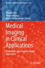
This volume comprises of 21 selected chapters, including two overview chapters devoted to abdominal imaging in clinical applications supported computer aided diagnosis approaches as well as different techniques for solving the pectoral muscle extraction problem in the preprocessing part of the CAD systems for detecting breast cancer in its early stage using digital mammograms. The aim of this book is to stimulate further research in medical imaging applications based algorithmic and computer based approaches and utilize them in real-world clinical applications. The book is divided into four parts, Part-I: Clinical Applications of Medical Imaging, Part-II: Classification and clustering, Part-III: Computer Aided Diagnosis (CAD) Tools and Case Studies and Part-IV: Bio-inspiring based Computer Aided diagnosis techniques.
Inhaltsverzeichnis

Frontmatter

Clinical Applications of Medical Imaging

Frontmatter
Abdominal Imaging in Clinical Applications: Computer Aided Diagnosis Approaches
Abstract
Computer aided diagnosis (CAD) is considered one of the main research subjects in medical image processing and diagnostic radiology. The development of CAD systems would provide anatomical knowledge coupled with image processing procedures to improve diagnosis/healthcare. For accurate diagnosis and treatment, researchers are interested with clinical image analysis. Since, the abdominal organs are characterized by complexity and high inconsistency. Thus, the identification of distinct algorithms to model the organs and abnormalities it is vital for understanding anatomy and disease. Moreover, a survey on CAD based abdominal image enhancement, segmentation, classification and fusion is included. Finally, challenging topics are addressed to explore new fields for development.
Amira S. Ashour, Nilanjan Dey, Waleed S. Mohamed
An Overview of Pectoral Muscle Extraction Algorithms Applied to Digital Mammograms
Abstract
Substantial numbers of patients are reaching to a progressive breast cancer stage due to increase in the false negatives coming out of cumbersome and tedious job of continuously observing the mammograms in fatigue. Hence, the early detection of cancer with more accuracy is highly expected to reduce the death rate. Computer Aided Detection (CADe) can help radiologists in providing a second opinion increasing the overall accuracy of detection. Pectoral muscle is a predominant density area in most mammograms and may bias the results. Its extraction can increase accuracy and efficiency of cancer detection. This work is intended to provide the researchers a systematic and comprehensive overview of different techniques of pectoral muscle extraction which are categorized into groups based on intensity, region, gradient, transform, probability and polynomial, active contour, graph theory, and soft computing approaches. The performance of all these methods is summarized in tabular form for comparison purpose. The accuracy, efficiency and computational complexities of some selected methods are discussed in view of deciding a best approach in each of the categories.
Suhas Sapate, Sanjay Talbar
Magnetic Resonance Brain Imaging Segmentation Based on Cascaded Fractional-Order Darwinian Particle Swarm Optimization and Mean Shift Clustering
Abstract
A digital image can be partitioned into multiple segments, which is known as image segmentation. There are many challenging problems for making image segmentation. Therefore, medical image segmentation technique is required to develop an efficient, fast diagnosis system. In this paper, we proposed a segmentation framework that is based on Fractional-order Darwinian Particle Swarm Optimization (FODPSO) and Mean Shift (MS) techniques. In pre-processing phase, MRI image is filtered, and the skull stripping is removed. In segmentation phase, the output of FODPSO is used as input to MS. Finally, we make a validation to the segmented image. The proposed system is compared with some segmentation techniques by using three standard datasets of MRI brain. For the first dataset, proposed system was achieved 99.45 % accuracy, whereas the DPSO was achieved 97.08 % accuracy. For the second dataset, the accuracy of the proposed system is 99.67 %, whereas the accuracy of DPSO is 97.08 %. Proposed system improves the accuracy of image segmentation of brain MRI as shown in the experimental results.
Hala Ali, Mohammed Elmogy, Eman El-Daydamony, Ahmed Atwan, Hassan Soliman
3D Brain Tumor Segmentation Based on Hybrid Clustering Techniques Using Multi-views of MRI
Abstract
3D medical images segmentation is a very difficult task. It may not be accurate and takes extreme time. In this chapter, we accurately detect the brain tumor from 3D MRI image with less time. The 3D image consists of multiple 2D slices. Segmenting each 2D slice by using the 2D techniques gives more accuracy rather than segmenting the whole 3D image. The integration between K-Means and Particle Swarm Optimization was proposed to segment the 2D MRI slices of the 3D MRI image. We solved the time problem of segmenting all 2D slices of the 3D image. The experiments emphasized the effectiveness of our proposed system in segmenting the 2D and 3D medical images. It achieved 100 % accuracy for the tested 3D dataset and 98.75 % average accuracy for all tested 2D and 3D datasets. The proposed integration reduced time by a mean of 10 min for the tested 2D and 3D datasets.
Eman A. Abdel Maksoud, Mohammed Elmogy

Classification and Clustering

Frontmatter
Comparison of CAD Systems for Three Class Breast Tissue Density Classification Using Mammographic Images
Abstract
It is well known that the changes in the breast tissue density are strongly correlated with the risk of breast cancer development and therefore classifying the breast tissue density as fatty, fatty–glandular and dense–glandular has become clinically significant. It is believed that the changes in the tissue density can be captured by computing the texture descriptors. Accordingly, the present work has been carried out with an aim to explore the potential of Laws’ mask texture descriptors for description of variations in breast tissue density using mammographic images. The work has been carried out on the 322 mammograms taken from the MIAS dataset. The dataset consists of 106 fatty, 104 fatty–glandular and 112 dense–glandular images. The ROIs of size 200 × 200 pixels are extracted from the center of the breast tissue, ignoring the pectoral muscle. For the design of a computer aided diagnostic system for three class breast tissue density classification, Laws’ texture descriptors have been computed using Laws’ masks of different resolutions. Five statistical features i.e. mean, skewness, standard deviation, entropy and kurtosis have been computed from all the Laws’ texture energy images generated from each ROI. The feature space dimensionality reduction has been carried out by using principal component analysis. For the classification task kNN, PNN and SVM classifiers have been used. After carrying out exhaustive experimentation, it has been observed that PCA–SVM based CAD system design yields the highest overall classification accuracy of 87.5 %, with individual class accuracy values of 84.9, 84.6 and 92.8 % for fatty, fatty–glandular and dense–glandular image classes respectively. These results indicate the usefulness of the proposed CAD system for breast tissue density classification.
Kriti, Jitendra Virmani
Ensemble Classifiers Construction Using Diversity Measures and Random Subspace Algorithm Combination: Application to Glaucoma Diagnosis
Abstract
Glaucoma is a group of eye diseases caused due to excessively high intraocular pressure within the eye. Ensemble classifier construction has attracted increasing interest in the field of pattern recognition and machine learning. Diversity among the classifiers is important factor for each ensemble to be successful. The most widely generation techniques are focused on incorporating the concept of diversity by using different features or training subsets. a classifier selection process becomes an important issue of multiple classifier system by choosing the optimal subset of members that maximizes the performance. The main goal of this study is to develop novel automated glaucoma diagnosis system which analyze and classify retinal images using a novel classification approach based on feature selection and static classifier selection schemes. Experimental results based on RIM-ONE dataset are very encouraging.
Soraya Cheriguene, Nabiha Azizi, Nilanjan Dey
Motor Imagery Classification Based on Variable Precision Multigranulation Rough Set and Game Theoretic Rough Set
Abstract
In this work classification of motor imagery BCI based on Variable Precision Multigranulation Rough Set and Game theoretic Rough Set is proposed. The efficient classification of motor imagery movements of patients can lead to accurate design of Brain Computer Interface (BCI). Data set are collected from BCI Competition III dataset 3a and BCI competition IV data set I. During acquisition there are several noises that affect classification of Electroencephalogram (EEG) Signal, so pre-processing is carried out with Chebyshev type1 filter between 4–40 Hz in order to remove the noises that may exist in signal. The Daubechies wavelet is used for extraction of features from EEG Signal. Variable Precision Multigranulation Rough Set is applied for classification of EEG Signal. Game theoretic Rough set is applied to determine best combination of \( \alpha \) and \( \beta \) are based on accuracy of Variable Precision Multigranulation Rough Set. An experimental result depicts higher accuracy with Variable Precision Multigranulation Rough Set and Game Theoretic rough set compared to existing technique.
K. Renuga Devi, H. Hannah Inbarani
Computer Aided Diagnosis System for Mammogram Abnormality
Abstract
In this chapter, computer aided detection and diagnosis for Breast tumor detection and classification in Mammography is proposed. The proposed system based on four phases. In the first phase, mammogram image is segmented to sub images with size 64 × 64 pixel then high intensity value of pixel is defined in this sub image and specifies this intensity as a seed point to region growing (RG) algorithm which used to specify the ROI. In the second phase, texture features were extracted using gray-level co-occurrence matrix (GLCM) and combined with shape features to characterize region of interest (ROI) to normal, benign or malignant. In the third phase, malignant ROIs are diagnosed and specified to aided doctor for decision taking. Finally, different methods for evaluating classifier are used using confusion matrix, kappa coefficient and response receiver operating characteristic curve (ROC). The effectiveness of the proposed system was measured using 322 mammogram images from the mammographic image analysis society (MIAS) database. From experimental results show that, the accuracy obtained from ROC curve analysis is AUC 94 % with standard error 0.11. The experimental results shows that the proposed system can accurately segment the breast region in a large range of digitized mammograms.
Ahmed M. Anter, Aboul Ella Hassenian
Automated Segmentation and Classification of Hepatocellular Carcinoma Using Fuzzy C-Means and SVM
Abstract
For successful classification of Hepatocellular Carcinoma (HCC) in ultrasound (US) images, effective preprocessing steps are highly desirable. Most of Computer Aided Diagnostic (CAD) systems miss the most important steps of image preprocessing and image segmentation. In such a framework, only some texture features, which are obtained directly from the images or ROIs, are used as inputs of classifiers. Image preprocessing and segmentation of US images are useful for better judgment of normal and cancerous cases. Although, there are many studies on the classification of medical images, the fully automatic classification is still a difficult task. In this work, we propose an automated classification of US liver tumors using SVM with the aid of Fuzzy c-means (FCM) and level set method. A large number of features were extracted by using statistical, textual, and histogram-based features to discriminate the HCC maximally by developing an SVM classification system. SVMs work on maximizing the margin between the separating hyperplane and the data to minimize upper bound of the generalization error. The proposed Fuzzy C-SVM based system is compared with the K-Nearest Neighbor (KNN) based approach. Experimental results demonstrated that the Fuzzy C-SVM based system greatly outperforms KNN-based approach.
Mai R. Ibraheem, Mohammed Elmogy

Computer Aided Diagnosis (CAD) Tools and Case Studies

Frontmatter
Ultrasound Based Three Dimensional Computer Aided Diagnosis (CAD) Tool for the Diagnosis of Anterior Talofibular Ligament
Abstract
Ultrasound imaging is an investigative tool to imagine the internal organisms of human beings. Ultrasound imaging has benefits such as low cost, portability, non-ionization and real time nature. However, ultrasound imaging has limitations also like homogenous intensity regions, homogeneous textures and low contrast regions. To overcome these investigated problems, this research is developed a Computer Aided Diagnosis (CAD) system that helped in the achievement of efficient segmentation and three dimensional reconstruction of anterior talofibular ligament to enhance the diagnosis. The developed CAD system would provide the information about the input dataset, segmented results and statistical analysis of injured anterior talofibular ligament. The analysis based on the obtained results indicates the improved performance of the developed CAD system with more than 94 % accurate results. In addition, this research opens new research dimensions for efficient musculoskeletal ultrasound modelling that makes it useful in clinical settings with accurate and cost effective diagnosis of anterior talofibular ligament injuries.
Vedpal Singh, Irraivan Elamvazuthi, Varun Jeoti, John George, Norashikin Yahya, Dileep Kumar
Electroanatomical Mapping Systems. An Epochal Change in Cardiac Electrophysiology
Abstract
In the last two decades new mathematical and computational models and systems have been applied to the clinical cardiology, which continue to be developed particularly to quantify and simplify anatomy, physio-pathological mechanisms and treatment of many patients with cardiac arrhythmias. The Authors report our large experience on electroanatomical mapping systems and techniques that are currently used to quantify and analyze both anatomy and electrophysiology of the heart. In the last 15 years the Authors have performed more than 15,000 invasive catheter ablation procedures using different non-fluoroscopic three-dimensional (3D) electroanatomical mapping and ablation systems (CARTO, Ensite) to safely and accurately treat many patients with different cardiac arrhythmias particularly those with atrial fibrillation with a median age of 60 years (IQR, 55-64). The Authors have also developed and proposed for the first time a new robotic magnetic system to map and ablate cardiac arrhythmias without use of fluoroscopy (Stereotaxis) in >500 patients. Very recently, epicardial mapping and ablation by electroanatomical systems have been successfully performed to treat Brugada syndrome at risk of sudden death in a series of patients with a median age of 39 years (IQR, 30-42). Our experience indicates that electroanatomic mapping systems integrate several important functionalities. (1) Non-fluoroscopic localization of electrophysiological catheters in three-dimensional space; (2) Analysis and 3D display of cardiac activation sequences computed from local or calculated electrograms, and 3D display of electrogram voltage; (3) Integration of ‘electroanatomic’ data with non-invasive images of the heart, such as computed tomography or magnetic resonance images. The widespread use of such 3D systems is associated with higher success rates, shorter fluoroscopy and procedure times, and accurate visualization of complex cardiac and extra-cardiac anatomical structures needing to be protected during the procedure.
Carlo Pappone, Carmine Garzillo, Simonetta Crisà, Vincenzo Santinelli
Image Quality Assessment: A Case Study on Ultrasound Images of Supraspinatus Tendon
Abstract
With the advancement of technologies on visual contents and the rate at which data is being captured, viewed, stored, and shared, the importance of assessment of quality of the contents has major importance. Image quality assessment has remained a topic of research over the last several decades for optical as well as medical images. User oriented image quality assessment is playing a key role in the assessment of visual contents. Studies are conducted to imitate the accuracy of human visual system for assessment of images. This chapter details about the approaches for development of methods for image quality assessment followed by brief introduction on existing image quality assessment methods. Later in the chapter, challenges for validation and development of image quality assessment metric for medical images are briefly discussed followed by the case study for assessment of ultrasound images of supraspinatus tendon.
Rishu Gupta, I. Elamvazuthi, J. George
Development of EOG and EMG-Based Multimodal Assistive Systems
Abstract
This study discusses a human-computer interface (HCI)- based novel approach for designing a computer-aided control and communication system using electrooculogram (EOG) and electromyogram (EMG) signals for people with severe hindrance to motor activities and communication. The EOG and EMG signals were attributed to eye movements and voluntary eye blinks, respectively. The acquired signals were processed and classified in a MATLAB-based graphical user interface (GUI) to detect different eye movements. A couple of Hall-effect sensors were conditioned to be used concurrently with multidirectional eye movements or voluntary eye blinks to generate multipurpose serial commands to control the movement of a robotic vehicle (representative assistive aid) and communications support systems. The user details were registered and the system operability was monitored in the same GUI. Due to multitasking and ease of use of the proposed device, the quality of life of the incapacitated individuals can be improved with greater independence.
Biswajeet Champaty, D. N. Tibarewala, Biswajit Mohapatra, Kunal Pal
Theory of Parallel MRI and Cartesian SENSE Reconstruction: Highlight
Abstract
Magnetic resonance imaging (MRI) is a well-known medical imaging technique, that exclusively uses the response of the hydrogen nucleus which is abundant in the human body. In recent years, parallel MRI techniques have been developed to accelerate image acquisition. A notable development in parallel MRI was the introduction of SMASH by Sodicksen and Manning. Since then, great progress in the development and improvement of parallel imaging reconstruction methods has taken place. The Sensitivity Encoding (SENSE) proposed by Preussmann and Weiger is the most widely used image-domain parallel MR image reconstruction technique. SENSE uses an initial estimate of the coil sensitivity in combination with an SNR optimized noise inversion to obtain the final reconstructed image. This chapter starts with a brief history of the parallel imaging, discusses the estimation of sensitivity and SENSE reconstruction.
Joseph Suresh Paul, Raji Susan Mathew, M. S. Renjith
Applications of Bio-molecular Databases in Bioinformatics
Abstract
Discovery of genome as well as protein sequencing aroused interest in bioinformatics and propelled the necessity to create databases of biological sequences. These data are processed in useful knowledge/information by data mining before storing into databases. This book chapter aims to present a detailed overview of different types of database called as primary, secondary and composite databases along with many specialized biological databases for RNA molecules, protein-protein interaction, genome information, metabolic pathways, phylogenetic information etc. Attempt has also been made to focus on drawbacks of present biological databases. Moreover, this book chapter provides an elaborate and illustrative discussion about various bioinformatics tools used for gene prediction, sequence analysis, phylogenetic analysis, protein structure as well as function prediction, molecular interactions prediction for several purposes including discovery of new gene as well as conserved regions in protein families, estimation of evolutionary relationships among organisms, 3D structure prediction of drug targets for exploring the mechanism as well as new drug discovery and protein-protein interactions for exploring the signaling pathways.
Archana Kumari, Swarna Kanchan, Rajeshwar P. Sinha, Minu Kesheri
Statistical Methods for Managing Missing Data: Application to Medical Diagnosis
Abstract
This paper presents a work consisting in realizing a decision support system based on the technique of case-base reasoning and dedicated to the diagnosis of a very dangerous pulmonary pathology: lung cancer. The system is realized for the oncology department of Ibn roch hospital of Annaba (Algeria) and will help young oncologist physicians in their activity by providing them with the experience of experts in the same domain. The principle issue in this work is the missing data in the system memory relating to the patient’s state. Indeed, missing values prevent the achievement of the diagnosis process. The problem is treated by proposing two statistical approaches in addition to re-evaluate in this new domain some ones which have been already proposed and evaluated in a previously domain. The validation is made on a base of 40 real cases collected from the archive of oncology department. Cases are collected as paper documents.
Souad Guessoum, Hadjer Zaayout, Nabiha Azizi, Nadjet Dendani, Hayet Djellali
Advances in Soft Computing Approaches for Gene Prediction: A Bioinformatics Approach
Abstract
The flooding of gene sequencing projects lead to the deposition of large amount of genomic data in public databases. These public databases contribute in genome annotation which helps in discovering the new genes and finding their function. Due to lack of genome annotation and high-throughput experimental approaches, computational gene prediction has always been one of the challenging areas for bioinformatics/computational biology scientists. Gene finding is more difficult in eukaryotes as compared to prokaryotes due to presence of introns. Gene prediction in very crucial especially for disease identification in human, which will help a lot in bio-medical research. Ab intio gene prediction is a difficult method which uses signal and content sensors to make predictions while homology based method makes use of homology with known genes. This chapter describes various gene structure prediction programmes which are based on individual/hybrid soft computing approaches. Soft computing approaches include Genetic algorithm, Hidden Markov Model, Fast Fourier Transformation, Support vector Machine, Dynamic programming and Artificial Neural Network. Hybrid soft computing approaches combine the results of several soft computing programs to deliver better accuracy than individual single soft computing approaches. Moreover, various parameters for measuring the accuracy of gene prediction programs will also be discussed.
Minu Kesheri, Rajeshwar P. Sinha, Swarna Kanchan

Bio-inspiring Based Computer Aided Diagnosis Techniques

Frontmatter
Artificial Bee Colony Based Segmentation for CT Liver Images
Abstract
The objective of this paper is to evaluate an approach for CT liver image segmentation, to separate the liver, and segment it into a set of regions of interest (ROIs). The automated segmentation of liver is an essential phase in all liver diagnosis systems for different types of medical images. In this paper, the artificial bee colony optimization algorithm (ABC) aides to segment the whole liver. It is implemented as a clustering technique to achieve this mission. ABC calculates the centroid values of image clusters in CT images. Using the least distance between every pixel value and different centroids will result in a binary image for each cluster. Applying some morphological operations on every binary clustered image can help to remove small and thin objects. These objects represent parts of flesh tissues adjacent to the liver, sharp edges of other organs and tiny lesions spots inside the liver. This is followed by filling the large regions in each cluster binary image. Summation of the clusters’ binary images results in a reasonable image of segmented liver. Then, the segmented image of liver is enhanced using simple region growing technique (RG). Finally, one of ABC algorithm or watershed is applied once to extract the lesioned regions in the liver, which can be used by any classifier to determine the type of lesion. A set of 38 images, taken in pre-contrast phase, was used to segment the liver and test the proposed approach. Testing the results is handled using similarity index to validate the success of the approach. The experimental results showed that the overall accuracy offered by the proposed approach, results in 93.73 % accuracy.
Abdalla Mostafa, Ahmed Fouad, Mohamed Abd Elfattah, Aboul Ella Hassanien, Hesham Hefny
Nature Inspired Optimization Algorithms for CT Liver Segmentation
Abstract
Nature inspired optimization algorithms have gained popularity in the last two decades due to their efficiency and flexibility when they applied to solve global optimization problems. These algorithms are inspired from the biological behavior by swarms of birds, fish and bees. In this chapter, we give an overview of some of nature inspired optimization algorithms such as Artificial Bee Colony (ABC), Cuckoo Search (CS), Social Spider Optimization (SSO) and Grey Wolf Optimization (GWO). Also, we present the usage of ABC and GWO algorithms for CT liver segmentation. The experimental results of the two selected algorithms show that the two algorithms are powerful and can obtain good results when applied to segment medical images.
Ahmed Fouad Ali, Abdalla Mostafa, Gehad Ismail Sayed, Mohamed Abd Elfattah, Aboul Ella Hassanien
Optimized Multi Threshold Brain Tumor Image Segmentation Using Two Dimensional Minimum Cross Entropy Based on Co-occurrence Matrix
Abstract
The present chapter proposes an automatic segmentation method that performs multilevel image thresholding by using the spatial information encoded in the gray level co-occurrence matrix (GLCM). The 2D local cross entropy approach that has been designed by extending the one dimensional (1-D) cross entropy thresholding method to a two dimensional (2D) one using the GLCM, serves as a fitness function. The use of conventional exhaustive search based implementations for multilevel thresholding are computationally expensive. Under such conditions evolutionary algorithm like particle swarm optimization (PSO) has been used. The effectiveness of this method was tested on brain tumor MR images and comparison was done with seven other level set based segmentation algorithms, using three different measures (1) Jaccard, (2) Dice and (3) Root mean square error (RMSE). The results demonstrate that average metric values were equal to 0.881902, 0.936394 and 0.070123 for proposed approach, which were significantly better than existing techniques.
Taranjit Kaur, Barjinder Singh Saini, Savita Gupta
Bio-inspired Swarm Techniques for Thermogram Breast Cancer Detection
Abstract
Bio-inspired swarm techniques are a well-established paradigm with current systems having many of the characteristics of biological computers and capable of performing a variety of tasks that are difficult to do using conventional techniques. These techniques involving the study of collective behavior in decentralized systems. Such systems are made up by a population of simple agents interacting locally with one other and with their environment. The system is initialized with a population of individuals (i.e., potential solutions). These individuals are then manipulated over many iteration steps by mimicking the social behavior of insects or animals, in an effort to find the optima in the problem space. A potential solution simplifies through the search space by modifying itself according to its past experience and its relationship with other individuals in the population and the environment. Problems like finding and storing foods, selecting and picking up materials for future usage require a detailed planning, and are solved by insect colonies without any kind of supervisor or controller. Since 1990, several collective behavior (like social insects, bird flocking) inspired algorithms have been proposed. The objective of this article is to present to the swarms and biomedical engineering research communities some of the state-of-the-art in swarms applications to biomedical engineering and motivate research in new trend-setting directions. In this article, we present four swarms algorithms including Particle swarm optimization (PSO), Grey Wolf Optimizer (GWO), Moth Flame Optimization (MFO), and Firefly Algorithm Optimization (FA) and how these techniques could be successfully employed to tackle segmentation biomedical imaging problem. An application of thermography breast cancer imaging has been chosen and the scheme have been applied to see their ability and accuracy to classify the breast cancer images into two outcomes: normal or non-normal.
Gehad Ismail Sayed, Mona Soliman, Aboul Ella Hassanien
Metadaten
Titel
Medical Imaging in Clinical Applications
herausgegeben von
Nilanjan Dey
Vikrant Bhateja
Aboul Ella Hassanien
Copyright-Jahr
2016
Electronic ISBN
978-3-319-33793-7
Print ISBN
978-3-319-33791-3
DOI
https://doi.org/10.1007/978-3-319-33793-7