2012 | Buch

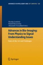
Advances in Bio-Imaging: From Physics to Signal Understanding Issues

State-of-the-Art and Challenges

herausgegeben von: Nicolas Loménie, Daniel Racoceanu, Alexandre Gouaillard

Verlag: Springer Berlin Heidelberg

Buchreihe : Advances in Intelligent and Soft Computing

insite
SUCHEN

Über dieses Buch

Advances in Imaging Devices and Image processing stem from cross-fertilization between many fields of research such as Chemistry, Physics, Mathematics and Computer Sciences.

This BioImaging Community feel the urge to integrate more intensively its various results, discoveries and innovation into ready to use tools that can address all the new exciting challenges that Life Scientists (Biologists, Medical doctors, ...) keep providing, almost on a daily basis.

Devising innovative chemical probes, for example, is an archetypal goal in which image quality improvement must be driven by the physics of acquisition, the image processing and analysis algorithms and the chemical skills in order to design an optimal bioprobe.

This book offers an overview of the current advances in many research fields related to bioimaging and highlights the current limitations that would need to be addressed in the next decade to design fully integrated BioImaging Device.
Inhaltsverzeichnis

Frontmatter

Biology

Frontmatter
Intravital Multiphoton Imaging of Immune Cells
Abstract
Intravital multiphoton microscopy (MP-IVM) is a powerful imaging approach that allows direct visualization of cells within their native environment in real time. Multiphoton imaging of immune cells has been performed in different tissues, and these studies have revealed intriguing insights into the unique migratory and interactive behavior of immune cells in the steady-state and during disease conditions. Here we provide an overview of a MP-IVM model of the mouse ear skin, as well as the benefits, limitations and pitfalls of this approach. We also discuss the prospects of intravital imaging in the areas of image analysis, data management and standardization.
Jo Keeble, Chi Ching Goh, Yilin Wang, Wolfgang Weninger, Lai Guan Ng
Functional MRI of Neural Plasticity and Drug Effect in the Brain
Abstract
Recent advances in magnetic resonance imaging (MRI) have opened up new perspectives for understanding brain function and its plasticity after damage or even in the process of learning and memory. Using functional MRI (fMRI), reorganization of the cortical representation can be detected after the peripheral nerves deafferentation or digit amputation. To detect the more trivial changes during learning and memory, we established two techniques. One is to use manganese as a contrast agent to detect minute reorganization of hippocampal mossy fiber after training with hidden platform in Morris water maze. The other technique detects the synchrony in fMRI signal among neural areas that represents functional connectivity. We demonstrated the spatial memory network can be visualized in water maze trained animal. Furthermore, we showed that synchrony rather than activity in the brain can be modulated by receptor targeted pharmaceuticals, which indicate a different drug mechanism. The translation of these methods will facilitate our understanding of brain plasticity, early diagnosis of dementia, and evaluation of drug efficacy.
Kai-Hsiang Chuang, Fatima A. Nasrallah
Automated Identification and Analysis of Visual Micro-experiments on Cellular Microarray
Abstract
This paper is an overview of the computer-based tools I designed at Institut Pasteur Korea in order to analyse a large quantity of microscopy cell based experiments. This development consisted in designing algorithms and software for automatically localize, identify and analyze cells on spots of cellular microarray at high resolution. We believe the applications of this work are numerous from genome wide loss of function screens to drug target deconvolution assays and diagnostic.
Auguste Genovesio

Physics and Chemistry

Frontmatter
Persistent Luminescence Nanoparticles for Bioimaging
Abstract
Optical imaging is a rapidly developing field of research aimed at noninvasive monitoring of disease progression, evaluating the effects and pharmacokinetic of a drug, or identifying pathological biomarkers . To this end, it requires the development of targeting and highly specific contrast agents . In fluorescence imaging, an external light of appropriate wavelength is used to excite the fluorescent molecule, followed almost immediately by the release of longer wavelength, lower energy light for imaging. Fluorescence is increasingly used for imaging and has provided remarkable results. However this technique presents several limitations, especially due to tissue autofluorescence under external illumination and weak tissue penetration of low wavelength excitation light. To overcome these drawbacks, we have developed an innovative technique using persistent luminescence nanoparticles (PLNP) for optical imaging in small animal. Such nanoparticles can be excited before systemic injection, and their biodistribution monitored in real-time for dozen of minutes without the need for any external illumination source. This review article will focus on recent works undertaken in our laboratory on the synthesis of PLNP, their surface modifications and applications for bioimaging.
Cyrille Richard, Thomas Maldiney, Quentin le Masne de Chermont, Johanne Seguin, Nicolas Wattier, Gabriel Courties, Florence Apparailly, Michel Bessodes, Daniel Scherman
Bioimaging Probes Development by DOFLA (Diversity Oriented Fluorescence Library Approach) for in Vitro, in Vivo and Clinical Applications
Abstract
Due to the remarkable development of bioimaging probes and equipment during the last decades, we are able to see a variety of biological systems with a resolution ranging from centimeters to subnanometers. Bioimaging is now an indispensable tool for basic research and clinical diagnosis. Particularly, the application of fluorescence in optical imaging has enabled us to investigate molecular events as well as the structures in living cells and tissues. Among the fluorescent molecules, low molecular weight chemicals have great potentials to be developed as highly specific and versatile bioimaging probes. Target-specific fluorescent probes have been developed conventionally by a hypothesis-driven approach in which fluorophores are conjugated to already developed molecules such as antibody, peptide or small molecule drug. However, the fluorescence-labeled macromolecules may not be used for the detection of intracellular molecules in living cells and tagging small molecule without affecting its property is relatively challenging. To overcome these problems, we have developed Diversity Oriented Fluorescence Library (DOFL) by exploring the diverse chemical space directly around fluorophores using combinatorial chemistry. By screening DOFL in various platforms such as in vitro, cell, tissue and whole organism, we have successfully developed bioimaging probes which interact specifically with the targets. In this article, we discuss how bioimaging contributes to the development of biomedical science, why the development of new bioimaging probes is necessary and what can be achieved by DOFL approach (DOFLA).
Seong-Wook Yun, Young-Tae Chang
In Vivo Electron Paramagnetic Resonance and Imaging in Biomedical Science
Abstract
Electron Paramagnetic Resonance (EPR) is aiming to non-destructively and non-invasively characterize and quantify paramagnetic species such as superoxyde, nitric oxide, dioxygen, ascorbyle radical, or metallo-enzymes. Due to the growing interest in these molecules in biomedical science, EPR and EPR imaging are important tools that are used to obtain information on paramagnetic species, molecules which can not be directly studied using other conventional spectroscopy or imaging methods used in biology.
Yves-Michel Frapart
Intra-Operative Ex-Situ and In-Situ Optical Biopsy Using Light-CT
Abstract
The field of digital pathology is evolving very quickly: there is an increasing implication of laboratories and imaging companies in the biomedical field of histology. For our part we propose a digital imaging tool based on optical interferometry with short coherence length light sources (OCT). This instrument provides, without preparation or contrast agents , virtual sections of biological tissues with a field of view (1cm 2) and spatial resolution (approximately 1 micron in 3-D) suitable for histology. We show the relevance of this approach by comparing our images obtained in minutes during surgery with histological sections obtained after a few days. In these studies we worked on fixed tissue, fresh tissue in vivo or ex vivo. Finally, an endoscopic probe based on the same principle has been validated ex-vivo for use inside the bodies of patients.
Martine Antoine, Brigitte Sigal, Fabrice Harms, Anne Latrive, Adriano Burcheri, Osnath Assayag, Bertrand de Poly, Sylvain Gigan, A. Claude Boccara
Photothermal Laser Material Interactions - From the Sledgehammer to Nano-GPS
Abstract
In this chapter we will summarize the main photothermal, photoacoustic and photomechanical effects of coupling a laser beam into a material from the absorption of the laser light to the deactivation of vibrationally and electronically excited states. Some methods to estimate the resulting temperature rise will be discussed and the resulting pressure increase in the heated area explained. The relaxation of both pressure and thermal transients will be explored and several methods described, such as pump-probe spectroscopy and imaging techniques, which can be used to investigate the dynamics of the relaxation pathways. We will explain how photothermal effects can manifest as optical effects. Finally, we will describe how we can harness photothermally induced optical changes to provide a new methodology in bioimaging involving indestructible 5-10 nm noble metal nanoparticles that can be observed using photothermal tracking microscopy for unprecedented periods of time in live cell imaging.
Jonathan Hobley, David Paramelle, Paul Free, David G. Fernig, Shinji Kajimoto, Sergey Gorelik
Dual Axes Confocal Microendoscope
Abstract
Biomedical research truly needs new advances in imaging. Existing modalities of in vivo imaging, such as magnetic resonance imaging or ultrasound, lack the spatiotemporal resolution required to image the fundamental building block of living tissue. By contrast, existing high-resolution techniques for imaging cells and their sub-cellular features are technologies that are best suited for in vitro experiments in tissue slices. Yet, the ability to make direct connections between human pathological symptoms/behavior and the underlying cells and molecules responsible for such behavior requires in vivo techniques that can image cellular constituents. Our group research aim is to develop novel high-resolution optical endoscopes to satisfy unmet needs in the clinical environment. These differ from medical endoscopes, which are generally larger and designed to image macroscopic abnormalities. Most importantly, this novel optical endoscopic imaging might suggest new approaches to disease diagnosis and treatment. This talk will be focused on the development a novel confocal imaging modality integrated with microelectromechanical systems (MEMS) technology and their imaging applications. Confocal microscopy is an attractive tool for three-dimensional (3-D) imaging due to its optical sectioning property. Conventional single-axis confocal (SAC) microscopes have a tradeoff between resolution, field of view, and objective lens size, since a high numerical aperture (NA) lens is needed for sufficient resolution, and a long focal length is needed for a large FOV and working distance. A dual-axes confocal (DAC) microscope architecture has been proposed utilizing two overlapping low NA beams, which effectively decouples these tradeoffs. The other important advantage is the ability to achieve a much superior optical sectioning compared to the SAC design. The microscopes are miniaturized into two form factors (5 mm and 10 mm diameter). The imaging demonstrations of the probes were on both ex vivo and in vivo from mice and human for cancer oncology and genetic research.
Wibool Piyawattanametha

Digitized Histopathology

Frontmatter
Ontology-Enhanced Vision System for New Microscopy Imaging Challenges
Abstract
Artificial intelligence and computer vision have long been separate fields basically because the data structures to work with and to reason about were rather distinct and non permeable. Ontology-driven systems may have the ability to build a bridge between these two fundamental topics involved in intelligent system design. We provide preliminary insights about this powerful synergy in the field of digitized pathology as a brand new topic in which, like currently for satellite imaging, the amount of raw data and high-level concepts to handle give no other choice but to innovate about the low-level image image processing machine and the knowledge modeling framework integration. Above all, the end-user who is most of the time naive about signal, image and algorithmic issues can thence play the key role in the design of such enhanced vision system.
Nicolas Lomenie, Daniel Racoceanu
Computational Approaches for the Processing of Cerebral Histological Images of Small Animals
Abstract
Histological sections of tissue have been studied for many decades and constitute one the most prevalent means of information on biological processes and functions for animals. With the introduction of digital images in medicine, image processing techniques derived from medical imaging were adapted to scanned histological sections in order to improve their visualization and analysis. More recently, the introduction of virtual microscopy yet increased the interest of analyzing histological sections on a computer screen and opened up a whole branch of biomedical image processing dedicated to the extraction of information contained in histological sections at very high magnification. In this work we present three novel approaches to study histological sections of the brain in small animals: 1) the alignment of histological sections to create a 3D image, 2) the processing of very large microscopy sections and 3) the correlation of histological sections with 3D in vivo images acquired on medical imaging devices.
Julien Dauguet

Image Analysis

Frontmatter
Liver Workbench: A Tool Suite for Liver and Liver Tumor Segmentation and Modeling
Abstract
Robust and efficient liver and tumor segmentation segmentation tools from CT images are important for clinical decision-making in liver treatment planning and response evaluation. In this work, we report recent advances in an ongoing project Liver Workbench which aims to provide a suite of tools for the segmentation segmentation, quantification and modeling of various objects in CT images such as the liver, its vessels and tumors. Firstly, a liver segmentation segmentation approach is described. It registers a liver mesh model model to actual image features by adopting noise-insensitive flipping-free mesh deformations. Next, a propagation learning approach is incorporated into a semi-automatic classification method for robust segmentation segmentation of liver tumors based on liver ROI obtained. Finally, an unbiased probabilistic liver atlas construction technique is adopted to embody the shape and intensity variation to constrain liver segmentation segmentation. We also report preliminary experimental results.
Jiayin Zhou, Wei Xiong, Feng Ding, Weimin Huang, Tian Qi, Zhimin Wang, Thiha Oo, Sudhakar Kundapur Venkatesh
A Bag-of-Words Model for Cellular Image Segmentation
Abstract
Cellular segmentation in microscopy images is an important step in modern biological research. Microscopy image segmentation is known to be a difficult problem, as illustrated in the paper, in many scenarios the microscopic images become a real challenge for existing methods to accurately segment these cellular objects of interest. In this paper we propose a learning based approach using a bag-of-words model and dedicated feature design to deal with this problem. By introducing the recent machine learning and computer vision techniques including sparse coding, superpixel representation, our approach is shown to achieve good performance in practice.
Li Cheng, Ning Ye, Weimiao Yu, Andre Cheah
Knowledge Based and Statistical Based Approaches in Biomedical Image Analysis
Abstract
Biomedical Imaging has grown significantly for the past twenty years, as it is considered as a unique method for visualizing biological processes within living organisms in a non-invasive manner. Although works in biomedical image analysis rely on underlying biological problems, scientists are just beginning to embrace the idea that these works will benefit from multidisciplinary interactions. Moreover, within the computer vision community, time has come for a more holistic and integrated approach in order to articulate statistical/machine learning and knowledge-based approaches. In this paper we present studies based on these two classic approaches and show how their complementarity may benefit biomedical imaging.
Florence Cloppet, Thomas Hurtut
Backmatter
Metadaten
Titel
Advances in Bio-Imaging: From Physics to Signal Understanding Issues
herausgegeben von
Nicolas Loménie
Daniel Racoceanu
Alexandre Gouaillard
Copyright-Jahr
2012
Verlag
Springer Berlin Heidelberg
Electronic ISBN
978-3-642-25547-2
Print ISBN
978-3-642-25546-5
DOI
https://doi.org/10.1007/978-3-642-25547-2